Do Animal Cells Have flagella: How, Why And Insights

Anatomy and physiology of animals A sperm 1

Animal cells are fascinating structures that make up the tissues and organs of animals. They are the building blocks of life, carrying out various functions necessary for an organism’s survival. One intriguing feature found in some animal cells is the presence of flagella. Flagella are whip-like structures that enable cells to move and propel themselves through their environment. In this article, we will explore the question: do animal cells have flagella? We will delve into the different types of animal cells, their structures, and whether or not flagella are a common feature. So, let’s dive in and unravel the mysteries of animal cell biology!

Key Takeaways

Anatomy and physiology of animals A sperm 1
Wikimedia
  • Animal cells can have flagella, which are whip-like structures that help with cell movement.
  • Flagella are composed of microtubules and are found in certain types of animal cells, such as sperm cells.
  • The presence of flagella in animal cells allows for various biological processes, including cell locomotion and the movement of fluids within the body.

Do Animal Cells Have Flagella and Cilia?

Presence of Flagella in Animal Cells

download 1
Wikimedia

Flagella are whip-like appendages that are responsible for the movement of cells. While flagella are commonly found in bacteria and protists, their presence in animal cells is relatively rare. However, there are certain types of animal cells that do possess flagella.

One example of an animal cell that has flagella is the sperm cell. Sperm cells are specialized for locomotion, and their flagella enable them to swim towards the egg for fertilization. The flagellum in sperm cells is a long, slender structure composed of microtubules. This arrangement of microtubules allows for the whip-like movement that propels the sperm forward.

It is important to note that not all animal cells have flagella. In fact, the presence of flagella in animal cells is limited to specific cell types that require motility. Most animal cells rely on other mechanisms for movement and do not possess flagella.

Presence of Cilia in Animal Cells

Cilia, on the other hand, are short, hair-like structures that are found on the surface of many animal cells. Unlike flagella, which are typically singular and longer, cilia are usually present in large numbers and are shorter in length.

Cilia play a crucial role in various cellular processes. They are involved in the movement of fluid across the surface of cells, which is important for processes such as the clearance of mucus in the respiratory tract. Cilia also aid in the movement of eggs through the fallopian tubes and the movement of embryos during early development.

Similar to flagella, the structure of cilia is composed of microtubules. These microtubules are arranged in a specific pattern that allows for coordinated movement. The beating motion of cilia is facilitated by the sliding of microtubules against each other, powered by ATP.

In summary, while flagella are relatively rare in animal cells, they can be found in specialized cell types such as sperm cells. On the other hand, cilia are more common in animal cells and play important roles in various physiological processes. Both flagella and cilia are composed of microtubules and are involved in cellular movement, but they differ in terms of their structure and function.

Where Do Animal Cells Have Flagella?

Flagella, which are whip-like appendages responsible for cell movement, can be found in various organisms, including animal cells. While not all animal cells possess flagella, they are present in specialized cells such as sperm cells and certain unicellular organisms. Let’s explore the location and function of flagella in animal cells.

Flagella found in specialized cells like sperm cells and certain unicellular organisms

In animal cells, flagella are most commonly found in specialized cells, such as sperm cells. Sperm cells rely on flagella for motility, allowing them to swim towards the egg for fertilization. The flagellum in sperm cells is a long, slender structure that propels the cell forward through its whip-like movement. This enables the sperm to navigate through the female reproductive tract in search of the egg.

Flagella are also present in certain unicellular organisms, such as some protozoa. These organisms use flagella for locomotion, allowing them to move through their environment in search of food or to escape from predators. The presence of flagella in these organisms is crucial for their survival and reproductive success.

Location and adherence of flagella to the cell surface

In animal cells, the flagellum is anchored to the cell surface by a structure called the basal body. The basal body serves as an anchor point for the extension and movement of the flagellum. It is located at the base of the flagellum and is connected to the cell membrane.

The flagellum itself is composed of microtubules, which are long, hollow tubes made up of proteins. These microtubules are arranged in a specific pattern, known as the 9+2 arrangement, which is characteristic of flagella and cilia. The 9+2 arrangement consists of nine pairs of microtubules surrounding a central pair, providing the flagellum with structural support and allowing it to move in a coordinated manner.

Basal body as an anchor point for flagellum extension and movement

The basal body plays a crucial role in the extension and movement of the flagellum. It acts as a scaffold for the assembly of the microtubules that make up the flagellum. The microtubules grow outwards from the basal body, extending the length of the flagellum. This extension allows the flagellum to generate the whip-like motion necessary for cell movement.

The movement of the flagellum is facilitated by the sliding of the microtubules against each other. This sliding is made possible by the action of specialized motor proteins, which use ATP (adenosine triphosphate) as a source of energy. As the microtubules slide, the flagellum bends and propels the cell forward or moves fluid past the cell.

In conclusion, while not all animal cells possess flagella, they are found in specialized cells like sperm cells and certain unicellular organisms. The flagellum is anchored to the cell surface by the basal body, which serves as an anchor point for flagellum extension and movement. Understanding the presence and function of flagella in animal cells provides valuable insights into cell motility and the diverse mechanisms that organisms employ for movement.

Do All Animal Cells Have Flagella?

Flagella are whip-like structures that protrude from the surface of certain cells and are responsible for their movement. While flagella are commonly found in many organisms, including bacteria, protists, and some plant cells, not all animal cells possess flagella. The presence of flagella in animal cells depends on the specific cell type and its functional requirements.

Not all animal cells possess flagella

Unlike plant cells, which commonly have flagella, animal cells exhibit a greater diversity in terms of their structural and functional characteristics. While some animal cells do possess flagella, many others lack this appendage. Instead, animal cells have evolved various other mechanisms for movement and locomotion.

Flagella presence depends on the specific cell type and its functional requirements

The presence or absence of flagella in animal cells is determined by the specific cell type and its functional requirements. For example, sperm cells, which are responsible for fertilization, possess flagella that enable them to swim towards the egg. The flagellum in sperm cells is a long, whip-like structure that propels the sperm forward, allowing it to reach its destination.

On the other hand, certain animal cells, such as red blood cells and muscle cells, do not possess flagella. Red blood cells, also known as erythrocytes, are responsible for transporting oxygen throughout the body. They lack a nucleus and other organelles, including flagella, to maximize their capacity for oxygen transport. Muscle cells, which are involved in contraction and movement, rely on a different mechanism for their function and do not require flagella.

Examples of non-flagellated animal cells like red blood cells and muscle cells

  1. Red blood cells: Red blood cells are specialized cells that lack a nucleus and organelles, including flagella. Their main function is to transport oxygen from the lungs to the body’s tissues and remove carbon dioxide. The absence of flagella allows red blood cells to adopt a biconcave shape, increasing their surface area and facilitating efficient gas exchange.

  2. Muscle cells: Muscle cells, also known as myocytes, are responsible for contraction and movement in the body. They contain specialized proteins, such as actin and myosin, which interact to generate force and movement. Muscle cells rely on the coordinated contraction of these proteins rather than flagella for their function.

In conclusion, not all animal cells possess flagella. The presence or absence of flagella in animal cells depends on the specific cell type and its functional requirements. While some animal cells, like sperm cells, have flagella for movement, others, such as red blood cells and muscle cells, rely on different mechanisms for their respective functions. This diversity in cellular structures and functions contributes to the complexity and adaptability of animal organisms.

Why Do Animal Cells Have Flagella?

Flagella are whip-like appendages found on the surface of certain animal cells. They play a crucial role in various cellular processes, including locomotion and fluid movement. Let’s explore the functions of flagella in animal cells and their significance in different biological contexts.

Functions of Flagella in Animal Cells

Flagella are primarily responsible for the movement of animal cells. They act as propellers, allowing cells to swim through fluid environments. This motility is particularly important for cells that need to navigate through complex environments, such as sperm cells. Sperm cells rely on their flagella to propel themselves towards the egg for fertilization.

Apart from locomotion, flagella also facilitate fluid movement within animal cells. They create a flow of fluid over the cell surface, aiding in the exchange of nutrients and waste products. This fluid movement is crucial for maintaining cellular homeostasis and ensuring proper functioning of the cell.

Role of Flagella in the Respiratory System and Other Cellular Processes

In addition to their role in locomotion and fluid movement, flagella are involved in specific cellular processes. In the respiratory system, for example, cilia (a specialized type of flagella) line the airways and help to move mucus and trapped particles out of the lungs. This mechanism, known as mucociliary clearance, is essential for maintaining healthy respiratory function and preventing infections.

Flagella also play a role in sensory perception. In certain animal cells, such as those found in the olfactory system, flagella act as sensory antennae, detecting and responding to chemical signals in the environment. This allows animals to sense and respond to their surroundings, aiding in survival and reproduction.

Exceptions of Non-Flagellated Animal Cells and Alternative Mechanisms for Movement

While flagella are common in many animal cells, it is important to note that not all animal cells possess flagella. Some cells rely on alternative mechanisms for movement. For example, muscle cells contract and relax to generate movement, while amoeboid cells use a process called amoeboid motion, which involves the extension and retraction of cellular protrusions called pseudopods.

Additionally, some animal cells may have structures similar to flagella, called cilia, which are shorter and more numerous. Cilia have a similar structure to flagella but serve different functions. They are involved in various processes, such as sensory perception, fluid movement, and the coordination of cellular activities.

In conclusion, flagella are important organelles found in certain animal cells. They enable locomotion, facilitate fluid movement, and play a role in various cellular processes. While not all animal cells possess flagella, alternative mechanisms for movement exist. Understanding the functions of flagella in animal cells contributes to our knowledge of cell biology and helps us appreciate the complexity of life at the microscopic level.

Do Plant Cells Have Flagella?

Plant cells, unlike animal cells, do not possess flagella. Flagella are whip-like appendages that protrude from the surface of certain cells and are responsible for their movement. While flagella are commonly found in animal cells, they are absent in plant cells. This absence can be attributed to the structural characteristics of plant cells and the alternative mechanisms they employ for movement and dispersal.

Differences between Animal and Plant Cells in Terms of Flagella Presence

One of the key distinctions between animal and plant cells lies in the presence or absence of flagella. Animal cells, particularly sperm cells, rely on flagella for motility and movement. These long, slender structures are composed of microtubules and are capable of propelling the cell forward through their rhythmic beating motion. The presence of flagella in animal cells allows for efficient locomotion and enables sperm cells to reach their target.

On the other hand, plant cells lack flagella. This is due to the unique structural characteristics of plant cells. Plant cells have a rigid cell wall that surrounds the cell membrane, providing support and protection. This cell wall acts as a barrier, preventing the formation of flagella on the cell surface. Additionally, plant cells have a large central vacuole and chloroplasts, which are essential for photosynthesis and maintaining the cell’s shape. These organelles further restrict the presence of flagella in plant cells.

Absence of Flagella in Plant Cells Due to Their Structural Characteristics

The structural characteristics of plant cells play a crucial role in the absence of flagella. As mentioned earlier, the rigid cell wall surrounding plant cells prevents the formation of flagella on their surface. The cell wall is composed of cellulose, a complex carbohydrate that provides strength and rigidity to the cell. Unlike animal cells, which have a flexible plasma membrane, plant cells have a more rigid cell wall that restricts the movement of appendages like flagella.

Furthermore, plant cells have a unique organelle called the plasmodesmata. Plasmodesmata are channels that connect adjacent plant cells, allowing for the exchange of nutrients, water, and signals. These channels traverse the cell wall and are lined with plasma membrane, which also hinders the formation of flagella on the cell surface.

Alternative Mechanisms for Movement and Dispersal in Plant Cells

Although plant cells lack flagella, they have evolved alternative mechanisms for movement and dispersal. One such mechanism is the use of cilia. Cilia are similar to flagella in structure but are shorter and more numerous. They can be found on certain specialized cells in plants, such as the cells lining the reproductive structures of flowers. Cilia in plant cells aid in the movement of fluids and facilitate the dispersal of pollen.

Another mechanism employed by plant cells for movement and dispersal is through the use of specialized structures called trichomes. Trichomes are hair-like projections that can be found on the surface of leaves, stems, and other plant parts. They serve various functions, including reducing water loss, providing protection against herbivores, and aiding in the dispersal of seeds. Trichomes can be either glandular or non-glandular and play a crucial role in the survival and reproduction of plants.

In conclusion, while animal cells possess flagella for movement and locomotion, plant cells do not have flagella due to their structural characteristics. The rigid cell wall and the presence of other specialized organelles in plant cells prevent the formation of flagella. However, plant cells have evolved alternative mechanisms such as cilia and trichomes for movement and dispersal. These adaptations allow plant cells to thrive and fulfill their biological functions without the need for flagella.

Why Do Plant Cells Not Need Flagella?

Plant cells are fascinating structures that differ in many ways from animal cells. One notable difference is the absence of flagella in plant cells. Let’s explore the reasons behind this intriguing phenomenon.

Lack of movement in plant cells due to the rigid cell wall

One of the primary reasons why plant cells do not require flagella is the presence of a rigid cell wall. Unlike animal cells, which have a flexible plasma membrane, plant cells are surrounded by a sturdy cell wall composed of cellulose. This cell wall provides structural support and protection to the plant cell, but it also restricts movement.

The rigid nature of the cell wall prevents plant cells from changing their shape and moving freely. While animal cells can alter their shape and move through their environment, plant cells are firmly anchored in place. Therefore, the need for flagella, which are whip-like appendages responsible for cell movement, is eliminated in plant cells.

Alternative mechanisms for communication and coordination in plant cells

Although plant cells lack flagella for movement, they have evolved alternative mechanisms for communication and coordination. Plant cells communicate through plasmodesmata, which are channels that connect neighboring cells. These channels allow for the exchange of nutrients, hormones, and signaling molecules between plant cells.

Additionally, plant cells rely on specialized structures called plasmodesmata to coordinate their activities. Plasmodesmata are microscopic channels that traverse the cell walls, connecting the cytoplasm of adjacent plant cells. Through these channels, plant cells can share resources, such as water and nutrients, and coordinate their growth and development.

Role of other organelles in plant cells, such as chloroplasts and central vacuoles

Plant cells possess unique organelles that fulfill essential functions and compensate for the absence of flagella. Two such organelles are chloroplasts and central vacuoles.

Chloroplasts are responsible for photosynthesis, the process by which plants convert sunlight into energy-rich molecules. These organelles contain chlorophyll, a pigment that captures light energy and initiates the production of glucose. By harnessing the power of photosynthesis, plant cells can generate the energy they need for growth and survival.

Central vacuoles, on the other hand, play a crucial role in maintaining cell turgor pressure and storing various substances. These large, fluid-filled sacs occupy a significant portion of the plant cell‘s volume. By regulating the osmotic balance within the cell, central vacuoles help maintain cell shape and provide support to the surrounding tissues.

In conclusion, plant cells do not possess flagella due to the rigid cell wall that restricts movement. However, they have evolved alternative mechanisms for communication and coordination, such as plasmodesmata. Additionally, plant cells rely on specialized organelles like chloroplasts and central vacuoles to fulfill essential functions. These adaptations allow plant cells to thrive and perform their unique roles in the plant kingdom.

Explanation on Structure of an Animal Cell – The Sperm Cell

The sperm cell is a specialized animal cell that plays a crucial role in sexual reproduction. Let’s explore the structure of a sperm cell and understand how it functions.

Introduction to the Sperm Cell as a Specialized Animal Cell

The sperm cell, also known as a spermatozoon, is the male reproductive cell. It is responsible for fertilizing the female egg during sexual reproduction. Sperm cells are produced in the testes of male animals, including humans.

Three Parts of the Sperm Cell: Head, Mid-Piece, and Tail

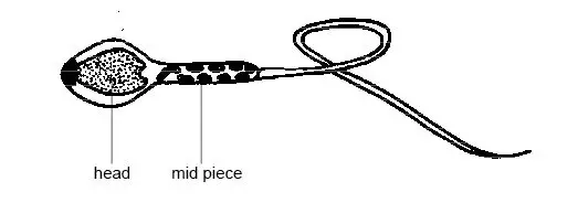
The structure of a sperm cell consists of three main parts: the head, mid-piece, and tail. Each part has its own unique function.

  1. Head: The head of a sperm cell contains the genetic material, including the DNA. It is covered by a cap-like structure called the acrosome, which contains enzymes that help the sperm penetrate the egg during fertilization.

  2. Mid-Piece: The mid-piece of a sperm cell is located just behind the head. It is packed with mitochondria, which provide the energy needed for the sperm’s movement. The mid-piece also contains a centriole, which plays a role in cell division.

  3. Tail: The tail, also known as the flagellum, is the longest part of the sperm cell. It is responsible for the sperm’s movement. The tail is made up of a whip-like structure called microtubules, which are arranged in a 9+2 pattern. This arrangement allows the flagellum to move in a wave-like motion, propelling the sperm forward.

Function of the Flagellum in Sperm Cell Movement

The flagellum, or tail, of the sperm cell is essential for its movement. It allows the sperm to swim through the female reproductive tract and reach the egg for fertilization. The flagellum’s whip-like motion propels the sperm forward, enabling it to navigate through the fluid environment.

The movement of the flagellum is made possible by the coordinated sliding of microtubules within the tail. Dynein, a motor protein, helps generate the force required for this movement. As the microtubules slide past each other, the flagellum bends and flexes, allowing the sperm to swim in a specific direction.

In conclusion, the sperm cell is a specialized animal cell with a unique structure that enables it to fulfill its reproductive function. The flagellum plays a crucial role in the movement of the sperm, allowing it to swim towards the egg for fertilization. Understanding the structure and function of the sperm cell provides valuable insights into the process of sexual reproduction in animals.
Conclusion

In conclusion, animal cells do not typically have flagella. While flagella are commonly found in many types of cells, such as bacteria and certain types of algae, they are not a characteristic feature of animal cells. Instead, animal cells rely on other structures and mechanisms for movement and locomotion. These include cilia, which are shorter and more numerous than flagella, and are found on the surface of many animal cells. Cilia play important roles in various cellular processes, such as the movement of fluids and particles across cell surfaces. Additionally, animal cells can also use pseudopodia, which are temporary extensions of the cell membrane, to move and engulf particles. Overall, while flagella are not present in animal cells, these cells have evolved other efficient mechanisms to carry out their functions and ensure their survival.

Do animal cells have flagella?

Yes, animal cells can have flagella, which are whip-like structures that enable cell movement. Flagella are found in various types of cells, including certain types of animal cells. To learn more about the different types and functions of flagella, check out our comprehensive guide on Types of flagella: an in-depth guide.

Frequently Asked Questions

1. Why do animal cells have flagella and cilia?

Animal cells have flagella and cilia to facilitate movement and locomotion. These structures enable the cells to propel themselves and navigate their environment.

2. Where do animal cells have flagella?

Flagella in animal cells are typically found on the surface of the cell. They extend outward from the cell membrane and can be found in various locations depending on the cell type.

3. Do all animal cells have flagella?

No, not all animal cells have flagella. The presence of flagella varies among different types of animal cells. Some cells may have flagella, while others may not.

4. Do animal cells contain flagellum?

Yes, animal cells can contain flagellum. The flagellum is a long, whip-like structure that protrudes from the cell and aids in cell motility.

5. Do plant cells have flagella?

No, plant cells do not have flagella. Instead, they have other structures, such as cilia or root hairs, that help with movement and other functions.

6. Do animal cells need flagella for movement?

Flagella are not the only means of movement for animal cells. While some animal cells rely on flagella for locomotion, others may use other mechanisms, such as cilia or pseudopodia, to move.

7. Why do cells have flagella?

Cells have flagella to enhance their motility and enable them to move towards or away from stimuli. Flagella play a crucial role in cellular processes such as reproduction and response to the environment.

8. Where do cells have flagella?

Flagella can be found in various types of cells, including animal cells, bacteria, and protists. In animal cells, flagella are typically located on the cell surface.

9. Do only animal cells have flagella?

Eukaryotic flagellum 1
Wikimedia

No, flagella are not exclusive to animal cells. Bacteria and some protists also possess flagella, which serve similar functions of movement and locomotion.

10. Do cells need flagella?

Not all cells need flagella. While flagella are beneficial for certain cells that require motility, many other cells do not possess flagella and rely on alternative mechanisms for movement.

Also Read:
Flagella Structure: Detailed Component Wise Structure

bacteria gf3ad23eda 1280 1 300x169 1

Flagella aids in the motility or locomotion or movement of the organisms. It is adhered to the surface of the cell preferably to the cell membrane or cytoplasm and promotes motility.

The flagella structure is simple in prokaryotes and it has basically three parts in them. In the case of eukaryotes they are complex in structure but the ultimate role of flagella remains the same in all flagellated organisms.

Flagella structure in Bacteria:

The flagella has a spiral or helical morphology that comprises proteins called the flagellin protein in bacteria.

The flagella structure is separated into three parts:

flagella structure
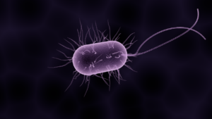
A flagellated cell (tail)
Image credits- Pixabay

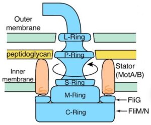
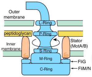
Basal Body

Basal Body is the region that is adhered to the cell membrane or cytoplasmic membrane or to the outer membrane of the cell basically. 

It consists of ring- like structures which are closely packed by proteins in pairs called MotB

The rings are

  • L-ring: L- ring is the outer or the exterior ring attached in the exterior lipo-polysaccharide layer, which are mostly found in gram positive bacteria.
  • P-ring: Attached in the peptidoglycan layer of the cell.
  • C-ring: Attached in the cytoplasm of the cell.
  • M-S ring: Attached in the cytoplasmic membrane of the cell.

Filament

  • The minute hair strand-like structure that comes out from the hook region.

Hook

  • The hook is a wider region that is present at the end of the filament.
  • It connects the filament to the protein in the tip or base. 
  • Gram positive bacteria have longer hooks in comparison with others.
flagella structure
Basal body- Flagella
Image credits- Wikimedia

Eukaryotic flagella structure:

We know that eukaryotic organisms are complex in structures.

Characteristics of Eukaryotic flagella structure:

  • The flagella structures of the eukaryotic are larger and complex by nature. 
  • The eukaryotic flagella are surrounded by a cell membrane for protection.
  • The eukaryotic flagella are made up of tubulins.
  • The locomotion in eukaryotic flagella is driven by the ATP that is produced by the membrane bound mitochondria- the powerhouse of the cell.
  • The locomotory mechanisms in eukaryotic flagella are called the bending movement or mechanisms as they move in the bending motion (S shaped).
  • In eukaryotes, the flagella is subjected to a 9+2 arrangement of microtubules.
  • The best example for eukaryotic flagellated structure is Sperm Cell.

Read More on Eukaryotic cell Vs Bacterial cell

Flagella filament structure:

  • The flagella basically have 3 parts in them- the basal body, filament and hook.
  • The flagella filament structures are stiff in nature. 
  • They are made up of complex amino acid structures, that is they are made up of protein.
  • Though flagella is considered to have minute thread like structures they have a hollow core in them.
  • The filament has a cap protein at the terminal part which is called as FliD.
  • When the cap protein is absent in the cell, they fail to develop the flagella and remain as an impaired locomotion cell.

Flagella internal structure:

The prokaryotic flagella is so simple in structure but when it comes to eukaryotic flagella, it is complex in nature.

The flagella internal structure in eukaryotes has the axoneme.

Cross section of axoneme:

  • Axoneme has 2 types of tubules in 9+2 fashion, that is 9 pairs of tubules.
  • One tubule is the locomotory and the Second one is the B tubule which is called the doublet microtubule.
  • In the core region that is the middle region there are 2 tubules which are not in pair. It is called the central microtubule that is attached to the inner sheath.

Chlamydomonas Flagella Structure:

Chlamydomonas is a single celled green algae which lives in water and moist spots.

Chlamydomonas has 2 flagella for swimming. It is called the apical flagella as it is present on the upper surface in the cervical spine. It originates from the basal grain located in the cytoplasm of the cell.

E. coli flagella structure:

Escherichia coli or also called E.coli is a gram-negative bacterium commonly found on the lower intestines of certain animals.

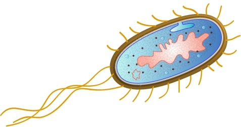
E. coli is said to have Peritrichous flagella.

Bacterial flagellate configurations 2
Bacterial flagella
Image credits- Wikimedia

Peritrichous flagella

When the flagellum is present all over that is present fully on the surface of the cell, it comes under the category of Peritrichous flagella.

It has

Basal Body

  • Basal Body of E. coli is the region that is adhered to the cell membrane or cytoplasmic membrane or to the outer membrane of the E. coli cell basically. 
  • The basal body of E. coli consists of ring- like structures which are closely packed by proteins in pairs called MotB

The details on these rings are already discussed in this article.

The rings are L- ring, P-ring, C- ring, M-S ring.

Filament

The minute hair strand-like structure that comes out from the hook region.

Hook

  • The hook is a wider region that is present at the end of the filament.
  • It connects the filament to the protein in the tip or base. 

The flagella in E.coli is one of the responsible factors for its virulence factor.

OSC Microbio 03 03 BaFlagella
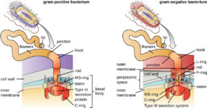
Flagella structure of Gram Positive and Gram Negative bacteria
Image credits- Wikimedia

Also Read:

13 Types Of Flagella: Exhaustive Categories And Detailed Explanations

Bacterial flagellate configurations 300x213 1

Flagella aids in the locomotion or movement of the organisms. It is like a strand of hair that is attached to the surface of the cell and promotes motility.

The following are the types of flagella that we will discuss in this article.

What are mastigonemes?

Flagella are usually simple and plain. But few types of flagella have small hair-like structures on the lateral sides of them.

The tiny and minute hairs present on the sides of the flagella are called mastigonemes.

types of flagella
A flagellated cell (tail)
Image credits- Pixabay

Types of flagella in algae:

Algae are eukaryotic organisms that mostly live in water, they are photosynthetic organisms and has a nucleus inside them.

There are 2 types of flagella seen in algae.

Whiplash flagella:

A smooth thin tail like structure or projection that comes out of the cell. It is called as acronematic as the flagella do not have any minute hairs from it, that is the flagella is hairless. So the whiplash flagella of the algae is acronematic.

Tinsel flagella:

A minute thin hair like projection with minute hairs in them on both the sides and this is why tinsel flagella are also called pantonematic.

Read more on Eukaryotic cell examples

Types of flagella in Protozoa

Protozoa are single celled organisms which are eukaryotic organisms.

Usually 4 types of flagella is seen in protozoa

Stichonematic:

When the mastigonemes are present only at one side as a row, it is called a stichonematic flagellum.

Pantonematic:

When the mastigonemes are present on both the sides, it is called a pantonematic flagellum.

Acronematic:

When there is no hair-like structure that is no mastigonemes are present, they appear to have a smooth flagella and the terminal part have a fiber-like structure it is called acronematic flagella.

Simple flagellum :

Absence of mastigonemes and no fiber like structure found in the terminal part, it is a simple flagellum.

Types of flagella in Fungi

Fungi are eukaryotic organisms that are present in soil, water and dead matter. They are single celled or multicellular fungi are also present. They have flagella similar to algae.

There are 2 types of flagella seen in fungi.

Whiplash flagella:

A smooth thin tail like structure or projection that comes out of the cell. It is called as acronematic as the flagella do not have any minute hairs from it, that is the flagella is hairless. So the whiplash flagella of the algae is acronematic.

Tinsel flagella:

A minute thin hair like projection with minute hairs in them on both the sides and this is why tinsel flagella are also called pantonematic.

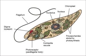
Types of flagella in Euglena

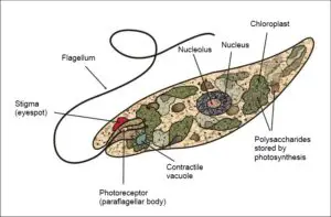
Euglena are small  protists organisms that are mostly present in fresh water habitats. They are flagellated eukaryotic organisms.

Two flagella are seen in a euglena.

  • The longer one is equal to the structure of the complete euglena, which is called the locomotory flagellum that is used for locomotion or movement.
  • The non-emergent one, which is very short and does not protrude out of the cell.
Euglena diagram
Euglena with Flagella
Image credits- Wikimedia

Types of flagella in Bacteria

Bacteria are microscopic prokaryotic organisms that is present everywhere on this earth.

Bacterial flagella is of 5 types

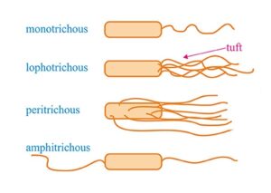
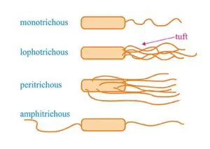
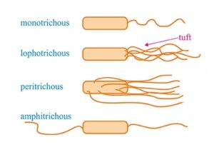
Monotrichous:

Mono- Single; A single flagella present at any end or pole of the cell is called Monotrichous, which is also called Polar flagella.

Example: Vibrio cholera, Campylobacter spp., Caulobacter crescentus etc.

Peritrichous:

When the flagellum is present all over that is present fully on the surface of the cell, it comes under the category of Peritrichous flagella.

Example: E. coli,  Bacillus subtilis, Salmonella, and Klebsiella.

Amphitrichous:

This is very similar to monotrichous flagella. One Flagellum is present at the both end or both poles.

Example: Alcaligenes faecalis

Lophotrichous:

When a bunch or group or cluster of flagella is present at one end it falls under the category of Lophotrichous flagella.

Example: Spirillum

Atrichous:

When there is no flagella present in the cell that flagella is absent, it is called Atrichous.

Example: Lactobacillus, Pasteurella.

Bacterial flagellate configurations
Bacterial flagella
Image credits- Wikimedia

Type of flagella in E.coli

Escherichia coli or also called E.coli is a gram-negative bacterium commonly found on the lower intestines of certain animals.

Peritrichous:

When the flagellum is present all over that is present fully on the surface of the cell, it comes under the category of Peritrichous flagella.

Escherichia coli by togopic
E. coli Flagella
Image credits- Wikimedia

Type of flagella in Treponema pallidum

Treponema pallidum is a spirochete bacterium that is spiral in shape. It caused diseases like syphilis, yaws etc..

Treponema pallidum have a periplasmic flagella which is called the axial filament. It is present in between the outer and inner membrane that is on the periplasmic space and aids in motility like a corkscrew motion.

Type of flagella in Protists

Protists are single celled organisms that are diverse in nature. The flagella in protists are the one that is present in euglena.

Two flagella are usually seen.

  • The longer one is equal to the structure of the complete protist sp, which is called the locomotory flagellum that is used for locomotion or movement.
  • The non-emergent one, which is very short and does not protrude out of the cell.

Now we know about the types of flagella, we need to understand the structure of flagella.

Structure of a Flagella:

Flagella is a tail-like or hair-like structure that aids in movement.

Bacterial Flagella Structure

The flagella has a spiral or helical morphology that comprises proteins called the flagellin protein. 

The flagella structure is separated into three parts:

Basal Body

  • Basal Body is the region that is adhered to the cell membrane or cytoplasmic membrane or to the outer membrane of the cell basically. 
  • It consists of ring- like structures which are closely packed by proteins in pairs called MotB

The rings are

  • L-ring: Which is the outer ring attached in the outer lipo-polysaccharide layer, which are mostly found in gram positive bacteria.
  • P-ring: Attached in the peptidoglycan layer of the cell.
  • C-ring: Attached in the cytoplasm of the cell.
  • M-S ring: Attached in the cytoplasmic membrane of the cell.
types of flagella
Basal body- Flagella
Image credits- Wikimedia

Filament

  • The minute hair strand-like structure that comes out from the hook region.

Hook

  • The hook is a wider region that is present at the end of the filament.
  • It connects the filament to the protein in the tip or base. 
  • Gram positive bacteria have longer hooks in comparison with others.

What is the function or role of flagella in an organism?

  • The main function of flagella is that they aid in the movement of the organism. 
  • They perform other functions like they play the role of sensory organs like detection of temperature and changes in pH. 
  • Certain organisms use their flagella to increase their rates of reproduction mostly in eukaryotic organisms.
  • It aids in the virulence factor in flagellated pathogenic organisms.

Also Read:
Is Ribosomes Eukaryotic: Detailed Analysis And Facts

ribosome 1 300x158 1

Ribosomes are tiny particles or a component in the cytoplasm of the cell or present on the surface of other organelles, that has RNA and other connected or related proteins. So in which organism ribosomes are present? Let’s have a deeper view.

Is Ribosomes Eukaryotic, No. Ribosome is an organelle. Ribosomes are present in both eukaryotic organisms and prokaryotic organisms. Ribosome is the place where the synthesis of protein is being done. They can be in the cytoplasm or inside any other organelle.

We all know the importance of protein. 

They are basically the building blocks of a cell even if it’s for a smaller organism like bacteria etc or bigger animals like elephants etc. In order to gain new cells and repair the older or damaged cell, the body needs to synthesize protein. 

Now this synthesis of protein happens inside an organelle called ribosome

So, ribosomes are present in both eukaryotic organisms and prokaryotic organisms. 

Prokaryotic organisms have 70s (S- Svedberg unit; The unit of Density ) ribosomes.

Eukaryotic organisms have 80s (S- Svedberg unit; The unit of Density) ribosomes.

is ribosomes eukaryotic
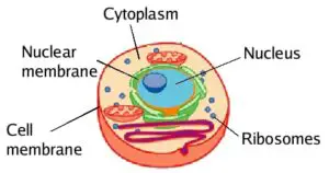
Cell with organelles
Image Credits- Wikimedia

Is Ribosomes Eukaryotic organelle specifically?

Ribosome is an organelle that is not specifically membrane bound.

So, ribosomes are present in both prokaryotes like bacteria, archaea and in eukaryotes like animals, fungi etc.

Which type of ribosomes are found in bacteria?

The bacteria have simple structure with 3 type of ribosomal RNA: [30S+ 50S]

16S rRNA:

16S rRNA or 16S ribosomal RNA, (where S= Sediment Coefficient- During centrifugation process, the amount of sediment it forms) is a portion in the prokaryotic subunit (30S). It has 21 proteins.

23 S rRNA and 5S rRNA:

23 S rRNA and 5S rRNA, where rRNA- ribosomal RNA (where S= Sediment Coefficient- During centrifugation process, the amount of sediment it forms) is a portion in the 50S prokaryotic subunit. It has 31 proteins.

Now, during protein synthesis these 30S and 50S subunits get connected or combined and become 70S subunits which are about 25 nanometer in diameter.

FACT: Bacteria has about 15000 ribosomes in it.

Which type of ribosomes are found in mitochondria?

Mitochondria being an organelle has ribosomes in it.

Mitochondria has 70S types of ribosomes inside their matrix. These ribosomes are called mitoribosomes or mitochondrial ribosomes.

These ribosomes aid in the production or synthesis of protein that is required by mitochondria for energy production (ATP) or any other organelles.

Read more on Adenosine nucleoside and nucleoside phosphoramidite | Overview of important aspects

Where are ribosomes found in a eukaryotic cell?

In eukaryotic cells,  ribosomes are found in many places based on their function and productivity.

Ribosomes in eukaryotic cell are present freely in the cytosol or cytoplasm or ribosomes are present on the surface of other membrane bound organelles like endoplasmic reticulum, mitochondria, plastids like chloroplasts.

Eukaryotic cells have complex mechanisms which require separate ribosomes for each one. This is the reason ribosomes are scattered like these for the synthesis of protein.

What type of ribosomes are found in prokaryotic cells?

Prokaryotic organisms like bacteria, archaea and cyanobacteria have the same type of ribosomes which is clearly discussed below.

Prokaryotic cell have 3 types of ribosomal RNA: [30S+ 50S]

16S rRNA:

16S rRNA or 16S ribosomal RNA, (where S= Sediment Coefficient- During centrifugation process, the amount of sediment it forms) is a portion in the prokaryotic subunit (30S). It has 21 proteins.

23 S rRNA and 5S rRNA:

23 S rRNA and 5S rRNA, where rRNA- ribosomal RNA (where S= Sediment Coefficient- During centrifugation process, the amount of sediment it forms) is a portion in the 50S prokaryotic subunit. It has 31 proteins.

Now, during protein synthesis these 30S and 50S subunits get connected or combined and become 70S subunits which are about 25 nanometer in diameter.

Which type of ribosomes are found in eukaryotic cells?

Eukaryotic cells perform complex cellular mechanisms and require protein or amino acid molecules to a great extent.

Eukaryotic cell on its own has an 80S subunit present on the cytosol and sometimes these ribosomes are attached in the surface of the other organelle like endoplasmic reticulum, chloroplasts, mitochondria.

Are ribosomes in prokaryotes and eukaryotes the same?

Prokaryotic organisms and eukaryotic organisms are entirely different in terms of their mechanisms and other factors.

 When it comes to the prokaryotic ribosomes and eukaryotic ribosomes they are not the same. They are different with very few similarities.

The purpose of the ribosome is same in prokaryotes and eukaryotes, that is they provide the site for synthesis of protein. They are composed of RNA and other ribosome associated proteins.

What are 30S 50S ribosomes?

Ribosomes have 2 subunits in them.

30S and 50S ribosomes are the prokaryotic ribosomes which in combination makes the complete 70S subunit of ribosome.

What are 60S and 40S ribosomes?

Ribosomes have 2 subunits in them.

60S and 40S ribosomes are the eukaryotic ribosomes which in combination makes the complete 80S subunit of ribosome.

Difference between prokaryotic and eukaryotic ribosomes?

Though ribosomes are organelle, they differ when they appear in prokaryotic organisms and eukaryotic organisms based on their requirements.

Prokaryotic and eukaryotic ribosomes differ in many ways like their subunit, they vary in size, location etc.

Prokaryotic ribosome:

  • Prokaryotic ribosomes are smaller- 70S ribosomes. 
  • It consists of 30S subunit and 50S subunit. 
  • Among these, 30S is the smaller subunit and 50S is the larger subunit. 
  • The mass is around 2700kd. 
  • The smaller subunit is composed of 16S RNA
  • The larger subunit is composed of 2 RNA that is  5S RNA and 23S RNA.
  • Generally in prokaryotic organisms, ribosomes are seen free floating in the cytoplasm.

Eukaryotic ribosome:

  • Eukaryotic ribosomes are larger- 80S ribosomes.
  • It consists of 40S subunit and 60S subunit.
  • Among these, 40S is the smaller subunit and 60S is the larger subunit.
  • The mass is around 4200kd.
  • The smaller subunit is composed of 18S RNA.
  • The larger subunit is composed of 3 RNA that is 5S RNA, 5.8S RNA, 28S RNA.
  • In eukaryotic organisms, ribosomes are present on the cytosol or cytoplasm and also on the surface of other membrane bound organelles such as endoplasmic reticulum, nucleus, mitochondria, chloroplasts etc.
is ribosomes eukaryotic
Ribosome structure
Image Credits- Wikimedia

What are 70S and 80S ribosomes?

Now we understand that ribosomes are nothing but a machine that aid in protein synthesis.

The 70S and 80S  are the prokaryotic and eukaryotic ribosomes respectively. In which 70S is the combination of 30S + 50S subunit and 80S is the combination of 40S+60S subunit of the ribosome.

Why do 60S and 40S make 80S not 100s?

The eukaryotic ribosome contains 60S and 40S subunits and in combination makes 80S subunits of the ribosome.

S is the svedberg unit which is the unit of sedimentation coefficient. During the centrifugation process, the contents will be pushed downward and upwards based on their weight. 

The exact total sum will not be retained due to the forces like centrifugal force and gravity. This is why 60S and 40S make 80S not 100s.

Why do 50S and 30S make 70S ribosomes?

The prokaryotic ribosome contains 50S and 30S subunits and in combination makes 70S  subunits of the ribosome.

S is the svedberg unit which is the unit of sedimentation coefficient. During the centrifugation process, the contents will be pushed downward and upwards based on their weight. 

The exact total sum will not be retained due to the forces like centrifugal force and gravity. This is why, 50S and 30S make 70S ribosome.

Also Read:
What is the Function of Ribosomes: Detailed facts

Ribosome 300x243 1

Ribosomes are tiny particles or a component in the cytoplasm of the cell that has RNA and other connected or related proteins. But what exactly is the role of ribosomes in the cell?

What is the function of ribosomes, we know that proteins are very essential molecules for any cellular mechanism. The main role or function of the ribosome as an organelle in the cell is they aid in protein synthesis. 

Now a question arises, how and when does the synthesis of protein take place. 

Each and every question will be solved in this article on  what is the function of ribosomes in cell, animal cell, plant cell, chloroplast and mitochondria.

What is the function of ribosomes in cells?

Ribosome is an organelle that is located either on the cytoplasm or within other organelles like endoplasmic reticulum, chloroplast, mitochondria of the cell.

Ribosomes aid in the conversion of genetic information into proteins. To be more precise, the organelle ribosome is like a space or site in which the synthesis of protein happens.

What is the function of ribosomes in animal cells?

The cellular components and their functions in animal cells are much more complex than other organisms.

In animal cells, The complex mechanisms and structures must be maintained by proteins. So the purpose of ribosomes in animal cells are to aid in the synthesis of protein processes.

Read More on Do humans have animal cell?

What is the function of ribosomes in plant cells?

Plant cell structures and mechanisms are unique in their own way

Ribosomes serve as a site of protein synthesis as they do in animals or other cells. The mRNA is inspected and translated into corresponding amino acids and thus forms the polypeptide chain that is the protein.

What is the function of ribosomes in mitochondria?

We know that mitochondria is an organelle that produces the energy molecule- Adenosine triPhosphate (ATP) in eukaryotic cells.

The ribosomes present inside the mitochondria are called Mitochondrial ribosomes. These mitochondrial ribosomes or the mitoribosomes aid in the synthesis of the membrane proteins that are seen in the mitochondria which is known as mitochondrial membrane proteins.

These exclusively produce only membrane proteins for mitochondria.

What is the function of ribosomes in bacteria?

Bacteria are simple prokaryotic organisms.

In bacteria, the ribosomes serve as the site for translation of mRNA and synthesis of protein. In Bacteria, the subunits are 50s(Large) subunits and 30s(Small) subunits.

What is the function of ribosomes in chloroplasts?

Chloroplasts are the organelle that has chlorophyll and gives a bright green color to the plants and organisms that have chloroplast in it.

Recent findings state that the ribosomes are present inside the matrix of chloroplast. Their main role is to aid in chloroplast gene expression.

What is the composition of a ribosome?

Ribosome is an organelle that consists of 2 ribosomal subunit.

The ribosomal Subunit- Small:

This subunit scrutinizes the mRNA strand.

The ribosomal Subunit- Large:

This subunit helps in the formation of the amino acid polypeptide chains.

In order to know more about the functions of ribosomes, we need to understand the structure of ribosomes.

Explanation on structure of ribosome:

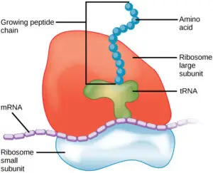
The ribosome has 2 RNA- Protein complexes which are known as subunits.

The structure of ribosomes can be compared to that of a sandwich (2 buns on the opposite side and the main ingredient in between them-Middle). The same way, the large subunit is placed on the top of the small subunit and the mRNA strand that needs to be translated is placed in the middle.

what is the function of ribosomes
Structure of Ribosome
Image Credits- Wikimedia

In case of eukaryotes, the nucleus directs the function to the ribosome.

In the case of prokaryotes, they have no nucleus. So the translation process starts immediately and does not wait for any orders.

The ribosomes are floating freely in the cytoplasm or present on the top of the other organelles like endoplasmic reticulum, which are called Rough endoplasmic reticulum or RER.

Do prokaryotes have ribosomes?

Yes, ribosomes are present in prokaryotes. They have 70s (S- Svedberg ) ribosomes.

Read More on Eukaryotic cell Vs Bacterial cell

Do eukaryotes have ribosomes?

Yes, ribosomes are present in eukaryotes. They have 80s (S- Svedberg ) ribosomes.

Read More on Eukaryotic cell examples

What exactly happens inside the ribosome?

Ribosome is a space where the synthesis of protein happens. This process is called translation.

Translation is a process in which the mRNA strand which is the end product of transcription process codes for the basic units of proteins, that is the amino acid and the synthesis of protein takes place.

The mRNA or the Messenger RNA is scrutinized or inspected. tRNA or the Transfer RNA adds the corresponding amino acid codons and thus forms the polypeptide chain.

Also Read:

Do Prokaryotes Have Telomeres: Why, How And Detailed Insights

mitosis g7c26f6df5 1920 300x169 1

A telomere is a region or a portion on the chromosome that is present at the end, that is the final region of the chromosome is the telomere part. So, do prokaryotes have telomeres in them?

Do prokaryotes have telomeres, No. Prokaryotic organisms are very basic organisms that do not have telomeres in their chromosomes. Telomeres are present only in the chromosomes of eukaryotic organisms.

But why do prokaryotic organisms not have telomeres in their chromosomes and what is their ultimate function in eukaryotes? Let’s see all the answers in this article.

What are telomeres?

Telomeres are the region in the chromosome.

Telomeres are the portion in the chromosome that does not carry any genetic information and present at the end. 

They are present only in eukaryotic organisms. Prokaryotic organisms lack telomeres in their chromosomes.

do prokaryotes have telomeres
Cell
Image credits- Pixabay

Why do prokaryotes not have telomeres?

We just saw that the telomeres are present at the end of chromosomes. In order to identify the end or start of the chromosome, the chromosome should be in a straight or linear form.

Prokaryotes have a single circular-shaped chromosome. Their replication process is much faster than eukaryotic organisms. As they are circular in shape, there is no end and telomeres are absent in prokaryotes.

Do prokaryotes have telomeres like any other structures?

No, in prokaryotes there is no structure like telomeres.

Why do eukaryotes have telomeres?

We just saw that prokaryotes have a round or circle shaped chromosome so there is no end for prokaryotic chromosomes to be identified.

In the case of eukaryotes, They have a linear or straight chromosome in which the end or the final part is identifiable. This is why telomeres are present only in eukaryotes. 

Telomeres play a significant role in protecting the chromosome from any damage, cell division process, cell aging process, DNA replication and transcription process.

What are the functions of telomeres in eukaryotic organisms?

  • The stability of chromosomes is due to the telomeres as they are present at the end.
  • There will be the destruction of chromosomes during the process of DNA replication. The non-coding sequences at the last of the chromosomes (Telomeres) are lost in this process.
  • Telomeres are present for the protection of genetic information.

Characteristics of nucleoid:

  • The shape of the Nucleoid is an irregularly shaped part in the cytoplasmic region of the prokaryotic cell that contains the genetic material or so-called genophore.
  • It is different from the nucleus as the nucleus has a membrane outside it. Nucleoids do not have membranes in them.
  • About 60% of the nucleoid is DNA (Genetic material that has the genetic information in it.
  • The other 40 % are other RNAs or proteins.
  • The main function of the nucleoid is to control the activity of the cell and its reproduction and maintain the genophore intact.

So how are the prokaryotic chromosome structure? Let’s see in detail.

Prokaryotic Chromosome structure:

  • Prokaryotes do not have a true or membrane bound nucleus in them. The chromosomes in prokaryotes are present in the region of cytoplasm where the genetic material embedded which is called nucleoid of the cell.
  • The chromosomes in prokaryotic cells are round or circular in shape. Mostly they are single. Few exceptions are present. They are highly coiled structures with double strands.
  • Only a single chromosome is present but the single chromosome has a large strand of DNA molecule that is highly coiled and placed inside it.
  • Prokaryotic DNA is wrapped with NAPs that are Nucleoid associated proteins which aids in the process of DNA replication and transcription.
  • Prokaryotes are haploid in nature. There are no pairs of chromosomes in prokaryotes.
  • Prokaryotes have a small portion or a molecule of DNA called plasmids which plays a vital role in genetic engineering.
  • So prokaryotes have chromosomal DNA and Plasmid DNA.

Read more on DNA Supercoiling

Also Read:

Do Prokaryotes Have Exons: Why, How And Detailed Insights

dna gbaa56591e 1920 300x169 1

Exons are the part or portion of the gene which codes for amino acids. Prokaryotes have a very simple cellular organization. So, Do Prokaryotes Have Exons?

Do Prokaryotes Have Exons, Yes. Prokaryotes have only exons in them. We know that prokaryotes are unicellular organisms and have a simple cellular organization. So, they lack complex mechanisms like post transcriptional modification etc. Thus they possess only exons in them.

Read more on DNA Structure

Why do prokaryotes only have exons ?

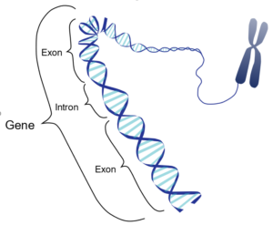
In eukaryotes, once the mRNA  is transcribed from DNA the introns in the mRNA will be cut and the exons will be combined together.

But, Prokaryotes are simple and have simple mechanisms evolving in their structure. So the introns are absent in them, they only have exons in their genes.

do prokaryotes have exons
Structure of a DNA
Image credits: Pixabay

Where are exons located in prokaryotes?

The exons are present in the gene of the DNA of the organism.

Do eukaryotes have exons in them?

Eukaryotes are complex organisms and undergo complex mechanisms for survival.

Yes, Eukaryotes have exons in them.

They also have introns which are cut off and the introns are connected together using a protein complex called spliceosome.

Do eukaryotes have more of introns or exons?

Eukaryotes have both in them ( introns and exons). 

Approximately, it is said that there are about 7.8  introns and 8.8 exons in a gene.

It depends on 1 organism to another based on their morphology complexity.

Why prokaryotes don’t have both introns and exons in their genes?

Prokaryotes have a straightforward mechanism in them.

Prokaryotes do not have enough space in them as well as in prokaryotes, post transcription the translation process immediately initiates or in some cases it happens simultaneously. Thus there is no time for intron splicing.

There is no mechanism of post transcriptional modification. So they do not require introns in them.

Read more on Bacterial DNA replication steps.

Simplified version of prokaryotic transcription:

Transcription is a process in which a portion of the DNA is copied into a mRNA strand which is newly synthesized with the help of RNA polymerase enzymes.

The prokaryotic transcription is performed in three steps.

Transcriptional Initiation:

The process initiates, in which the enzyme RNA polymerase recognizes the promoter region of the gene and unwinds the DNA.

Transcriptional Elongation: 

The new free nucleotides attach to the DNA strand and due to which the attachments elongate and the mRNA strand is being formed.

Transcriptional Termination

Once the enough strand is made, the enzyme and other cofactors that is associated in DNA complex, dissociates itself from the DNA and the process is terminated 

As the end product, newly synthesized mature mRNA is produced which is ready for translation.

What will happen to the mRNA post transcription?

In prokaryotes, the transcription process takes place in the cytoplasm.

After the mRNA is synthesized, a process called translation that is the protein synthesis takes place. 

In prokaryotes, the process of  transcription and the process of translation process are combined or coupled. The transcription completes and immediately the translation starts or in some cases the transcription and translation process happens simultaneously, which is called a coupled process.

Simplified version of prokaryotic translation:

Translation is a process in which the transcribed mRNA strand codes for amino acid and the synthesis of protein takes place.

The translation process in prokaryotes takes place in the cytosol organelle of the cell. The subunits such as large and small of the ribosome attach to the mRNA for translation that is synthesis of proteins process.

Prokaryotic translation also consists of 3 stages.

Translational Initiation: 

Assembly of the ribosomes ( large and small subunits) around the mRNA takes place. The first codon (start codon) is attached or binds to the initiator tRNA.

Translational Elongation: 

The same process of attachment of the codon is being done and all the peptides are combined to form polypeptides.

Translational Termination:

Finally the stop codon comes in and the process is terminated.

Also Read:

Do Eukaryotes Have Introns: Why, How And Detailed Insights

image 86 300x247 1

A portion or a part in the genetic material that is the DNA or RNA that does not code for proteins, is called introns.

Do eukaryotes have introns, Yes. Eukaryotic organisms have introns in them. Introns are present in between exons. Their main role is recombination in exons. They play a significant role in evolution by enhancing gene expression.

Why do eukaryotes have introns?

We know that eukaryotes are complex in structure and function.

Eukaryotes require diversity in proteins for satisfying this complex morphology. Though introns do not code for proteins, they involve themselves highly in gene expression and regulation. This is the reason the introns are present in eukaryotes.

do eukaryotes have introns
Image credits- Wikimedia

Do eukaryotes have intron splicing?

There are introns and exons in a gene. Introns are non-coding sequencing for proteins.

Yes, eukaryotes have intron splicing. The introns are detached from the sequence and the exons are spliced or connected together and then the protein synthesis takes place.

Do prokaryotes have introns?

Introns are present only in complex organisms.

Prokaryotes do not have introns in them (Absence of introns)

Read more on Eukaryotic cell Vs Bacteria cell

Why don’t prokaryotes have introns?

Prokaryotes are simple or basic organisms.

Introns are not present in prokaryotes because prokaryotes do not possess a mechanism called post-transcriptional RNA modification, in which the introns are detached and the exons are joined or connected together.

Types of introns:

Based on the genetic biochemical and sequence analysis, introns are divided as

Spliceosome introns

Spliceosome introns are the non-coding gene part that is detached or removed by the spliceosome during post-transcriptional modification.

Self- splicing group-I

Self- splicing group-I are detached by RNA catalysis

Self- splicing group-II

Self- splicing group-II are detached by RNA catalysis

tRNA introns

tRNA introns or the Transfer RNA introns are detached by proteins.

What is the splicing process?

In the process of transcription, RNA polymerase makes copies of the gene structure including exons and introns. This is called pre-mRNA or hnRNA (Heterogeneous nuclear RNA)

Now, the introns will not code for proteins thus they need to be removed or detached. The detachment of introns and splicing or combining of the exons is called as splicing process.

mRNA Splicing:

The transcription process and the formation of mature mRNA take place in the nucleus of a cell. After this process, the mature mRNA is then transported to the cytoplasm for the translation (protein synthesis) process.

We know that the gene has introns and exons. Introns are non-coding sequences whereas exons are coding sequences that code for amino acids in a protein.

Post transcription, the end product is a pre- mRNA transcript. It has introns and exons. The structure is like the intron is attached in between 2 exons. The removal of these introns must take place before the translation process.

This is done by a protein complex called the spliceosome. This protein complex removes or cuts off the introns on the sequences and attaches all the exons together. This is called a post-transcriptional modification. Post this process, the mature mRNA molecule is transported to the cytoplasm for protein synthesis (translation).

Read more about Genetic Diversity Examples, please click.

Summary of the splicing process:

  • Pre mRNA transcript is the product of the transcription process
  • The pre mRNA transcript has introns (non-coding sequence) and exons (coding sequence for proteins) in them.
  • For protein synthesis, that is the translation process, introns should be detached and the exons should be connected together.
  • The spliceosome is a protein complex that detaches the introns from the gene and connects the exons. This process is called post transcriptional modification.
  • Transcription and post-transcriptional modification take place in the nucleus of the cell.
  • After the detachment of introns and combination of the exons, the pre mRNA transcript is transformed into a new form called the mature mRNA which is completely ready for translation or the protein synthesis process.
  • This mRNA is transported to the cytoplasm for the translation process.

Also Read:

Are Protists Eukaryotic: Why, How And Detailed Insights And Facts

Euglena diagram 300x197 1

Protists are mostly single-celled or unicellular organisms mostly live in aquatic ambience.

Protists are eukaryotic mostly single-celled or unicellular organisms. Though they are relatable to other organisms they aren’t considered as true animals, plants or fungi this is why they fall into a different category.

Why are protists eukaryotic?

Now we know that protists are mostly single-celled organisms but still what makes them fall under the category of eukaryotes.

Protists have a nucleus and other complex organelles in them which makes it fall under eukaryotes.

Why are protists eukaryotes and not prokaryotes?

Protists are mostly single-celled organisms and still, they come under eukaryotes. We know that prokaryotes are simple and eukaryotes have complex structures. But protists are eukaryotes, how?

The presence of a nucleus inside them and other complex membrane-bound organelles make protists fall under eukaryotes.

Are Protists Eukaryotic
Image credits: Wikimedia

Are protists simple or complex organisms?

Protists, though they are mostly unicellular organisms, they are not simple organisms.

Yes, protists are complex organisms. They have complex organelles in them which are similar to plants, animals, fungi and other bigger organisms.

Are protists haploid or diploid?

Protists can be haploid or diploid.

Protists transform themselves from being haploid to diploid.

Most of the protists are unicellular which are haploid. Multicellular protists are diploid. This may change from one species of protists to another depending on their morphology and other components.

Are protists motile or nonmotile?

Motility or movement of small organisms is based on the presence of flagella, cilia, pseudopodia (False feet) in the protists.

Most of the protists have flagella and have the capability to move.

Eg: Flagellum- Euglena, Pseudopodia- Amoeba, Cilia- Paramecium.

Euglena diagram
Euglena
Image credits: Wikimedia

How do protists obtain energy?

Based on their nutrition uptake, protists are classified into 3 types

Ingestion protists:

This group of protists engulf the minute food particles and ingest them. The food particle is then digested in the organelle called vacuoles.

Eg: Amoeba

Figure B23 02 01
Ingestion protists
Image credits: Wikimedia

Absorption protists:

This group of protists are called decomposers. The food particle is absorbed into the organism by the process of diffusion.

Eg: spirogyra

Photosynthetic protists:

Photosynthetic protists are the group of protists that are able to prepare their own food by the process of photosynthesis in which light is used as a primary source. They are the producers and are majorly present in the aquatic system.

Eg: Algae, diatoms, dinoflagellates etc.

What protists are decomposers?

Protists are producers majorly. Few protists are decomposers too.

Slime moulds are decomposers. Other than slime moulds, chytrids, Oomycetes, labyrinthulomycetes are other examples of protists being decomposers.

Are protists aerobic or anaerobic?

Protists can be aerobic or anaerobic. Few protists are obligate aerobes. Few are anaerobic organisms

Characteristics of protists:

  • Protists are mostly single-celled organisms or commonly known as Unicellular organisms.
  • There are few multicellular protists too.
  • Protists are complex organisms as they have nuclei inside them.
  • Protists have other membrane-bound organelles like endoplasmic reticulum, mitochondria, vacuole etc
  • Protists are mostly motile. They have flagellum, cilia or pseudopodia which is commonly called “False feet”.
  • Protists require a moist environment to survive. So they are aquatic organisms.
  • Few protists have symbiotic relationships. They live like a parasite inside other organisms like plants, humans etc.
  • Based on their nutrition uptake, protists are classified into 3 types: Ingestion protists, Absorption protists, Photosynthetic protists.

Importance of protists in Ecology:

  • They involve themselves as a major part in food chain
  • Protists have the capability to consume/ ingest bacteria and other microbes. This way the microbe population will be under control.
  • The photosynthetic protists play a vital role in ecology as they aid in reducing atmospheric carbon dioxide.
  • The major protists, Phytoplankton is the basic component of marine food chain.
  • Protists aid in building coral reefs.
  • Protists aid in recycling nutrients in the ecosystem.

Importance of protists in the Economy:

  • By the process of photosynthesis, Protists release oxygen. This can be used for biofuel preparation.
  • Few protists like Red algae etc. have medicinal value.
  • Protists like seaweeds are consumed in many countries like Japan etc. They can also be used as a fertilizer as they are rich in potassium, nitrogen etc.
  • Diatoms are used in making paper, cement, dental impressions etc.
  • Agar agar is a red algae used in growth media in microbiology and as a thickening agent in food industries (Jams, jellies etc.)

Read more on Eukaryotic examples

Also Read:

Are Bacteria Prokaryotic Or Eukaryotic: Why, How And Detailed Insights And Facts

school gfc089f4a3 1280 300x244 1

Bacteria comprise a single cell and the single cell is considered a whole organism.

Bacteria are single-celled prokaryotic organisms. They do not possess any internal membranous organelles and their morphology or cellular organization is so simple, this differentiates them from being a eukaryote as they have complex structures. Bacteria have no nucleus and other complex organelles.

As the bacteria’s cellular organisation is very simple, this differentiates it from being a eukaryote as eukaryotic organisms have complex structures. Bacteria being a prokaryote has no nucleus and other complex organelles.

Are Bacteria Prokaryotic Or Eukaryotic?

Bacteria are prokaryotic organisms

We just saw the answer for, Are Bacteria Prokaryotic Or Eukaryotic? now, how are bacteria considered prokaryotes? what are the structures present inside the cell? All details regarding it will be discussed in this article.

Are Bacteria Prokaryotic Or Eukaryotic?
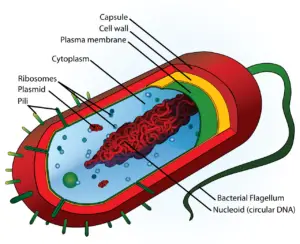
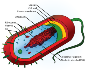
Are Bacteria Prokaryotic Or Eukaryotic- Bacterial structure (internal)
Image credits- Pixabay

Why are bacteria called prokaryotes?

We already know that the cell must be a prokaryote or eukaryote. So the chief difference between a prokaryote and a eukaryote is regarding the presence of a nucleus.

Bacteria do not have a nucleus to be more precise, they do not have a true nucleus so this concluded that bacteria is a prokaryotic organism.

Prokaryotic bacteria characteristics

  • Bacteria are single-celled organisms that are they are Unicellular structures.
  • They are very small that they can not be viewed by the naked eye. The microscope is used to visualize bacteria.
  • Bacteria do not have any membrane-covered organelles like Golgi bodies, mitochondria, chloroplast.
  • Bacteria do not have a well-defined nucleus.
  • Bacteria have circular DNA that is present freely in the cytoplasm of the cell. Circular DNA is adhered to the cell membrane/plasma membrane and is known as the nucleoid.
  • Bacterial cell size ranges from 0.1 to 5.0 micrometre.
  • The bacterial cell is made up of complex polysaccharides

Are all bacteria prokaryotes?

Yes, all bacteria are prokaryotes as their cellular organization is so simple and lacks a true nucleus.

Internal structures present in the bacterial cell

Though bacteria is a single-celled organism or unicellular organism, the single cell is able to perform and survive as a whole organism with the aid of certain internal structures

There are a few components present inside the bacterial cells. It is not mandatory that all bacteria must possess these components. Depending upon their type and survival, they may or may not have certain components.

The components present inside the bacterial cells are: 

  • Cell wall:The main use of a cell wall is to protect the contents of the cell and give a rigid structure to it. It is composed of mucopeptides and mucopolysaccharides. Based on the cell wall, the bacteria is categorized into 2 types, when the bacteria has a thick peptidoglycan layer on their cell wall it is called Gram-Positive bacteria. When the peptidoglycan layer content is low on the cell wall, it is Gram- Negative bacteria.
  • Cell membrane: Cell membrane is made up of two layers of phospholipid matrix. The major role of cell membrane is transport of molecules like nutrients etc. 
  • Cytoplasm: Cytoplasm is like a semisolid fluid in which all other components of the cell stay intact.
  • Chromosome: The chromosome consists of the genetic material. This contains the major information of the cell to thrive.
  • Flagella: Flagella is a tail-like structure that makes the bacteria motile.

Frequently asked questions:

1. How can bacteria survive without a nucleus?

The main component in a eukaryotic nucleus is the genetic material that is DNA or RNA that is embedded inside the membrane. 

In bacteria, they have a component called the nucleoid. This component is devoid of the membrane. They have the genetic material and plasmid which is a small round/ circle shaped extra genetic portion that floats freely in the cytoplasm.

2. How do the prokaryotes, in specific- Bacteria reproduce?

Every organism (be it prokaryotes or eukaryotes) should reproduce to survive and sustain on this living earth.

Bacteria reproduce by a mechanism called Binary fission, in which a single bacteria divided itself into two new daughter cells.

Read more on Bacterial replication.

3. Are Bacteria considered immortal?

As we just saw, the mature bacteria divides itself into 2 new daughter cells.

As the single bacteria symmetrically divides itself and the same cell replication process goes on. Bacteria are considered immortal.

Also Read: