9 Pathogenic Fungi Examples: Detailed Facts

Candida Gram stain 300x200 1

The study of fungi that causes diseases to humans is known as the medical mycology. While, the study of fungi those are pathogenic or that causes diseases to plants is known as the plant pathology. In the world, there are around 300 fungi that are till now known to cause diseases to human being.

  • Candida albicans
  • Aspergillus acidus
  • Cryptococcus neoformans
  • Histoplasma capsulatum
  • Pneumocystis jirovecii
  • Stachybotrys alternans
  • Talaromyces marneffei
  • Blastomyces dermatitidis
  • Emmonsia pasteuriana
  • Sporothrix schenckii

Dimorphic fungi that are important pathogens of humans include other animals such as Paracoccidioides brasiliensis and Coccidioides immitis/posadasii.

Pathogenic fungi are fungi that are responsible for causing diseases in plants, humans, and different organisms. Though fungi are eukaryotic in nature, there are several pathogenic fungi that are microorganisms.

We will discuss a few pathogenic fungi examples here. These are species of different pathogenic fungi that has different genus:

1. Candida albicans

Candida albicans is an aggressive kind of pathogenic yeast. It is popular member found in the gut flora of humans. It can also exist independently of the human body.

In roughly 40 percent to 60 percent of healthy persons, Candida albicans is found in the gastrointestinal tract as well as the mouth. This species belongs to the genus named Candida that is responsible in causing Candidiasis which is a human infection.

This is the result of the fungus when there is an overgrowth. Candidiasis is one example that is often observed in patients who are HIV-infected.
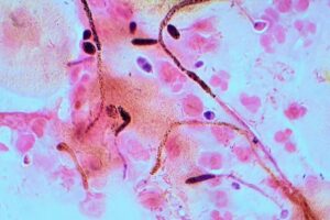
Candida Gram stain
Image Credit : Candida albicans Wikipedia

Candida albicans is usually used as a model organism in the case of fungal pathogens. It’s known as a dimorphic fungus because it can grow both as yeast and as filamentous cells.

2. Aspergillus acidus

Aspergillus is the genus of moulds that can be found in a variety of conditions around the world. It consists of species that can be a few hundred mold.

Aspergillus acidus is one of the species that belong to this genus. This species, Aspergillus acidus, is used in the form of food fermentation for tea. The species of Aspergillus are necessary both medically and commercially. Some animals and humans can become infected with some of the species.

The term “aspergillosis” describes that group of diseases that are caused by the source of the fungus Aspergillus. Fever, cough, chest pain, or shortness of breath are all symptoms that can occur with a variety of other disorders, making diagnosis challenging. In humans, the fungus Aspergillus can cause some of the major forms of disease. They are as follows:

  1. Patients who suffer from allergic bronchopulmonary aspergillosis (ABAS)-this affects patients who suffer from respiratory diseases such as asthma, sinusitis, and cystic fibrosis.
  2. Acute invasive aspergillosis: this is a form of disease that grows into surrounding tissues. This disease is generally common in people who have weak immune systems, such as patients suffering from AIDS and also cancer patients who are undergoing chemotherapy.
  3. Disseminated invasive aspergillosis – this is an infection that spreads widely throughout the body.
  4. Aspergilloma – this is a “fungus ball” that has the ability to form within cavities such as the cavities of the lungs.
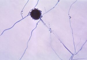
800px Aspergillus niger 01
Image Credit : Aspergillus – Wikipedia

3. Cryptococcus neoformans

Cryptococcus neoformans is a soil-borne obligate aerobe that can live in both plants and animals.It belongs to the class named Tremellomycetes of encapsulated yeasts.

It’s frequently discovered in bird faeces in the form of yeast. Cryptococcus neoformans can infect both immunocompetent and immunocompromised hosts, causing illness. Cryptococcus neoformans reproduces by budding and develops as a form of yeast which is unicellular in nature.

Cryptococcosis is the result of a Cryptococcus neoformans infection. The majority of Cryptococcus neoformans illnesses originate in the lungs. Cryptococcus neoformans is transmitted to humans through the intake of aerosolized basidiospores, and it can move to the central nervous system, causing meningoencephalitis.

The infection begins in the lungs and spreads through the bloodstream to the meninges and eventually to other areas of the body. Phagocytosis is inhibited by capsules. In general, diabetic and hosts with weakened immune systems, it can produce a systemic infection.

It may include deadly meningitis known as meningoencephalitis. Fluconazole is a solo therapy that can be used to treat cryptococcosis which does not put any impact on the central nervous system.

4. Histoplasma capsulatum

Histoplasma capsulatum is a kind of species that belong to the dimorphic fungus. Its reproductive form is known as the Ajellomyces capsulatus.

Pulmonary and disseminated histoplasmosis can be caused by Histoplasma capsulatum. Histoplasma capsulatum is commonly found in the Midwest and East of the United States. It is connected by Central and South America, and also some other parts of the planet.

Histoplasma capsulatum is spread all over the world, except in Antarctica. Most typically this species is associated with river basins. Histoplasma capsulatum is especially common in the areas of Ohio and regions of Mississippi River.

Histoplasmosis can spread through the bloodstream to affect the internal organs and tissues. This, however, occurs in a limited number of cases.

Half or more of these instances involve hosts that have weakened immune systems. Endocarditis and peritonitis are the two more diseases that are seldom related with the species, Histoplasma capsulatum.

5. Pneumocystis jirovecii

Pneumocystis jirovecii is a type of fungus which has yeast like characteristics. It belongs to the genus named Pneumocystis.

Pneumocystis pneumonia is caused by this bacterium, which is a serious human infection, especially in people who are immunocompromised. Pneumocystis pneumonia is a significant disease of immunocompromised humans, especially in patients with HIV.

It is also necessary to patients, whose immune system is critically terminated for several other reasons, such as, as a result of a bone marrow transplant. It is a very prevalent asymptomatic severe disease with a strong immune system in human beings.

The drugs that can be used to treatments are Trimethoprim or sulfamethoxazole, pentamidine, dapsone. The majority of instances in HIV patients happen whenever the CD4 level falls under 200 cells per microliter.

P. jirovecii is considered to have both an androgynous phase and stages where both the parents are involved in its life cycle. Binary fission is most likely how haploid cells multiply asexually. Primary homothallism, also known as self-fertilization, seems to be the method of reproduction where both the parents are included. The reproductive phase occurs in the lungs of the host.

6. Stachybotrys chartarum

The microfungus Stachybotrys chartarum, often considered as black mould or toxic black mould, generates its conidia or spores in slime bodies. It can be seen in soil and grain such as wheat, corn, cereal.

The bacteria Stachybotrys chartarum has recently been linked to a condition is known as sick building syndrome. Stachybotrys chartarum has two chemotypes: one that generates trichothecene mycotoxins such satratoxin H and another that forms atranones.

S. chartarum is a mold that usually grows slowly mould that has a tough time competing with other moulds. It is infrequently encountered in nature, and it rarely encounters the kind of living environment that human occupation can occasionally provide.
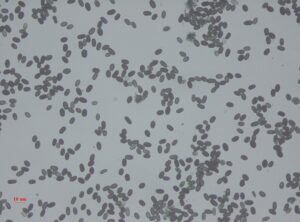
pathogenic fungi examples
Image Credit : Stachybotrys spores – Wikipedia

Most indoor air contaminants are considered infrequent. But the conidia are commonly present in cellulose-rich construction materials from wet or water-damaged structures, for example, wallpaper and drywall that are gypsum-based.

Stachybotrys chartarum takes time to grow. That means it is a slow-growing fungus and it does not take part in any competition against any other molds. This kind of fungus is hardly found in nature.

It is infrequently encountered in nature, and it rarely experiences the kind of living situation that human occupation can occasionally provide. Most indoor air contaminants are considered infrequent.

7. Talaromyces marneffei

In 1956, Talaromyces marneffei, which is formerly known as Penicillium marneffei, was discovered. The organism is indigenous to Southeast Asia, where it is a leading source of opportunistic diseases living with HIV/AIDS.

T. marneffei causes human diseases known as talaromycosis. They have also been recorded in HIV-positive patients present in Australia, Europe, Japan, the United Kingdom, and the United States.

Except for one, all of the patients had already visited Southeast Asia. The condition is thought to be an AIDS-defining sickness. Treatment for talaromycosis varies depending on the severity of immunosuppression and involvement of organ.

However, most Talaromyces marneffei isolates have low MICs. They have low MICs for posaconazole, amphotericin B, voriconazole, and itraconazole.

8. Blastomyces dermatitidis

Blastomycosis is a type of disease that is caused by the fungus named Blastomyces dermatitidis. It causes an aggressive and frequently fatal fungal sickness in humans and other animals in endemic areas.

This fungus is often present in soil, decaying wood which is wet, in places those are close to water bodies for example stream, lake or river.  Blastomyces dermatitidis can also be found in indoor areas such as in accumulated debris in moist sheds or shacks.

The fungus, Blastomyces dermatitidis, is native to the following areas:

  • Eastern North America, specifically boreal northern Ontario and southeastern Manitoba
  • Quebec south part of the St. Lawrence River,
  • Few regions from the US Appalachian Mountains        
  • Eastern mountain chains that are interconnected
  • The west coast of Lake Michigan, Wisconsin, and the entire area of Mississippi Valley
  • Valleys from few important tributaries such as the Ohio River.

It is considered to grow as a form of cottony white mould in the environment, which is same to the growth present in artificial culture at a temperature of 25 °C or 77 °F.

9. Emmonsia parva

Emmonsia parva, E. crescens, and E. pasteuriana altogether form the genus named Emmonsia. But still they show different ecological features of their own.

Emmonsia parva, also known as Chrysosporium parvum, is a saprotrophic fungus that has filaments, and this is one of three species that belong to the genus Emmonsia. The fungus is dimorphic in nature and grows in two distinct forms.

At ambient temperature, it develops as hyphae, but whenever conidia are heated to a temperature of 40 °C, they transform into bigger adiaspores. The fungus is well known for its link to adiaspiromycosis, a lung sickness that most typically affects small mammals but can potentially affect people.

While E. crescens is distributed all over the world, E. parva is only found in places like North and South America, Eastern Europe, Australia, and Asia. The fungus is predominantly a saprotroph, meaning it feeds on decaying matter.

10. Sporothrix schenckii

Sporothrix schenckii, a fungus prevalent in the environment all over the world, is named after medical student Benjamin Schenck, who was the first to isolate it from a human specimen in 1896.

The species can be found in soil, as well as live and decaying plant material like peat moss. It is the cause of sporotrichosis, sometimes also known as the “rose handler’s disease,” and can affect both humans and animals. Sporothrix schenckii can be present in either hyphal or yeast morphologies.

Sporothrix schenckii develops its hyphal morphology when growing in the nature or in the laboratories at a temperature of 25 °C or 77 °F. Colonies are wet, leathery to velvety, and have a highly creased surface when viewed under a microscope.

The color starts with white and gradually changes to cream and then to dark brown over time. This color is also known as “dirty candle-wax” color. When observed under microscope, hyphae are septate and have a diameter of 1 to 2 m.

At a temperature of 37 °C or 99 °F, Sporothrix schenckii develops its yeast nature either in the tissues of the host or in the laboratories. When observed macroscopically, the form of the yeast seems as clear as white or off-white colonies. Yeast cells are 2 to 6 m long and have an extended cigar-shaped appearance under the microscope.

Non pathogenic fungi examples

The following are some of the most common nonpathogenic fungus strains identified to cause ISR in crop plants:

  1. Mycorrhiza,
  2. Trichoderma sp.
  3. Penicillium sp.
  4. Fusarium sp.
  5. Phoma sp., etc.

Innate immunity has been shown to be triggered by a variety of signalling pathways, including salicylic acid, jasmonic acid, and ethylene.

Conclusion

There are different ways in which fungi can cause diseases. They are: replication of the fungus where fungal cells occupy tissues and disturb their functions, Immune response with the help of immune cells or antibodies Competitive metabolism by consuming the source of energy and nutrients stored for the host.

Also Read:
11 Single Cell Plant Examples: Detailed Explanations,Images

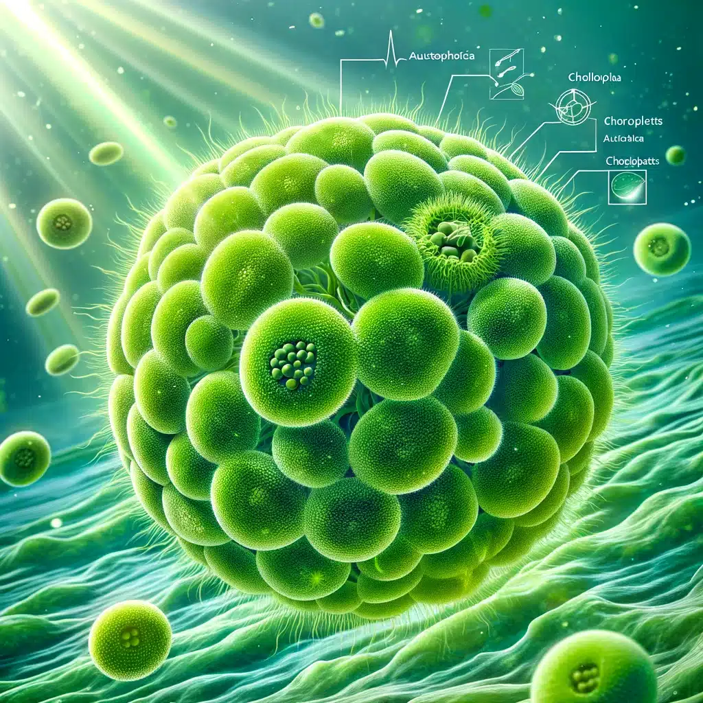
Chlorella autotrophica

Unicellular organism is also defined as the single celled organism. It is a creature that comprises of only a single cell which is different from a multicellular creature that contains many or multiple cells.

A plant single cell is referred to as that one plant cell that consists of unique traits as compared to its surrounding cells, which depend on its developmental stage, specific reaction towards environmental stresses, molecular heterogeneity, etc.

These single cell plants are basically the green algae that have chloroplast in them. We will discuss a few single cell plant examples here. These are species of different single celled plants that has different genus:

The above mentioned organisms fall under the division of Chlorophyta of the group of green algae. The single celled plants only fall under the category of green algae.

Living creatures on this plant is categorized into major groups- prokaryotic organisms and eukaryotic organisms. All the prokaryotes are unicellular in nature and are divided into classes like bacteria and archaea. Whereas eukaryotes are mainly multicellular in nature.

But eukaryotes can also be unicellular sometimes, for example, unicellular fungi, protozoa, and unicellular algae. Many multicellular creatures have a life-cycle stage that is unicellular in nature. Gametes, for example, are reproductive unicells for multicellular organisms.

Below we will discuss the different types of single cell plant examples.

Single cell plant examples:

Chlamydomonas reinhardtii

Chlamydomonas reinhardtii fall under the genus of Chlamydomonas. It is a green colored alga which has a only one cell and has a diameter of around 10 micrometres that can swim with the help of two flagella. It consists of a cell wall which is made up of glycoproteins those are rich in hydroxyproline. The cell wall also contains a large chloroplast that is of cup shape, a pyrenoid which is also large in size and an eyespot which helps in sensing the light.

The vegetative cells present in this species are haploid in nature. It has 17 tiny chromosomes. During the process of nitrogen starvation, these vegetative cells divide to form gametes that are haploid as their character. There are two types of mating procedures. Both the procedures are similar in emergence and thus are called isogamous. These are defined as mt+ and mt-containing regions that have the ability to fuse together to give rise to a zygote that is diploid in nature.

single cell plant examples
Image Credit: Cross section of Chlamydomonas reinhardtiiWikipedia

The zygote does not consist of any flagella. In the soil, it acts as a hibernating or dormant version of the species, and in the presence of light, the zygote goes through the process of meiosis and delivers four flagellated haploid cells that help in resuming the vegetative life cycle.

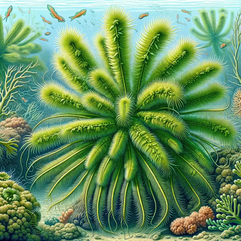
Caulerpa taxifolia

Among all the green algae, Caulerpa is the name of genus of seaweeds that belong to the family of Caulerpaceae. They are somewhat different because though they contain a single cell but comprises of several nuclei. This chararcteristic feature makes the one of the largest single cells on this planet. Caulerpa species may include- Caulerpa prolifera, Caulerpa taxifolia and Caulerpa racemosa.

Caulerpa taxifolia

Caulerpa taxifolia is a type of green seaweed. This kind of algae belongs to the genus Caulerpa. It can be found in the tropical seas of the Pacific Ocean, Caribbean Sea, and Indian Ocean. The likeness of its leaf-like lobes to those of the yew gave origin to the name of the species taxifolia, for example in Taxus.

Caulerpa taxifolia has a light green color. It consists of stems called stolons and is found on the floor of the sea. They may rise to a height of around 20 cm to 60 cm. C. taxifolia may sometimes get tangled in fishing gear and boat propellers. Mechanical removal, chlorine poisoning, or salt treatments are all options for controlling C. taxifolia.

Acetabularia acetabulum

Acetabularia acetabulum is the name of a species of green alga that belongs to the family of Polyphysaceae. These species can be found in the Mediterranean Sea at a depth of about one to two metres. This alga sticks to the sea floor, which helps of its root like structures called rhizoids.

This organism is made up of only one cell and has a single nucleus, which is located at the bottom of the stem. It consists of a cap which, when expanded, the nucleus separates once by the process of meiosis. This takes place when the nucleus has gone through the division several times by the procedure of mitosis, thus giving rise to numerous “secondary” nuclei that are haploid in nature.

This is a unicellular alga whose every frond is developed from a large single cell that contains numerous million of chloroplasts. In the daytime, they are continuously moving so that they can expose themselves to receive maximum amount of light, thus the green stems occur to be darker. But at night, they form clusters and the green stems appear to be pale.

In developmental biology, Acetabularia acetabulum has been used as a model organism. Acetabularia acetabulum’s principal reserve polysaccharide is starch, which is found in the form of granules within the stroma of the chloroplast. The sea slug Elysia timida is one of the predators of Acetabularia acetabulum.

Tetraselmis suecica

Tetraselmis is the name of the genus of phytoplankton. This is also green algae, which falls under the order of Chlorodendrales. They are identified by their extremely green chloroplasts. They have flagella in their cell bodies; pyrenoid is present inside the chloroplast and a scale-produced thecal wall.

Species that belong to this genus are mainly found in both the fresh water and marine ecosystems all around the globe. Because of their photosynthetic character, their habitat area is primarily restricted by water depth.

As a result, if adequate resources and light are accessible for net photosynthetic production, they may exist in a variety of aquatic situations. The species of Tetraselmis have been shown to be very useful for both industrial and research purposes.

Tetraselmis suecica is one of its species, which is a marine green alga. It consists of single cells that are mobile. These cells are visible under the observation of a light microscope up to a range of concentrations of one million cells per milliliter. This species can be cultivated for aquaculture in the form of foodstock. It is a motile chlorophyte and contains a high amount of lipid content.

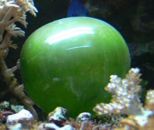
Valonia ventricosa

Valonia ventricosa are also known as sailor’s eyeballs or bubble algae. It is a species of algae that can be seen in the oceans of both tropical and subtropical areas all over the world. This species belongs to the phylum of Chlorophyta. It is one of the largest known unicellular organisms.

As it belongs to the phylum Chlorophyta, it is obvious that they contain chlorophyll in their cells, and thus they are able to perform photosynthesis. Valonia ventricosa has a coenocytic structure along with numerous chloroplasts and nuclei.

This organism comprises a large vacuole that is located in the centre and has a multilobular structure, that is, lobules extending from the centre of a spheroid area. It has a peripheral cytoplasm and its membrane is covered by the cell wall, which has a thickness of about 40 nm only. Valonia ventricosa usually grows individually, but sometimes, in rare cases, it can also grow in groups.

single cell plant examples
Image Credit: Valonia ventricosaWikipedia

They usually appear in the tropical and subtropical regions of the tidal zones, such as south to Brazil, the Indo-Pacific, north through Florida and the Caribbean. They can be found in every ocean on the planet, and they often live in coral shards. The maximum depth of viability that has been observed is around 80 metres, that is, 260 feet.

Read more about Single Cell Protein Example

Chlorella autotrophica

Chlorella autotrophica, or Chorella sp., is a species of euryhaline, which is a unicellular microalga that falls under the division of Chlorophyta. They are mostly present in brackish waters.

Chlorella autotrophica

The species is classified as an exclusive autotroph since it is unable to utilise organic carbon material as a food source. It’s sometimes mistaken for a Chlorella vulgaris variation.

C. autotrophica can be used for many purposes. The species has been used in rearing bivalves as a source of feedstock; it can also be fried in aquaculture, which is a source of the amino acid named L-Proline.

Due to its propensity to collect triglycerides during nitrogen starvation, the algae species is also a viable feedstock for biodiesel production.

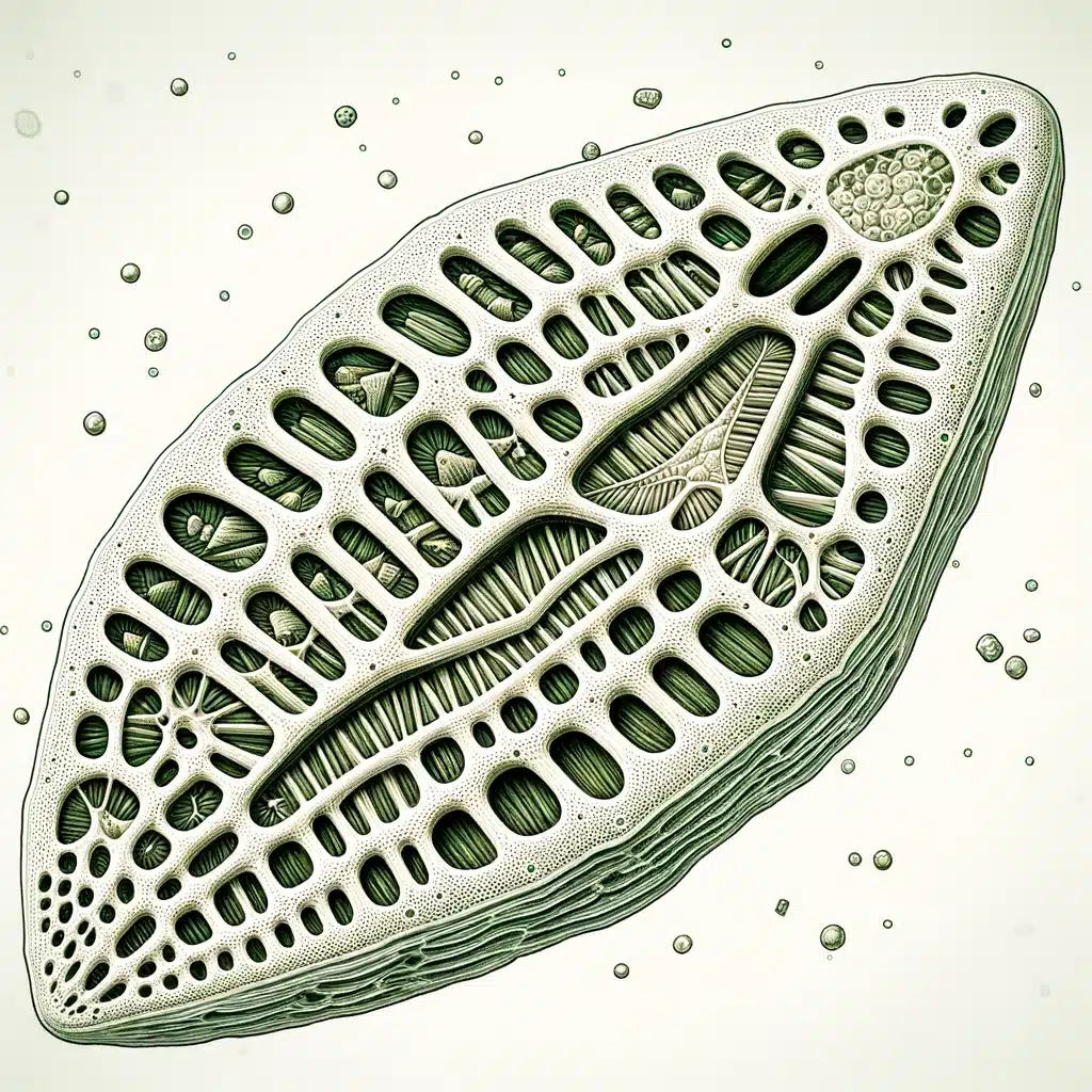
Pinnularia opulenta

Pinnularia is the name of the genus of algae that is more specifically, a type of diatom. It is primarily a freshwater alga and is generally present in moist soil and in ponds. They can also be seen in estuaries, sediments, oceans, and springs. The most widely used measure of this genus’s members is 40 cm (that is, 1.25 feet) under water at a temperature of 5 °C (that is, 41 °F).

Pinnularia opulenta

Pinnularia are unicellular creatures with elongated elliptical shapes. Pectic compounds on a stiff silica framework make up the majority of their cell walls. Thecae are two sections that make up their walls (or less formally, valves). The epitheca refers to the bigger outer valve, while the hypotheca refers to the smaller inner valve. A mucilaginous layer surrounds the cell.

The cytoplasm is structured in layers that mimic the structure of the cell walls. The nucleus is supported in the middle of a huge central vacuole with the help of a transverse cytoplasmic bridge.

Two chloroplasts are found that line the sides of the cells. They consist of two chlorophylls, namely, chlorophyll a and chlorophyll c, and also some pigments such as fucoxanthin and beta-carotene. The cytoplasm also comprises chrysolaminarin and a few volutins.

Peridinium

Peridinium is a genus of motile, marine and freshwater dinoflagellates. Peridinium species may include- Peridinium balticum (Levander), Peridinium digitale and Peridinium cinctum.  

Their morphology is regarded as typical of armoured dinoflagellates, and their shape is frequently depicted in dinoflagellate structural illustrations. Most of the Peridinium are either circular or oval in shape. They have a range of colors that starts from green to yellow or brown. Their size may vary from around 0.01 to more than 0.1 mm (that is, 0.0004–0.004 inch) in diameter.

The cell consists of a theca, which is the tough outer covering of the cell. This is classified into two parts, an epitheca and a hypotheca, which are more or less the same in size and are divided by a huge cutting which is known as the cingulum, also called girdle.

Peridinium is a huge genus that includes small to medium dinoflagellates, some of which are photosynthetic, but not all of them. Apparently a few photosynthetic species are capable of forming large blooms (also known as the “red tides”). Some peridinium seem to be entirely autotrophic, relying solely on photosynthesis for nutrition and energy.

Navicula is the name of a genus that has diatom algae that are in a boat shape. There are around 1,200 species of Navicula. The ability of Navicula diatoms is that they are able to creep about on one another and on rough surfaces like microscope slides. Navicula species may include Navicula bullata, Navicula tripunctata, and Navicula oblonga.

Navicula diatoms can reproduce without their parents being fused together. They reproduce by forming two new valves inside the frustules of the parent. The protoplast of each new cell is now trapped within one old and one new valve as the contents of the cell divide and separate.

Valves are lanceolate and circular, with slightly subrostrate apices and unprotracted apices. The axial region is smaller. The core portion is asymmetrically shaped and tightly expanded. The raphe is filiform and linear, with proximal ends that are minimally extended.

Bacillaria paxillifer

Bacillaria is a diatom genus that belongs to the family of Bacillariaceae. Bacillaria species may include- Bacillaria paradoxa and Bacillaria paxillifer.

Bacillaria paxillifer is a colonial diatom species that belongs to the family named Bacillariaceae. Colonies of this diatom have the ability to move. Members consist of long axes that are parallel to each other. The members glide against one another in a coordinated manner, allowing the structure to stretch and compress.

This genus is photosynthetic in nature and reproduces both with and without the parents getting fused to one another. In stacked colonies, cells are flattened and have the ability to move and slide along one another.

In girdle view, when present in colonies, cells are rectangular in shape, and in valve view, they are lanceolate in form. Raphe goes from pole to pole and is somewhat keeled.

There are two huge plate-like chloroplasts, one at either end of the cell. The nucleus is in the centre of the cell. The color of the cells is yellow-brown. Fibulae are tough, and the valve surface is coated in transverse parallel structures, which are known as striae.

Read more about Bioindicator Examples

Chaetoceros furcellatus

The genus Chaetoceros was first discovered by Ehrenberg in the year 1844. Chaetoceros species may include Chaetoceros furcellatus, Chaetoceros angularis, and Chaetoceros intermedius.

The Arctic neritic diatom, Chaetoceros furcellatus, belongs to the Chaetoceros genus. Finding the extremely distinctive resting spores is the easiest way to identify this species. In the Barents Sea, C. furcellatus is a popular and essential species. Cells are linked together to form long, slightly rounded strands.

Although Chaetoceros is mainly a marine genus, there have been reports of species in American inland waters. It is a kind of centric diatom with a silica-based frustrule or cell wall that consists of a long, thin spine known as setae.

The spines link the frustules collectively, forming a cell colony. Colonies of cells can create looped, coiled, linear, or curved chains. The size of a cell can vary from 10 um to 50 um.

Also Read:

9+ Seedless Plant Examples: Detailed Explanations And Images

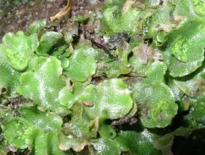
800px Lunularia cruciata 300x227 1

A magnificent diversity of plants that reproduce without seeds automatically creates the planet’s surface.

Plants that reproduce without seeds constitute only a small part of the plants in our terrestrial landscape. Around three hundred million years ago, seedless plants took control of the landscape and started growing in a vast area of swampy forests in the Carboniferous period.

The dead bodies of these plants form a large blanket of coal that we mine in present days. We will discuss a few seedless plant examples here. Modern-day seedless vascular plants may include the following:

According to existing evolutionary theory, all plants, including green algae and land plants (land dwellers), are monophyletic, meaning they are all the descendants of a single ancestor.

The plants had to struggle enough to go through the transition of water plants to land plants. They had to develop their characteristics of avoiding drying out without water, had to scatter reproductive cells in air, had to develop strength and support for different structure and also had to be different than water plants for sunlight to be captured and filtered.   

Another difference between seed and seedless plants is that seed plants evolved modifications that permitted them to thrive in even the driest environments on the planet, but many seedless plants nevertheless need to be kept wet. Not all plants have achieved complete water independence.

Some seedless plant examples:

Bryophytes

The nonvascular plants known as bryophytes are the closest living relatives of ancient terrestrial plants. The oldest bryophytes made their appearance some 490 million years ago, during the Ordovician epoch.

As they are nonvascular plants, they lack lignin like vascular plants. Lignin is a strong polymer found in the cell walls that are present on the stems of vascular plants.

Due to these parts and further resistant structures, the possibility of bryophytes forming their fossils is greatly reduced. Although some spores comprised of sporopollenin have been identified, they have been linked to ancient bryophytes.

There are about 18,000 species of bryophytes, which thrive mostly in damp habitats, although some grow in deserts. In a bryophyte, all the conspicuous vegetative organs belong to the haploid organism, or gametophyte. The diploid sporophyte is barely noticeable.

Bryophytes consist of about 18,000 species of its own species that are mainly found in the swampy areas, though some bryophytes can also grow in the desert habitats.

In a bryophyte, all the responsible vegetative organs belong to the organism that is haploid in nature, or gametophyte. But the sporophyte which is rarely noticeable is diploid in nature.

The bryophytes form gametes that can swim with the help of flagella. Bryophytes contain sporangium, a multicellular structure that helps in reproduction. The embryo present remains connected with the parent plant, so that it can help in the nourishment of the embryo. The land plants comprise this characteristic feature.

Liverworts

Liverworts that belong to the phylum Marchantiophyta may be considered as plants that belong to a close relation to the ancestor plants that shifted to the terrestrial. Liverworts have populated many domains in the environment and more than 6,000 existing species have been added to the mix.

Lobate are the green structures that are formed by some gametophytes. The shape of the lobes is comparable to that of the liver, which is where the common name comes from for this division.

The cycle of the reproduction system begins when there is a release of haploid spores from the sporangium that has developed on the sporophyte. These spores are then dispersed by water or wind so that they can germinate into a flattened structure called a thalli, which is joined to the substrate by means of filaments that are thin and single celled.

Male and female gametangia develop on separate, individual plants. When a spore is released, the male gametes swim towards the female gametangium or the archegonium with the help of their flagella, and thus fertilization takes place.

800px Lunularia cruciata
Image Credit : Liverwort (Lunularia cruciata)- Wikipedia

The zygote begins to develop into a small sporophyte which still remains connected to the parent gametophyte. During the process of meiosis, it gives rise to the spore of the next generation.

Liverwort plants can also reproduce without parents being interacted with each other. This process is done by the branches that are broken or by scattering of the fragments of the leaves known as gemmae.

The gemmae are the small, whole, complete bits of plant generated that are found on the surface of the thallus in the form of a cup. Their process of reproduction is something different. While there is rain, raindrops splatter these out of the cup-like structure. Thus the gemmae get somewhere nearby and further develop into gametophytes.

Hornworts

The hornworts that belong to the phylum Anthocerotophyta have a populated diversity of domains on land, despite the fact that they are rarely far from a supply of water. Hornworts have almost 100 species of their own varieties. The short, blue-green gametophyte is the most common period in the life of hornworts.

The sporophyte is the group’s defining trait. It’s a long, narrow pipe-like structure that sprouts from the parent gametophyte and continues to grow throughout the plant’s life.

seedless plant examples
Image Credit: Life cycle of a typical hornwort PhaeocerosWikipedia

Stomata are present in the hornworts and the sporophyte has a lot of them. Photosynthetic cells that are present in the thallus comprises of a single chloroplast.

Meristem cells that are found at the bottom of the plant continue to divide and add to the height of the plant. Several hornworts develop symbiotic relationships with cyanobacteria that help in the fixation of nitrogen from the environment.

Tracheophytes

Tracheophytes are the vascular plants that form the controlling and most noticeable part of terrestrial group of plants. Earth’s 90 percent of the total vegetation consist of the tracheophytes that represent more than 260,000 of its species. Their success and capacity to spread to all habitats is due to a number of evolutionary advances.

In the vascular plants that are seedless, the sporophyte which is diploid in nature is the dominant phase of the lifecycle. The gametophyte has evolved into a quiet, self-contained organism. In the dominant period of the lifecycle, there has been a clear reversal of roles throughout plant history.

The seedless vascular plants are still dependent on water during the process of fertilization so that the sperm can swim on a layer of moisture with the help of flagella towards the egg. This step in the reproductive process explains why ferns and their companions thrive in moist conditions.

Club mosses

The club mosses belong to the phylum Lycopodiophyta. On the forest floor, horsetails with their jointed stalks and wiry leaves grow on tree trunks, while mosses thrive on tree trunks.

The club mosses that belong to the phylum Lycopodiophyta are the most ancient category of vascular plants that are seedless in nature. They inhabited the Carboniferous terrain, expanding to be tall trees and establishing vast wetland forests.

The phylum Lycopodiophyta has almost 1,200 species, which includes other plants like the quillworts (Isoetales), the club mosses (Lycopodiales), and spike mosses (Selaginellales). None of these are bryophytes or true mosses. Lycophytes can be either homosporous or heterosporous.

Horsetails

Horsetails, whisk ferns and ferns all belong to the same phylum named Monilophyta. But horsetail is placed in the different category, that is, Class Equisetopsida.

The genus named Equisetum is the sole survivor of the Arthrophyta, a vast group of plants that produced gigantic trees and extensive swamp forests during the Carboniferous. The plants are frequently found in marshes and moist regions.

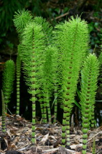
Equisetopsida
Image Credit: Horsetails (Equisetopsida)- Wikipedia

The leaves and branches come out in the form of coils from the joints that are equally spaced. The process of photosynthesis is mostly carried out by the green part present on the stem. The leaves are not able to take part in photosynthesis because of their needle-shape.

The stiffness of horsetail plants is due to the presence of silica that is collected in the cells of the epidermis. Rhizomes are the names of the stems that grow under the ground that anchor the plant to the ground. Nowadays, these horsetails are homosporous in nature, and they produce gametophytes that are bisexual.

Ferns

Ferns are believed to be the most modern vascular plants that are seedless and they show the features that are commonly seen in plants that consist of seeds. Ferns form large leaves and branching roots.

Ferns can be easily recognized as seedless vascular plants due to the presence of their enormous fronds. The stems of the ferns are often considered rhizomes, notwithstanding the fact that rhizomes are present only in a few species that grow underground. Ferns are terrestrial plants.

A few species of ferns can grow up to a height of 20 metres, that is, 66 feet tall, for instance, Cyathea brownii found on Norfolk Island and Cyathea medullaris present in New Zealand. New leaves usually grow into stalks by unwrapping a tight spiral known as crozier or fiddlehead. Leaves of ferns are categorized into two groups namely- sporophylls and tropophylls.

Spores are produced by sporophylls, whereas tropophylls do not produce any spores. The underground organisms present do not perform photosynthesis, and they receive their nutrients and water from the soil. They are usually fibrous in nature, and their structure is extremely similar to that of the roots present in seed plants.

Ferns are found in a wide range of habitats, from the tropics to temperate woods. Even if some species can thrive in arid conditions, most ferns prefer wet, shady areas.

Whisk ferns

Psilotum nudum, known as the whisk fern, is a plant that is similar to the ferns. Just like the other species present in the order Psilotales, it does not contain roots. The name, Psilotum nudum, suggests “bare naked” in Latin, as it is deprived or appears to lack many of the organs that are found in typical vascular plants, as an outcome of evolutionary reduction.

Whisk ferns are also known as the psilophytes as they lack both leaves and roots in them, which can be a consequence of evolutionary reduction. Photosynthesis is carried out by the green part of the stem of the whisk fern. The embryo consists of only two parts namely- distal shoot apex and proximal foot.

The shoot apex gives rise to a rhizome where the roots are absent. This is because P. nudum and its other relatives may also be considered epiphytes sometimes. Rather than roots, the rhizome will gradually generate drastically diminished leaves. Auxin concentrations have a big impact on these processes.

Sporangia are present at the top of the branch where tiny yellow knobs like structures are found. Whisk ferns are categorized beyond the true ferns. Recent studies of DNA illustrates that this group of ferns and whisk ferns both lost their roots as well as their vascular tissues during the evolutionary period.

The plant, that grows wild in the southern part of Japan, was once cultivated in a large amount in the Japanese gardens in the form of an ornamental plant.

P. nudum can be found in the tropical areas of Africa, tropical Asia, central America, the southern part of America, tropical and subtropical North America, Australia, Hawaii, the southern part of Japan, the Island of Lord Howe, New Zealand, and a few remote areas in South-West Europe.

Algae

Algae, whose singular form is alga, are the members of a category named Kingdom Protista. They are the predominantly aquatic organisms that can carry out the process of photosynthesis. The study of algae is known as phycology, and the person who studies about algae is known as a phycologist.

Their pigments that help carry out photosynthesis are very much different from those found in normal plants. So, they have cells whose traits do not match those of other plants and animals. Algae is necessary economically because it is the source of crude oil. Sometimes they are also consumed as food products. Algae has several pharmaceutical and industrial uses for humans.

Just like the plants mentioned above, algae is also deficient in true roots, leaves, and stems. They share traits that are similar to those of the avascular lower plants, such as hornworts, mosses, and liverworts.

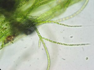
Stigeoclonium sp zugespitzte seitenzweige
Image Credit: Green algae- Wikipedia

In the beginning of the 1830s, algae were categorized into some important classes based on their color, such as green, red, and brown. These colors are basically the reflection of various pigments in the chloroplast like chlorophyll, carotenoids, and anthocyanins, as well as some water-soluble proteins such as phycobiliproteins.

The size of algae has a different range of magnitude. For example, sometimes algae may be of a single cell organism, while the largest size of algae may comprise of millions of cells. According to the morphology of their vegetative, growth, or mode, algae can be classified into numerous categories:

1. Filamentous forms that have cells where they are strung together like beads, for example in Spirogyra.

2. Parenchymatous forms, or tissue like formations, such as in Macrocystis, which is the giant kelp that can be several metres in length.

3. Algae that are coenocytic, like the green seaweed named Codium, can grow to quite enormous sizes without developing discrete cells. Coenocytic algae are generally multinucleated in nature, unicellular algae with no cell walls that divide the protoplasm (cytoplasmic and nuclear components of a cell).

Few algae comprises of flagella so that they can swim through the water.

Mushroom

A mushroom, often known as a toadstool, is a fungus’ fleshy, spore-bearing fruiting body that grows on the surface of earth, on ground, or on its food supply.

A mushroom grows from a nodule, or pinhead, with a diameter of less than two millimetres, known as a primordium, which is often located on or near the substrate’s surface. The mycelium, or mass of threadlike hyphae that makes up the fungus, produces it.

Mushrooms can also be edible sometimes. Edible mushrooms may include some fungal species that are either cultivated or wildly harvested. Mushrooms that are cultivated simply and some mushrooms that are commonly found in the wild are frequently used for economic purposes and are accessible in markets. A few examples of these kinds of mushrooms are as follows: morel, prized truffle, and matsutake. Certain preparations may make certain deadly mushrooms safe to eat.

Before we are about any mushroom which is wild that it can be consumed or not, it be passed by some process of identification. The only safe method to guarantee edibility and protect against probable accidents is to accurately determine and properly identify a species.

Please click to learn more on Single Cell Plant Examples.

Also Read:

5 Independent Assortment Example: Detailed Explanations

peas 300x264 1

The Law of Independent Assortment states that alleles for separate traits are passed independently of one another.

The biological evolution of one allele for a particular trait is not related to the biological evolution of one allele for any other trait. Mendel proved this law with the help of his experiments that he performed on the dihybrid cross. That is to say, an allele received by a gamete for a specific gene does not disturb any other allele that other gene receives.

Here are some independent assortment example –

Independent assortment takes place during the procedure of meiosis. Meiosis is the same as the process of mitosis. Their end products are gamete cells. These gamete cells contain half the DNA of the regular diploid cells and thus are considered haploid. This part is important for reproduction between two parents because it permits the two gametes to combine with each other to form a zygote that is diploid in nature and contains all the DNA that is required to produce a new organism.

The law of independent assortment

The law of segregation helps us estimate how a single trait linked to a single gene will be passed down.

We need to know if the two genes are inherited separately or not in order to produce a precise forecast. So, we need to determine whether they “neglect” one another while sorting into gametes or if they “stay together” and are inherited as a group.

Gregor Mendel discovered that several genes acceded to each other independently, and this is what is known as the law of independent assortment. Mendel’s law of independent assortment states that the alleles of two (or more) different genes get sorted into gametes independently of one another. That is, an allele obtained by a gamete for a specific gene does not interfere with any other alleles received by that gene.

The law of assortment explains the random inheritance of the genes that take place from paternal and maternal sources. This law states that various genes and their corresponding alleles are acceded to separately or independently within the organisms that can reproduce sexually. While the procedure of meiosis takes place, chromosomes get separated into various gametes. When there is crossing over, the genes present on a chromosome get interrelated so that they can reorganize themselves. As a result, each gene is transmitted autonomously.

Independent assortment in alleles

In a Mendelian system, alleles that are located at loci on distinct chromosomes must be acquired separately since each homologue will assort individually.

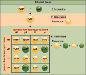
The biological evolution or biological selection of one allele for one trait is not related to the biological evolution of another allele for any trait. With the help of his experiments with the dihybrid cross, Mendel discovered this law. In his experiments where he found monohybrid crosses, he achieved a theoretical result of 3:1 ratio between the phenotypes of dominant and recessive.

Whereas in his other experiments where there were dihybrid crosses, he found the ratio to be 9:3:3:1. This proves or demonstrates that one of the pairs of alleles is acquired separately from the other, with each having a 3:1 phenotypic ratio.

Independent assortment in meiosis

IN RABBITS:

Let us talk about a hypothetical population of the rabbits that are said to have only two traits that are visible: firstly, the fur color, that can either be black or white; and secondly, the eye colors, which can either, be green or red. The black fur allele (denoted as B) acts dominant over the white (denoted as b), whereas on the other side, the green eye allele (denoted as G) acts dominant over the red (denoted as g).

In this hypothetical example, two bunny rabbits that are hybrid in nature are mixed. This means that, both the bunny rabbits will have black colored fur and their eyes will be green in color, but still, their genotype will be heterozygous in nature. Thus, their genotype will be denoted as BbGg. All of the rabbits in this population of two share the same mix of features. To put it another way, they’re all black and have green eyes.

Before breeding starts, each bunny rabbit is required to create gametes. While this process continues, not only that the alleles are divided (the law of segregation), but each copy of each chromosome is assigned to a different gamete at random. This means though the parental phenotypes are black fur with green eye color, the offspring or the babies can acquire various combinations or sequences of these features or traits.

For example, a single baby can receive a genotype of bbgg, where its phenotype will be white fur and red eyes. On the other hand, an offspring can also have Bbgg as its genotype, where its phenotype will be black fur and red eye color. This shows the Law of Independent Assortment.

Independent assortment in chromosomes

While meiosis takes place after the discovery of chromosome, it became easy to explain the independent assortment as a result of the independent movement of every pair of homologous chromosomes and also their mode of action in the process of meiosis.  For every gene independent assortment is necessary so that they can create new genetic sequences and combinations that will help increasing the genetic diversity, also known as genetic variations, within the organisms.

Mendel’s law of independent assortment defines that the resultant chromosomes are classified in a random manner by fusing the chromosomes of maternal and paternal sources. Finally, the zygote forms a mixture of chromosomes rather than a distinct set of features from each parent.

Meiosis is a kind of cell division the decreases the chromosome number of a parent by half of its original number to create four reproductive cells which are known as gametes. In humans, diploid cells contain 46 chromosomes; from these 46 numbers of chromosomes, half of the number of chromosomes is acquired from maternal (mother) source, that is, 23 chromosomes from female gamete. The other half of the number of chromosomes is from paternal (father) source, that is, 23 chromosomes from male gamete. Pairs of these similar chromosomes are known as the homologous pair of chromosomes.

During the process of meiosis, the homologous pairs of chromosomes are divided in half to give rise to haploid cells, and this division or assortment of the homologous pairs of chromosomes is random in nature. This indicates that almost all the chromosomes from a maternal source will not be grouped together in one cell, whereas almost all the chromosomes from a paternal source will be grouped together in another. Alternatively, after the process of meiosis takes place, every haploid cell consists of a mixture of genes from each parent (mother and father) of the organism.

Independent assortment of plants

PEA COLOR AND PEA SHAPE:

Gregor Mendel executed various experiments that involve breeding of pea plants. He studied how “units of heredity” used to function in their own way, which gradually became to be known as the genes after when DNA was discovered and confirmed that they are the material that codes for genetic information.

Mendel discovered the Law of Independent Assortment after he did his experiment of breeding the two various pea plants with two separate characteristics. His experiment was to breed a yellow pea plant that has round peas with that of a wrinkled plant that has green colored peas. As because the yellow plant with round peas acts dominant over the winkled plant with that of green peas, thus all the offspring or the next generation species or peas will be of yellow and round shaped.

When this first generation was crossbred with each other in a dihybrid cross, the second generation had a lot of variation. Peas were no longer merely yellow and round or green and wrinkled; some were green and round and others yellow and wrinkled.

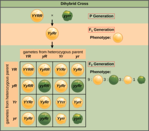
Moreover, in a ratio of 9:3:3:1, the offspring displayed their traits or characteristics. This means, nine peas were round in shape and yellow in color, three peas were round in shape but green in color, other three peas were wrinkled in shape and yellow in color and only one pea was wrinkled in shape but green in color.

This ratio remained constant for every other dihybrid that was crossed afterwards. Mendel observed 9 yellow round: 3 yellow wrinkled: 3 green round: 1 green wrinkled pea.

independent assortment example
Independent Assortment in Peas- Wikipedia

This kind of variation took place because only one allele was passed on to their offspring by each of the parent. Because the yellow plant with round peas has a dominating role over the winkled plant with green peas, all progeny or subsequent generations of peas will be yellow and round.

Mendel’s experiment indicated that the alleles for round or wrinkled peas were inherited independently from the alleles for yellow or green peas because the plants were not only round and yellow or green and wrinkled. They are now known to exist on distinct chromosomes, allowing them to be jumbled up while the process of meiosis takes place.

Independent assortment of animals

IN Trypanosoma brucei:

Trypanosoma brucei is a zoonotic protozoan parasite species complex that are transferred by tsetse flies and categorized into three subspecies-

  1. Trypanosoma brucei gambiense
  2. Trypanosoma brucei rhodesiense
  3. Trypanosoma brucei brucei

In human, the first two subspecies Trypanosoma brucei gambiense and Trypanosoma brucei rhodesiense cause sleeping sickness. On the other hand, the third Trypanosoma brucei brucei, causes cattle illness. Trypanosoma brucei brucei is not infective to humans.

Because no chromosomal condensation has been observed in any life cycle stage and no gamete-like phases have been identified, traditional genetic analysis has been used to determine if this parasite has a reproduction cycle and undergoes meiosis.

T.brucei is diploid in nature and the offspring of the cross would be expected to be heterozygous in nature for markers that are homozygous in nature and the difference between the parents, but would only acquire one allele from each locus where the parents are heterozygous. In a Mendelian system, every heterozygous locus’s two traits are transmitted in a 1:1 ratio, whereas those on separate chromosomes are acquired separately.

What phase is independent assortment?

Independent assortment takes place during the procedure of meiosis. Mitosis and meiosis are similar processes. Gamete cells are the end result of their work. These haploid gamete cells contain half the DNA of typical diploid cells and are therefore classified as haploid.

In eukaryotes , the independent assortment occurs during meiosis in the metaphase l phase of meiotic division. It gives rise to a gamete that carries a mixture of chromosomes. Gametes consist of half the number of chromosomes that are present in a regular diploid somatic cell.

During the production of gamete, the regular number of chromosomes that is diploid in number gets reduced to half of its original number in the course of meiotic division to form gametes that contain only 23 chromosomes thus considered haploid in nature. While there is sexual reproduction, the female gamete and male gamete combine with each other to bring out a newly formed organism or a formation of diploid zygote. The independent assortment occurs in eukaryotes during the metaphase l phase of meiotic division during meiosis.

The Law of Independent Assortment studies the genetic inheritance that is passed randomly between both parents. We know that the normal count for human diploid cells is 46 chromosomes. From these 46 numbers of chromosomes, half of the number of chromosomes is acquired from maternal source, that is, 23 chromosomes from female gamete which is also specified as the ovum or the egg cell.

The other half of the number of chromosomes is from paternal source, that is, 23 chromosomes from male gamete, specifically known as the sperm cell. As mentioned in the Law of Segregation, during the meiotic division two of the homologous chromosomes part from one another.

During crossing over, the linked genes that could not be assorted into a random order permit those genes to reorganize themselves when meiosis takes place. During this procedure, the interchange of the chromosomes that are homologous in nature takes place both in the chromosomes of maternal and paternal sources to confirm that the independent assortment of the linked genes has taken place successfully.

The resultant chromosomes are ordered independently by fusing the chromosomes of maternal and paternal sources, according to Mendel’s law of independent assortment. Finally, the zygote forms a mixture of chromosomes rather than a distinct set of features inherited each parent. As a result, chromosomes are regarded to be independently arranged, resulting in a zygote with a mix of maternal and paternal chromosomes.

Is independent assortment the same as crossing over?

Independent assortment occurs only when the genes are placed on various chromosomes. If the two genes are located far from each other on the same chromosome, then crossing over necessarily separate the genes and thus the genes are now able to assort independently.   

The second law of Mendel is not applicable for all the genes. Genes are “linked” when they are close enough with each other on the same chromosome and travel methodically while meiosis occurs. Thus, independent assortment does not happen with linked genes. Only when the genes are spread across several chromosomes does independent assortment occur.

Independent assortment helps in the production of new combinations or new sequences of alleles. While meiosis l takes place, crossing over occurs during the process of prophase and independent assortment occurs during the process of anaphase. While these happen, new sequences of alleles occur for the sets of chromosomes. Random fertilization of the gametes produced during meiosis also introduces genetic diversity (genetic variation). There are various marks while genetic diversity can get increased during sexual reproduction.

Crossing over happens during prophase and independent assortment happens during anaphase during meiosis l. New allele sequences for the chromosomal sets emerge as a result of these events. Genetic variation is also introduced through random fertilization of gametes produced during meiosis. Any of the genetically unique sperm generated by a male may fertilize the genetically unique egg produced by a female.

While metaphase l takes place, the pairs of chromosomes that are homologous in nature are arranged along the plate of metaphase. The alignments of the pairs that are homologous are random in nature and vary for every cell that passes through the process of meiosis. One maternal and one paternal pair of sister chromatids are seen in each tetrad.

The tetrad corresponding to human chromosome 1 in one cell, for example, may align so that the paternal sister chromatids face one pole and the maternal sister chromatids face the other. In another cell, there is a 50% probability that the opposite orientation will occur during metaphase I.

Also Read:

5+ Multiple Alleles Example: Detailed Explanations

drosophila 300x132 1

A gene’s alternative form is known as the allele. These alleles are responsible for the variation in phenotypic expression of a particular trait, such as the difference between brown and green eyes.

Alleles are two or more versions of a gene. Each gene is passed down in two forms, one from each parent. As a result, two distinct alleles for a trait would be present. There are various multiple alleles example we can talk about.

What are multiple alleles?

The term allele refers to one of a gene’s variants. Genes are expected to show variations or diversity in genetics; alleles combine to generate the collection of genetic information that characterises a gene.

A variation of a similar nucleotide arrangement that encodes the blend of a quality item at a similar area on an extensive DNA particle is known as an allele. An allele can be founded on a solitary nucleotide polymorphism at its generally fundamental level (SNP). It can be based on tens of thousands of base pairs at higher levels.

Multiple alleles are defined as three or more different types of a single gene that are in the same area. In a single organism, however, only two of the alleles can exist. Individuals in a population or species of organisms often have numerous alleles at each locus. The quantity of alleles (polymorphism) present at a locus, or the negligible part of heterozygotes in the populace, is a proportion of allelic variety.

The expression “allele” is a short type of allelomorph, which was begat by British geneticists William Bateson and Edith Rebecca Saunders to portray variable variations of a quality found as different aggregates at the beginning of hereditary qualities. Allelo- is a Greek word that means “mutual,” “reciprocal,” or “each other.”

If we talk about the ABO blood group, it is generally accessed by the three alleles, out of which only two are found in one individual. The ABO gene, which has six common variations, is responsible for blood categorization (alleles). Pretty much every experienced human’s aggregate for the ABO quality is some blend of these six alleles, as per populace hereditary qualities.

A null allele is defined as an invalid allele as it has only one protein. The quality capacity of the null allele is generated as ordinary.

Multiple allele examples in plants

A diploid organism contains two alleles only in respect of one gene. But if there are more than two alleles in a single gene, the condition is known as multiple allelism.

Mendelian segregation in pea plant:

Multiple alleles refer to the condition when three or more genes are present at the same locus on an individual chromosome. In short, numerous alleles of a solitary quality are called different alleles. The concept related to multiple alleles is referred to as “multiple allelism”.

The traits of the pea plant were studied by Gregor Mendel. His studies conveyed simple and complete dominance. The pea plants had two alleles that influenced their contribution to the traits shown by the plant. Later, it was known that sometimes the same traits consist of more than two alleles, and they give codes for the phenotypes of the plant. This made it possible to see many more phenotypes for any given trait while still adhering to Mendel’s Laws of Inheritance.

A specific region of the chromosome was inhabited by 2 types of genes in Mendelian inheritance: a normal gene that denotes a round seed shape and a mutant recessive gene that denotes a wrinkled seed shape. It’s probable that, in addition to the wrinkled gene, there are many other mutations in peas. The typical allele and at least two freak qualities will involve this area.

Self-sterility in plants:

Tobacco’s self-sterility, Nicotiana longiflora, was described by Kolreuter in 1764. East was the one who devised the rationale. According to him, self-sterility is caused by a series of alleles known as s1, s2, s3, and s4, among others. The hybrids S1/S2 or S1/S3 or S3/S4 are self-sterile since pollen grains from these kinds could not mature. However, pollen from S1/S2 was effective and capable of fertilisation with S3/S4.

The genes that cause self-sterility in plants are thought to work by modulating the pollen tube growth rate. In favorable combinations, the pollen tube expands more and more swiftly as it approaches the ovule, but it slows down significantly in incompatible ones, causing the flower to wither away before fertilization may take place.

Multiple allele examples in animals

Wings of Dorsophila:

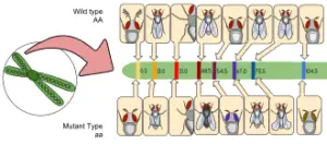
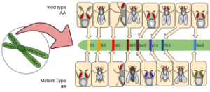
Drosophila wings are normally long. Two transformations happened at a similar locus in various flies, one coming about in minimal or decreased wings and the other in antlered or less created wings. Alleles with minimal and antlered alleles have an ordinary quality and are alleles of a similar typical quality. The letter “vg” stands for vestigial, while the letter “vga” stands for antlered wing. The normal allele is represented by the sign +.

Thus, there are three races of Drosophila:

(i) Long ++ (+/+)

(ii) Vestigial vg vg (vg/vg)

(iii) Antlered vga vga (vga/vga)

multiple alleles example
Image Credit- Drosophila – Wikipedia

The F1 hybrids produced by crossing a fly with vestigial wings with another fly with antlered wings had intermediate wing lengths, showing that neither mutant gene is dominant. The minimal antlered compound is a half and half that has two changed qualities at a similar locus.

In addition to the above traces and sacred wings, there are some other mutations in the same place, resulting in the loss of notched wings, strap wings, or  wings. They all have several alleles.

Coat color in rabbit:

A number of distinct alleles influence the colour of rabbit skin. Brown is the natural color of the skin. Apart from that, there are albino and Himalayan white mutant races. In comparison to the albino, the Himalayan has a darker nose, ear, foot, and tail.

Albino (a) and Himalayan (ah) are allelic mutant genes that share the same locus. The normal allele (+) is recessive in both albino and Himalayan people. In the F1 generation of a cross between an albino and a Himalayan, the result is a Himalayan, not an intermediate, as is the case with other multiple alleles.

Multiple alleles in blood groups

The agglutination test was used to divide a large number of people into these four groups, and the blood group distribution in the offspring of known blood group parents was explored.

These blood characteristics are determined by a series of three allelic genes, IA, IB and i,according to the evidence:

Blood Groups Genotype
AB IAIB
B IBIB or IBi
A IAIA or IAi
O ii

IA is an antigen A-producing gene, IB is an antigen B-producing gene, and i is a gene that generates neither antigen. Blood transfusion, contested percentage instances, and demographic description all benefit from the availability of these alleles in humans, as well as the ease with which blood groups may be determined.

The alleles of the genes, which control a variety of blood biochemical properties, work in such a way that each allele in the heterozygous compound IAIB has its own set of traits and effects. Both antigens A and B are present in the cells of the heterozygote. IA and IB, on the other hand, have complete domination over i, which lacks both antigens.

A table shows the possible blood types of children from parents of various blood groups:

PARENTS CHILDREN
Phenotypes Genotypes Phenotypes Genotypes
O x O ii X ii O ii
O x A ii X IAIA or IAi O, A ii, IAi
O x B ii X IBIB or IBi O, B ii, IBi
O x AB ii X IAIB A, B IAi, IBi
 A x A IAIA or IAi X IAIA or IAi A, O IAIA, ii
A x B IAIA or IAi X IBIB or IBi A, B, AB, O IA, IB,IAIB, ii
A x AB IAIA or IAi X IAIB A, B, AB IAIA, IBi, IAIB

Multiple alleles example blood type

Rhesus (Rh) factor is a protein located on the periphery of red blood cells that is inherited. One is Rh positive if the protein is present in their blood. Rh negative means when one person’s blood does not carry the protein in them.

Rh gets its name from the fact that rhesus monkey blood is used in the basic test for detecting the presence of the Rh antigen in human blood. Each person inherits two Rh factors, one from each parent, as part of their genetic makeup. Only when both parents have at least one negative component can a child be born with a negative blood type.

Rh + blood is the most common blood type. Levine’s research focused on erythroblastosis foetalis, a type of anemia that arises in newborns on rare occasions. It was discovered that most infants with this anemia are Rh-positive, as are their fathers; however, their mothers are Rh-negative.

The following is a description of the disease’s origin: Anti-Rh antibodies are formed in the mother’s bloodstream when the Rh+ foetus develops in the uterus of an Rh– mother. These antibodies build up in the mother’s blood to the point that they can assault the foetus’ red blood cells, especially if she has several Rh+ pregnancies in a row.

Haemolysis as well as anemia are caused by the response caused by the antibodies present in the body of the mother and the red cells of her unborn child. Sometimes these diseases can be critical, and they can cause the death of the newborn or the abortion of the foetus.

Are multiple alleles the same as polygenics?

Several allele inheritances occurs on the same DNA strand, whereas polygenic inheritance occurs on multiple DNA strands.

Multiple alleles are implicated in the determination of a single characteristic by complete dominance or codominance, whereas polygenic traits generate a particular trait in a population via codominance or incomplete dominance of every polygene.

A bunch of non-allelic genes help in regulating the polygenic features, while a varied number of alleles are found in different forms of a solitary gene. The number of DNA strands involved is the key distinguishing feature. The non-allelic genes generally regulate the polygenic features. On the other hand, several alleles include a gene and its different forms.

Environmental factors sometimes have a small effect on the identification of the traits while several alleles are being revealed. Environmental influences play a considerable impact on the characteristics that have been discovered in the polygenic inheritance.

Are multiple alleles and inheritance patterns related?

Multiple alleles are a form of non-Mendelian pattern of inheritance in which there are more than the usual two alleles that code for a species’ trait.

When numerous alleles are involved in a trait, a variety of dominance patterns might emerge. When one of the alleles is entirely recessive to the others, any of the others that are dominant to it will disguise it.

Different alleles may also be co-dominant and exhibit their qualities equally in the individual’s phenotype. When alleles are combined in a genotype, there are various circumstances where incomplete dominance occurs. An organism with this sort of inheritance linked to several alleles would have a blended phenotype, which combines the features of both alleles.

Can a gene have multiple alleles?

Any individual consist of only two alleles in the gene locus but still a particular gene may include more than two alleles in it.

In genetics, it is possible at population level that multiple alleles are taking place, despite the theory that humans can have only two alleles in one gene.As a result, numerous alleles play a significant role in generating variety within a species. It’s thought that the majority of human genes have more than two typical alleles.

Multiple allele characteristics are traits regulated by a single gene having more than two alleles. The ABO blood type is an example. The blood type of a person is determined by which antigens (proteins) are present on their red blood cells. This trait has three common alleles, which are denoted by the letters IA, IB, and i.

Also Read:

Chromosome Function in Cell: A Comprehensive Guide

chromosome function in cell

Chromosomes, the intricate structures that house our genetic material, play a pivotal role in the intricate dance of cellular processes. These thread-like entities are responsible for the storage, replication, and transmission of the genetic information that defines our very existence. Composed of DNA and histone proteins, chromosomes are the guardians of our genetic blueprint, ensuring the seamless functioning of cells across all domains of life.

Chromosome Structure and Organization

Chromosomes are not merely passive repositories of genetic information; their intricate structure and spatial organization within the nucleus are crucial for regulating gene expression and maintaining genomic integrity. Each chromosome is composed of a single, continuous DNA molecule, which is tightly wrapped around histone proteins to form nucleosomes. These nucleosomes then fold and coil into a higher-order chromatin structure, ultimately giving rise to the characteristic X-shaped appearance of chromosomes during cell division.

The organization of chromosomes within the nucleus is not random; rather, they occupy specific territories, with genes located in close proximity to one another more likely to be co-expressed. This phenomenon, known as the “chromosome territory” model, has been extensively studied using advanced imaging techniques, such as laser-UV-microbeam and fluorescence in situ hybridization (FISH).

Chromosome Dynamics during the Cell Cycle

chromosome function in cell

Chromosomes undergo remarkable structural changes as cells progress through the cell cycle. During interphase, when the cell is not actively dividing, chromosomes exist in a relatively decondensed state, allowing for gene expression and DNA replication. However, as the cell enters mitosis, the chromosomes undergo a dramatic transformation, condensing into the familiar X-shaped structures visible under a microscope.

This condensation process is crucial for the accurate segregation of chromosomes during cell division. Studies using isolated chromosomes have revealed that the elastic properties of chromosomes change during mitosis, becoming stiffer as they condense. Interestingly, this structural change is not driven by a scaffold of protein crosslinkers, but rather by a network of intertwined DNA molecules.

Chromosome Segregation and the Role of Topoisomerase II

Ensuring the proper separation of sister chromatids during cell division is another critical function of chromosomes. This process is facilitated by the enzyme topoisomerase II, which is responsible for decatenating, or untangling, the DNA strands. When topoisomerase II is inhibited, the chromosomes become decondensed and less defined, leading to drastic morphological changes along the entire length of the chromosome.

Chromosome Conformation and Gene Expression

The spatial organization of chromosomes within the nucleus not only influences gene expression but also plays a crucial role in coordinating transcriptional activity. Using single-cell imaging methodologies, researchers have found that individual genes adopt a constrained conformation and reposition toward the centroid of the surrounding chromatin upon activation. Interestingly, the physical distance between genes on individual chromosomes, rather than their genomic distance, is the primary factor driving co-bursting of gene expression.

By combining this analysis with live-cell imaging, researchers have been able to arrive at a corrected transcriptional correlation for genes separated by less than 400 nanometers (nm). This highlights the importance of understanding the three-dimensional organization of chromosomes and its impact on gene regulation.

Advances in Chromosome Research

The study of chromosomes has been greatly advanced by the development of cutting-edge imaging and genetic techniques. These tools have enabled researchers to quantitatively measure the distances between genomic DNA and subnuclear compartments, as well as analyze chromosome conformation and biological processes in single or living cells.

For example, a recent study using synthetic analysis of chromatin tracing and live-cell imaging revealed a physical property of human chromosomes, where individual genes adopt a constrained conformation and reposition toward the centroid of the surrounding chromatin upon activation. This finding provides valuable insights into the intricate relationship between chromosome structure and gene expression.

Conclusion

Chromosomes are the fundamental units of genetic information, responsible for the storage, replication, and transmission of our genetic blueprint. Their intricate structure, dynamic organization, and critical functions in regulating gene expression and ensuring proper chromosome segregation during cell division make them essential for the proper functioning of cells. As our understanding of chromosome biology continues to evolve, thanks to advancements in imaging and genetic techniques, we are gaining unprecedented insights into the complex interplay between chromosome structure and cellular processes.

References:

  1. Quantifying the large-scale chromosome structural dynamics during cell cycle. Royal Society Open Science. https://royalsocietypublishing.org/doi/10.1098/rsob.230175
  2. Novel insights into mitotic chromosome condensation. PMC. https://www.ncbi.nlm.nih.gov/pmc/articles/PMC4962293/
  3. Synthetic analysis of chromatin tracing and live-cell imaging reveals a physical property of human chromosomes. eLife. https://elifesciences.org/articles/81861
  4. Chromosome Territories | Learn Science at Scitable – Nature. https://www.nature.com/scitable/topicpage/chromosome-territories-the-arrangement-of-chromosomes-in-3025/
  5. Toward an Integrative View of Chromosome Structure and Function. ScienceDirect. https://www.sciencedirect.com/science/article/pii/S1097276519309517

Bacterial Chromosome Structure: What, How And Detailed Facts

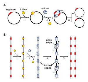
300px Origins of DNA replication Figure 1

A nucleoid, which has a well-defined cytoplasmic structure, contains bacterial chromosomes. Here, the double helix DNA is covered with proteins that are like histone.

The bacterial chromosome differs from the bacterial genome. The bacterial chromosome structure has a strong bond of protein-DNA-RNA that can differ in arrangement, DNA content, dimensions, and conditions related to growth. On the other hand, a genome is the carrier of genetic information for an organism.

Chromosomes of bacteria have clear cytoplasmic shapes that are present in nucleoid. Proteins have structures similar to histone that coat the double helix DNA in the nucleoid. Many bacteria may have chromosomes that are circular in shape, large in size, and single, but it is not the same for all bacteria. Other bacteria may also have multiple chromosomes just like Rhodobacter sphaeroides.

Rhodobacter sphaeroides has more than one chromosome, out of which one is of size 3.0 mb and another is of size 0.9 mb, whereas Burkholderia cepacia has three chromosomes and their sizes include 3.6, 3.2, and 1.1 mb, respectively. Some other species also have linear chromosomes, like the spirochete Borrelia burgdorferi and the gram-positive Streptomyces coelicolor.

Moreover, several bacteria have some extra-chromosomal components in them, like plasmids. The genome size of B. burgdorferi is higher than 0.56 mb as it is composed of approximately 0.9 mb of linear chromosomes and more or less 19 mb of linear and circular plasmids. Chromosome structure has a significant impact on chromosome replication. In contrast to eukaryotes, the initiation of replication takes place in a single location on bacterial chromosomes.

E. coli has a single chromosome with a circular shape, and its replication begins at the oriC site (origin of replication). In contrast to that of eukaryotes, the process of replication progresses in both directions in a manner similar to that of a semiconservative manner.

DNA replication is seen all over the chromosome, which is circular in shape until both of the replication forks link in the terminal ends, which makes up a hindrance to the progress of the replication fork.

bacterial chromosome structure
Image credit: Circular chromosome- Wikipedia

Are bacterial chromosomes circular or linear?

All bacteria do not have circular chromosomes. Out of several genera of bacteria, linear chromosomes are the most common, such as Borrelia, Streptomyces, and Agrobacteria

Few bacteria have more than one chromosome, whereas several bacteria have linear plasmids and chromosomes. When compared to the linear chromosomes present in eukaryotic cells, researchers found that the bacteria have circular chromosomes that are solitary and covalently closed.

The linear chromosomes are believed to have emerged from the circular ancestral chromosomes. To prove that the bacterial chromosome was circular, electron microscopy was used. This process was carried out in both gram-negative bacteria (such as Escherichia coli) and gram-positive bacteria (such as Bacillus subtilis). Bacterial plasmids have been discovered to be circular as well.

Chromosome replication is suggested to be a general mechanism of early fixation. The region of origin of replication has a gene organization. The genesis of replication region gene arrangement is evolutionarily consistent across several bacteria lineages (e.g., E. coli of the phylum Proteobacteria and gram-positive B. subtilis), implying that a generic mechanism for chromosome replication was fixed early.

Telomers are the last parts of the DNA molecules that are linear. They have two difficulties that do not suit the DNA molecules that are circular. Firstly, as we know that the loose ends of the double-stranded DNA are too delicate for degradation by the intracellular nucleases, there should be a procedure that can preserve the ends.

Secondly, the end portions of the DNA molecules that are linear have a particular process for the replication of DNA. These problems can be solved by the characteristics of telomers. There are two varieties of telomers that have been discovered in bacteria, namely, invertron telomers and hairpin telomers.

What does a chromosome look like in bacteria?

While bacterial chromosomes are circular in shape, human chromosomes have open ends. It means that the bacterial chromosomes are attached to each other.

Bacterial chromosomes have distinct cytoplasmic morphologies that are visible in the nucleoid. Proteins coat the double helix DNA in the nucleoid with structures similar to histone. Although many bacteria have circular chromosomes that are huge in size and single in number, this is not the case for all bacteria.

Bacteria are naturally distinct from human beings. Almost all bacteria have only a single chromosome. The reason why chromosomes can get fit in the bacterial cell is that they have folds in them. A nucleoid is where a chromosome can be found. This is more or less similar to the nucleus present in human cells, but it is not the same thing.

While the human nucleus has a membrane of its own, the nucleoid in the bacterial chromosome does not. Thus, the DNA does not break away from the cell. The DNA is enfolded around the DNA binding proteins. This is useful as it helps the chromosome fit into the cell due to the folds.

Current studies in the fields namely cell biology and microscopic methods disclosed that DNA of bacterial chromosome has folds like structure that help them occupying small space in the cell. Bacterial chromosome which is present in nucleoid is independently assembled in supercoiled loops known as domains.  

The shape of nucleoid is highly active as the arrangement of the domain permits the DNA chromosome to go through the changes of structure during various cellular processes such as segregation, replication and transcription that occurs in the cells of bacteria concurrently.

How are bacterial chromosomes arranged?

Bacterial chromosomes are structured in stereotyped configurations in daughter cells, which are consistently and vigorously recreated.

Bacterial chromosomes have spatial organization patterns that categorize under two broad classifications: where chromosome is oriented longitudinally in ori-ter pattern ad where chromosome occupy space in transverse configuration with two arms- left and right, called replichores which are present separately in cell halves in left-ori-right pattern.

The most common type of organization pattern in chromosome of bacteria includes the longitudinal positioning, also known as ori-ter organization. Here, the origin is found at or close to the pole of old cell and the terminus is located near the new cell. Between them are present the left arm and right arm that are situated next to each other. Before the evolution of bacterial cell biology, the pattern was first proposed in sporulating B. subtilis cells.

The longitudinal pattern of ori-ter has both the features of being uncomplicated and instinctive. Still, a methodical observation of E. coli that was growing slowly and experimented with long ago showed a spectacularly different orientation. At the initial stage of the replication of E. coli, the origin occupies its position in the middle of the cell, whereas the left and right arms are found to be in the cell halves separately. The terminus region has a size of nearly 300 kb and it helps in connecting the left and right arms to form a complete circle.

Therefore, the ori-ter axis is kept perpendicular to the long axis of cell that generates a transverse organization (left-ori-right (transverse) organization). As the origins get replicated, they are separated to the quarter regions of the cell. The left arm and the right arm that are replicated newly are separated to the either side so that it can regenerate the transverse pattern in the next origination or generation.

300px Origins of DNA replication Figure 1
Image credit: Origin of replication- Wikipedia

Is the bacterial chromosome structure double-stranded?

The bacterial chromosome is generally defined as a circular, solitary and double helix DNA components that is consist of nearly all genetic information of a cell.

Several bacteria contain a haploid genome. A single bacterial chromosome consists of a round, double helix DNA strands. The chromosome of bacteria is a genetic element which is a round DNA molecule that has the capability to self replicate.

When the bacterial genome gets replicated, each strand present in double helix DNA plays a role in the synthesis of the newly formed complementary strand. Every daughter DNA molecule, which is also a double helical molecule, contains an old strand of polynucleotide and a new strand that is synthesized. This particular kind of DNA replication is known as semiconservative.

The bacterial chromosome is a single molecule of DNA. This is a super coiled DNA molecule which is helical, double stranded. In several bacteria, the end parts of the double helix DNA molecules get bonded to each other covalently to give rise to a genetic and physical circle. The DNA molecules that are linear in bacteria are protected by two different kinds of telomeres such as invertron telomeres and palindromic hairpin loops.

The telomere named palindromic hairpin loops are secured by the absence of the loose ends that are double stranded, whereas, invertron telomeres are preserved by proteins that gets bind to 5 prime ends (5’). These two procedures are also useful for some eukaryotic viruses, phage and some eukaryotic plasmids.

Invertron telomeres are made up of a protein that is covalently bonded to the 5′ ends of DNA molecules. The 5′-terminal protein or TP for short, is the name given to this 5′ end. At the telomere, the DNA polymerase connects with the terminal protein, which promotes the formation of a covalent bond between a dNTP and a TP. The dNTP coupled to TP has a loose group of 3′-OH that acts as a precursor for chain elongation.

Bacteria are often thought of as inert cells that copy themselves without altering them. However, this is not always the situation. Bacteria are extremely adaptable microorganisms. Even if they’re from distinct species, many bacteria can exchange genetic information.

Also Read:

How Is Adenine Formed: When, Where, Detailed facts

Adenine.svg

Adenine and guanine are formed during purine metabolism. The nucleotide inosine monophosphate (IMP) is the source of both adenine and guanine.

The nucleotide which is called the inosine monophosphate is composed of atoms that are derived from amino acids, gluatamine, aspartic acid, Coenzyme teteahydrofolate and as well as glycine and is thus made from the already existing ribose phosphate. H2O and NH3 ice is mixed and thus how is adenine formed.

When is adenine formed?

Adenine is just like an amine product that is added to the purine after the addition of the initial amino group. Adenine is only formed when a mixture of H2O-NH3 ice is present.

In the structure of DNA, adenine gets combined to thymine with the help of two hydrogen bonds that will make the nucleic acid structures stable. Whereas in RNA, adenine gets to bind to uracil. RNA helps in the synthesis of protein.

Adenine forms a nucleoside called adenosine when it gets attached to ribose. When attached to deoxyribose, adenine forms deoxyadenosine. Adenine is joined with thymine in the structure of DNA by two hydrogen bonds, resulting in a stable nucleic acid structure.

Adenine forms a chemical bond with uracil in RNA. Amino acids are the building blocks of protein. Amino acids are synthesized by the four-letter code. These four-letter codes include the two purine and two pyrimidine nucleobases. Adenine (A) and thymine (T) with cytosine (C) and guanine (G) together form the code that helps in the cellular synthesis of amino acids.

The DNA and RNA in cells are made up of five different types of nucleotides. The purine derivatives of these five bases are adenine (A) and guanine (G). The other bases, such as thymine (T), uracil (U), and cytosine (C), are referred to as pyrimidine derivatives. Purines are found in animals and plants, and they are used to make adenine. Some organs and fishes are likewise high in purine. The pyrimidines are a different sort of nucleotide group. Pyrimidines are smaller than purines because they only have one nitrogen circle.

800px Nucleotides 1.svg
Image credit: Nucleotides-Wikipedia

Where is adenine formed?

The nitrogenous base adenine is present in DNA. It is the nucleotide building block of DNA, consisting of two joined rings. Thymine is invariably paired with adenine.

When DNA is joined, a covalent connection is formed. The deoxyribose sugar and nitrogen form this connection. The hydrogen atom is thus removed by the link created. The new structure formed is known as adenine residue, as a fragment of bigger molecule.

There are two types of purine nucleobases out of which adenine is useful for the formation of nucleotides which are present in the nucleic acids of DNA and RNA. The bits of RNA and DNA that are required for pairing are known as “nucleobases.” On the other hand, “nucleotides” are the chemical compounds that comprises of a heretocyclic base, a single or more than one phosphate groups and a sugar.

DNA and RNA present in the cells comprise five main bases. Out of these five bases, adenine (A) and guanine (G) are known as the purine derivatives, whereas the other bases, thymine (T), uracil (U) and cytosine (C), are known as the pyrimidine derivatives.

Purines are generally present in animals and plants, from where we can obtain adenine. Foods which are rich in purines include some organs like kidneys, liver and brain. Fish is also a major source of purine, for example mackerel, anchovies and herring.   

Structure of adenine:

Adenine is a chemical compound which comprises of nitrogen, atoms of hydrogen and carbon. The chemical formula of adenine is C5H5N5. A nucleotide is created when a purine like adenine gets linked up with phosphate and ribose.

DNA and RNA are made up of four nitrogenous bases, with adenine serving as the genetic code for living creatures. Adenine is a key component of adenosine triphosphate (ATP), which provides energy to cells.

how is adenine formed
Image Credit: Adenine-Wikipedia

The fundamental building blocks or small elements often make up the complex structures. For instance, the construction of a house comprises bricks, windows, and doors, which are generally smaller components. In a similar way, living creatures are also built up of molecules that consist of atoms and other smaller molecules.

On considering how is adenine formed, adenine is an essential and fundamental building ingredient required in life. The genetic code of all the biological beings like plants, humans, fungi and several microorganisms are stored in DNA (deoxyribonucleic acid) and RNA (ribonucleic acid). Both acids contain adenine, which aids in the stabilization of nucleic acid in molecules.

With regards to the question how is adenine formed we know that when a purine, such as adenine, is joined to phosphate and ribose, a nucleotide is formed. Adenine fits in the nucleotide family known as the purines. A purine’s fused structure combines a six-membered nitrogen circuit with a five-membered nitrogen circle. There is another type of nucleotide group known as the pyrimidines. These comprises of one nitrogen circle, thus the size of pyrimidines are less than purines. 

Detailed facts:

Several experiments were done in the purpose of studying the adenine production.

Erwin Chargaff, an Austrian biologist, provided another important piece of DNA structure information. Chargaff investigated the composition of A, T, C, and G bases in DNA from various animals.

Adenine was referred to as Vitamin B4 in the early literature. The reason behind it was that adenine is being produced inside the body and it was not necessary that adenine must be taken with diet. But later, the facts related to adenine’s being called a vitamin do not link up with the description given to the vitamin. Thus adenine has been removed from the group of Vitamin B.

Hermann Emil Fischer was one of earliest scientist to do research about adenine. He discovered that the two Vitamin Bs namely riboflavin and niacin, forms a chemical bond with adenine to bring the cofactors nicotinamide adenine dinucleotide (NAD) and flavin adenine dinuleotide (FAD) respectively.   

In 1960, Oró produced adenine in his first experiment. The procedure includes less than 1.0 M of ammonium cyanide (CH₄N₂). Adenine was manufactured in 0.5% yield by warming up the mixtures of ammonium cyanide at a temperature of 70 degree Celsius for many days. After the experiment was done the abiotic production of adenine from the HCN polymerization has been attained several times under various situations. 20% adenine is the highest yield that resulted from the reaction between HCN and liquid ammonia in a sealed-tube. 

In today’s world, the most popular way used in the industry for the production of adenine is an advanced form of formamide method. In this method the formamide is being heated up at a temperature which is lower than 120 degree Celsius in a sealed flask for about five hours to obtain adenine as a final product. Using phosphorus oxychloride (phosphoryl chloride) or phosphorus pentachloride, which works as an acid catalyst, and sunshine or ultraviolet conditions, the quantity of the response, is considerably increased.

Also Read:

Is Adenine Used In Dna Replication :How, Why And Detailed facts

282px Base pair GC.svg

The principle of Chargaff states that the semi-conservative mode of DNA replication gives rise to two helices that includes an old as well as a new DNA strand.

Adenine is used in DNA replication as it is one of those nitrogenous bases that are used in the formation of nucleic acids. In DNA, adenine makes a bond with thymine with the help of two hydrogen bonds to serve the balancing of nucleic acid patterns.

How is adenine used in DNA replication?

Inside the DNA structure, adenine bases are present on one strand to form chemical bonds with thymine bases present on the opposite strand.

DNA has four nucleobases, and adenine is one of them. The three other chemical bases found in DNA are cytosine (C), guanine (G), and thymine (T). Adenosine triphosphate is a molecular form of adenine that acts as a store of energy.

Inside the cell, the chemical reactions are taken care of by ATP. Adenine is one of the main building blocks out of four in the DNA. The characteristics of adenine include that it will always pair up with thymine, which is present on the opposite strand of DNA when they are in a double helix.

Among the purine nucleobases, adenine is used in the formation of nucleotides of the nucleic acids. Not only in DNA and RNA, it is also used all over in the cell. The cell receives its energy from a part of the adenine, that is, ATP. Thus, adenine plays double role inside the cell: buildings blocks of DNA and RNA; energy source of the cell.

The process when DNA makes a copy of its own is known as DNA replication. This happens in all living organisms, which is further essential for biological inheritance. DNA replication is necessary for the growth and repair of damaged tissues during cell division. The process also makes sure that each of the new cells formed receives a copy of their own DNA. The cell has its own characteristics of division that make DNA replication a unique process

Other bases of DNA include:

There are four other nucleotides or bases in DNA other than adenine (A). They are: cytosine (C), guanine (G), and thymine (T). These bases pair up with each other as A with T and G with C.

The five nucleobases of DNA are adenine (A), cytosine (C), guanine (G), thymine (T), and uracil (U). They are called primary or canonical. They give the genetic instructions. The bases A, G, C, and T are present in DNA, and A, G, C, and U are found in RNA.

Adenine and guanine have a structure similar to that of a fused-ring skeletal that originated from purine, and thus they are called the purine bases. These purine nitrogenous bases have characteristics like-they have one amino group (NH2) that is located at the C6 carbon of adenine and at the C2 carbon of guanine. Similar to this, cytosine, uracil, and thymine form a simple-ring structure that is derived from pyrimidine, and thus these three bases are known as the pyrimidine bases. 

Structure of bases :

At the edge of the structure of nucleic acid, the phosphate molecules bind two sugar-rings of two adjoining nucleotide monomers. As they connect with each other, they create a long chain of biomolecules.

The structure of DNA is a double helix and has two strands that are chemically located in opposite directions. This orientation allows the base pairing of the nucleotides. The pairing between the two bases is important for the DNA replication or transcription of genetic codes present in DNA.

The chain bonds of phosphates along with sugars (ribose or deoxyribose) form the “backbone” for the single or double helix structures of DNA. For each of the base pairs in the DNA strand, there is a purine and a pyrimidine. On considering the basis of the DNA, either it binds with t or C gets to bind with G. These pairs are referred to as purine pyrimidine bases, forming the basis of complement pairing and combining together to make a double helix. This structure is sometimes compared to the rungs of a ladder.

Both the forms of purines and pyrimidines pair up on their parts and are an outcome of dimensional constraints. This type of combination features geometry of permanent width for the helical structure of DNA. There is a presence of a double or triple bond of hydrogen between the carbonyl and the amine groups, which are based on C-G pairs and remain complemented as a base alternative to the DNA stands.

282px Base pair GC.svg
Image credit – Base pair – Wikipedia

Why is adenine used in DNA replication?

Inside the DNA strand, adenine forms chemical bonds with thymine using two hydrogen bonds that will help stabilize the nucleic acid composition.

Adenine, when combined with ribose, results in the formation of adenosine. On the other hand, when adenine reacts with deoxyribose, it produces deoxyadenosine. When adenosine is added to three phosphate groups, it gives rise to the formation of adenosine triphosphate (ATP).

Adenosine triphosphate carries out the chemical energy transfer between the chemical reactions and is one of the basic methods that are used in cellular metabolism. Adenine forms a chemical bond with uracil in RNA that is used for protein synthesis. Adenine is responsible for cell respiration and plays a key role in the synthesis of proteins.

Adenine is considered one of the most important organic bases that are needed for life. It is referred to as purine, and makes up a large portion of the genetic makeup of cellular life. It helps in the formation of nucleotides. A DNA strand is formed when adenine binds with thymine. The chemical formula of adenine is C5H5N5. In the world of chemistry, adenine plays different roles in the formation of either DNA or RNA.

Also Read:

Antiparallel Dna Strands: What, Why, Structure, Significance And FAQs

400px AT GC 300x164 1

Antiparallel DNA strands refer to the arrangement of the two strands in a DNA molecule. In this arrangement, the two strands run in opposite directions, with one strand running in the 5′ to 3′ direction and the other running in the 3′ to 5′ direction. This antiparallel orientation is crucial for DNA replication and transcription processes. The antiparallel arrangement allows for complementary base pairing between the strands, where adenine (A) pairs with thymine (T) and guanine (G) pairs with cytosine (C). This complementary base pairing is essential for maintaining the genetic code and ensuring accurate DNA replication and protein synthesis.

Key Takeaways

Fact Description
Antiparallel DNA strands Two DNA strands running in opposite directions
Direction of the strands One strand runs in the 5′ to 3′ direction, while the other runs in the 3′ to 5′ direction
Complementary base pairing Adenine (A) pairs with thymine (T), and guanine (G) pairs with cytosine (C)
Importance in DNA processes Crucial for DNA replication and transcription
Maintenance of genetic code Ensures accurate DNA replication and protein synthesis

Understanding the Structure of DNA

DNA, short for deoxyribonucleic acid, is a molecule that carries the genetic instructions for the development, functioning, and reproduction of all living organisms. It is often referred to as the “blueprint of life.” Understanding the structure of DNA is crucial in the field of molecular biology as it provides insights into how genetic information is stored and transmitted.

The Antiparallel Nature of DNA Strands

One of the key features of DNA is its antiparallel nature. This means that the two strands of DNA run in opposite directions. One strand runs in the 5′ to 3′ direction, while the other runs in the 3′ to 5′ direction. This antiparallel arrangement is essential for DNA replication and the accurate transmission of genetic information.

Structural Features of DNA

DNA has a double helix structure, resembling a twisted ladder. This structure is formed by two complementary strands of nucleotides held together by hydrogen bonds. The nucleotides consist of a sugar molecule (deoxyribose), a phosphate group, and one of four nitrogenous bases: adenine (A), thymine (T), cytosine (C), and guanine (G).

The base pairing between the nitrogenous bases is specific and follows the rules of complementary base pairing. Adenine always pairs with thymine, forming two hydrogen bonds, while cytosine always pairs with guanine, forming three hydrogen bonds. This base pairing ensures the stability and integrity of the DNA molecule.

The Watson-Crick model, proposed by James Watson and Francis Crick in 1953, provided the first accurate description of the DNA structure. According to this model, the two DNA strands are twisted around each other in a helical fashion, with the nitrogenous bases facing inward. This arrangement allows for efficient base pairing and easy access to the genetic information.

DNA replication is a fundamental process in which the DNA molecule is duplicated. It occurs during cell division and is essential for the transmission of genetic information to daughter cells. The replication process involves the action of various enzymes, including DNA polymerase, DNA helicase, and DNA ligase.

During replication, the DNA strands separate at specific sites called replication forks. DNA helicase unwinds the double helix, creating two single strands. DNA polymerase then adds complementary nucleotides to each single strand, following the 5′ to 3′ direction. The leading strand is synthesized continuously, while the lagging strand is synthesized in short fragments called Okazaki fragments. These fragments are later joined by DNA ligase to form a continuous strand.

DNA topology refers to the three-dimensional arrangement of DNA in space. DNA supercoiling is a common phenomenon in which the DNA molecule becomes twisted upon itself. DNA gyrase is an enzyme that helps relieve the tension caused by supercoiling, ensuring the proper functioning of DNA.

Understanding the structure of DNA and its various features is crucial for unraveling the mysteries of the genetic code. It provides insights into how genetic information is stored, replicated, and transmitted, paving the way for advancements in fields such as medicine, agriculture, and biotechnology.

The Antiparallel Arrangement of DNA Strands

The antiparallel arrangement of DNA strands is a fundamental characteristic of the double helix structure, which is the iconic shape of DNA. This arrangement refers to the orientation of the two strands running in opposite directions. In other words, while one strand runs from the 5′ to 3′ direction, the other strand runs from the 3′ to 5′ direction. This antiparallel arrangement plays a crucial role in DNA replication and other essential cellular processes.

How are DNA Strands Antiparallel?

The antiparallel arrangement of DNA strands is a consequence of the base pairing between nucleotides. Each nucleotide consists of a sugar molecule, a phosphate group, and a nitrogenous base. The two strands of DNA are held together by hydrogen bonds formed between complementary base pairs. Adenine (A) pairs with thymine (T), and guanine (G) pairs with cytosine (C).

In the Watson-Crick model of DNA, the two strands are oriented in opposite directions. One strand has its 5′ end (containing the phosphate group) at the top, while the other strand has its 3′ end at the top. This arrangement allows for the complementary base pairing to occur, ensuring the stability and integrity of the DNA molecule.

Why are DNA Strands Antiparallel?

400px AT GC
Wikipedia

The antiparallel arrangement of DNA strands is essential for DNA replication. During replication, the DNA molecule unwinds at specific sites called replication forks. DNA helicase enzymes separate the two strands by breaking the hydrogen bonds between the base pairs. As the strands separate, DNA polymerase enzymes synthesize new strands by adding complementary nucleotides.

The antiparallel arrangement allows DNA polymerase to synthesize new strands in the 5′ to 3′ direction. The leading strand is synthesized continuously in the 5′ to 3′ direction, following the replication fork. However, the lagging strand is synthesized discontinuously in small fragments called Okazaki fragments. These fragments are later joined together by DNA ligase.

The antiparallel arrangement also plays a role in DNA topology and supercoiling. DNA gyrase enzymes help relieve the tension caused by the unwinding of the DNA molecule during replication. They introduce negative supercoils by temporarily breaking and rejoining the DNA strands.

In summary, the antiparallel arrangement of DNA strands is a crucial aspect of the double helix structure. It facilitates base pairing, DNA replication, and other essential processes in molecular biology. Understanding this arrangement helps us unravel the mysteries of the genetic code and the intricate workings of life itself.

The Significance of Antiparallel DNA Strands

Antiparallel DNA strands play a crucial role in various biological processes, particularly in DNA replication and genetic information transfer. Understanding the significance of antiparallel DNA strands is essential for comprehending the intricate mechanisms that govern molecular biology.

Role in DNA Replication

During DNA replication, the double helix structure of DNA unwinds to expose the two complementary strands. The antiparallel nature of these strands is vital for the accurate replication of genetic material.

DNA replication occurs in a 5′ to 3′ direction, meaning that the new DNA strand is synthesized in the opposite direction to the parental template strand. The antiparallel arrangement of the DNA strands allows for the continuous synthesis of one strand, known as the leading strand, while the other strand, called the lagging strand, is synthesized in short fragments known as Okazaki fragments.

The process of DNA replication involves several key enzymes and proteins. DNA helicase unwinds the double helix, creating a replication fork where the two strands separate. DNA polymerase then adds nucleotides to the growing DNA strand, following the complementary base pairing rules. The antiparallel arrangement ensures that the DNA polymerase can synthesize the new strand in the correct direction.

To join the Okazaki fragments on the lagging strand, DNA ligase plays a crucial role. It seals the gaps between the fragments, resulting in a continuous DNA strand. The antiparallel nature of the DNA strands is essential for the proper functioning of DNA ligase and the seamless completion of DNA replication.

Importance in Genetic Information Transfer

Antiparallel DNA strands also play a vital role in the transfer of genetic information. The complementary base pairing between the two strands allows for the accurate transmission of the genetic code during processes such as transcription and translation.

During transcription, the DNA sequence is transcribed into RNA by RNA polymerase. The antiparallel arrangement ensures that the RNA molecule is synthesized in a complementary manner to the DNA template strand. This process allows for the faithful transfer of genetic information from DNA to RNA.

In translation, the mRNA molecule is used as a template to synthesize proteins. The antiparallel nature of DNA strands ensures that the mRNA sequence is complementary to the DNA template strand, allowing for the correct translation of the genetic code into amino acids.

Furthermore, the antiparallel arrangement of DNA strands also plays a role in DNA topology and supercoiling. DNA gyrase, an enzyme involved in DNA topology, helps relieve the torsional strain caused by the winding of DNA strands. The antiparallel arrangement allows DNA gyrase to efficiently manage the supercoiling of DNA, ensuring the proper functioning of the genetic material.

In conclusion, the significance of antiparallel DNA strands cannot be overstated in molecular biology. From DNA replication to genetic information transfer, the antiparallel arrangement ensures the accurate transmission and faithful replication of the genetic code. Understanding the role of antiparallel DNA strands provides valuable insights into the fundamental processes that govern life itself.

Exploring the Antiparallel DNA Structure

The double helix structure of DNA is a fundamental concept in molecular biology. It consists of two complementary strands that run in opposite directions, known as antiparallel strands. In this article, we will delve into the intricacies of the antiparallel DNA structure, including the labeling of antiparallel DNA strands and the potential disadvantages associated with them.

Labeling Antiparallel DNA Strands

DNA structure and bases
Wikipedia

Labeling antiparallel DNA strands is crucial for understanding the structure and function of DNA. The labeling process involves identifying the orientation of the strands and determining the 5′ to 3′ directionality. The 5′ end of a DNA strand has a phosphate group attached to the 5th carbon of the sugar molecule, while the 3′ end has a hydroxyl group attached to the 3rd carbon. By labeling the strands, scientists can analyze the nucleotide sequence and study the interactions between the strands.

To label antiparallel DNA strands, researchers often use fluorescent dyes or radioactive isotopes. These labels allow for visualization and tracking of the DNA strands during experiments. Additionally, specific techniques such as DNA sequencing rely on accurate labeling to determine the order of nucleotides in a DNA molecule.

Potential Disadvantages of Antiparallel DNA Strands

While the antiparallel DNA structure is essential for DNA replication and other cellular processes, it does present some potential disadvantages. One such disadvantage is the formation of DNA knots and tangles due to the topological properties of the antiparallel strands. DNA topology refers to the different ways in which DNA can be twisted, coiled, or knotted.

During DNA replication, the unwinding of the double helix by DNA helicase creates a replication fork. As the replication fork moves along the DNA molecule, the antiparallel strands can become tangled, leading to the formation of knots. These knots can impede the progress of DNA replication and cause errors in the genetic code.

To overcome these knots and tangles, cells employ enzymes like DNA gyrase, which can introduce temporary breaks in the DNA strands and relieve the tension. Another mechanism involves the synthesis of short DNA fragments, known as Okazaki fragments, on the lagging strand during DNA replication. These fragments are later joined together by DNA ligase.

In conclusion, the antiparallel DNA structure plays a crucial role in maintaining the integrity and stability of the genetic material. By labeling the antiparallel strands and understanding their potential disadvantages, scientists can gain valuable insights into the intricate workings of DNA and its role in molecular biology.

Frequently Asked Questions

Does DNA have Antiparallel Strands?

Yes, DNA does have antiparallel strands. This means that the two strands of DNA run in opposite directions. One strand runs in the 5′ to 3′ direction, while the other runs in the 3′ to 5′ direction. The antiparallel nature of DNA is an essential feature of its structure.

Why do DNA Strands need to be Antiparallel?

The antiparallel arrangement of DNA strands is crucial for several reasons. Firstly, it allows for the formation of the double helix structure. The complementary strands of DNA are held together by hydrogen bonds between the nitrogenous bases. The antiparallel arrangement ensures that the bases can pair up correctly, with adenine (A) always pairing with thymine (T) and guanine (G) always pairing with cytosine (C).

Secondly, the antiparallel nature of DNA is essential for DNA replication. During replication, the DNA strands separate, and each strand serves as a template for the synthesis of a new complementary strand. The 5′ to 3′ directionality of one strand allows for continuous replication, while the 3′ to 5′ directionality of the other strand leads to the formation of short Okazaki fragments on the lagging strand.

What does it mean that DNA is Antiparallel?

When we say that DNA is antiparallel, we mean that the two strands of the DNA molecule run in opposite directions. The 5′ end of one strand is aligned with the 3′ end of the other strand. This arrangement is known as the Watson-Crick model, named after the scientists who proposed the structure of DNA.

The antiparallel nature of DNA is crucial for the functioning of enzymes involved in DNA replication, such as DNA polymerase. These enzymes can only add nucleotides to the 3′ end of a growing DNA strand. Therefore, the antiparallel arrangement ensures that DNA replication can occur smoothly and accurately.

In addition to DNA replication, the antiparallel strands of DNA also play a role in DNA topology and supercoiling. The winding and twisting of DNA strands can result in the formation of knots and tangles. Enzymes like DNA gyrase help relieve these topological stresses by introducing temporary breaks in the DNA strands and allowing them to rotate.

In summary, the antiparallel nature of DNA strands is a fundamental aspect of DNA’s structure and function. It enables base pairing, DNA replication, and proper DNA topology. Understanding the antiparallel arrangement is essential for studying molecular biology and deciphering the genetic code.

Conclusion

In conclusion, antiparallel DNA strands play a crucial role in the structure and function of DNA. The antiparallel arrangement of the two strands allows for efficient replication and transcription processes. The complementary base pairing between the strands ensures the accurate transmission of genetic information during DNA replication and protein synthesis. Additionally, the antiparallel orientation of the strands contributes to the stability and integrity of the DNA molecule. Understanding the concept of antiparallel DNA strands is essential in comprehending the fundamental mechanisms of genetics and molecular biology.

References

In the field of molecular biology, understanding the structure and function of DNA is crucial. The discovery of the double helix structure by Watson and Crick in 1953 revolutionized our understanding of genetics and paved the way for further research in the field. This groundbreaking model explained how DNA’s base pairing and complementary strands allow for accurate DNA replication and the transmission of genetic information.

To comprehend the intricate process of DNA replication, it is essential to grasp the concept of the 5′ to 3′ direction. DNA replication occurs in this specific direction, where new nucleotides are added to the growing DNA strand. The nucleotide sequence is precisely maintained through base pairing, where adenine (A) pairs with thymine (T) and cytosine (C) pairs with guanine (G) through hydrogen bonds.

The Watson-Crick model of DNA replication elucidates the role of various enzymes in this process. DNA polymerase is responsible for synthesizing new DNA strands by adding nucleotides to the existing template strands. This enzyme ensures the accuracy of DNA replication by proofreading and correcting any errors that may occur.

During DNA replication, the double helix unwinds at specific sites called replication forks. DNA helicase plays a crucial role in this unwinding process by breaking the hydrogen bonds between the complementary strands. As the replication fork progresses, the leading strand is synthesized continuously, while the lagging strand is synthesized in short fragments known as Okazaki fragments.

To connect these Okazaki fragments and complete the synthesis of the lagging strand, an enzyme called DNA ligase is required. This enzyme seals the gaps between the fragments, resulting in a continuous DNA strand. Additionally, primer DNA is necessary to initiate DNA replication, providing a starting point for DNA polymerase to begin synthesizing new DNA strands.

DNA topology and supercoiling also play significant roles in DNA replication. DNA gyrase is an enzyme that helps relieve the tension and strain caused by the unwinding of the DNA double helix during replication. It achieves this by introducing temporary breaks in the DNA strands, allowing them to unwind and prevent the formation of knots or tangles.

In conclusion, the process of DNA replication is a complex and highly regulated mechanism that ensures the accurate transmission of genetic information. Understanding the key players, such as DNA polymerase, DNA helicase, DNA ligase, and the role of DNA topology, is essential in unraveling the mysteries of the genetic code and advancing our knowledge in the field of molecular biology.

What are the Detailed Explanations of the 5 Coliform Bacteria Examples?

Coliform bacteria explained in detail refers to the thorough explanations of the five examples of these bacteria. These include Escherichia coli, Enterobacter aerogenes, Klebsiella pneumoniae, Citrobacter freundii, and Enterobacter cloacae. Each bacterium has its own distinct characteristics, habitats, and potential health implications. Understanding these details helps in identifying and managing their presence in water, food, and other environments.

[]

Are antiparallel DNA strands and the pyrimidine nature of adenine connected?

Adenine, one of the four nucleobases found in DNA molecules, has long been recognized as a purine base. However, recent research has shed light on its pyrimidine nature as well. Adenine’s classification as both a purine and a pyrimidine base stems from its ability to form two different tautomeric forms. This discovery has interesting implications, particularly when considering the arrangement of DNA strands. The antiparallel orientation of DNA strands, where one strand runs in the opposite direction to its complementary strand, may interact with adenine’s pyrimidine nature in intriguing ways. To gain a deeper understanding of how these concepts intersect, delve into the article Adenine: Understanding Its Pyrimidine Nature.

Frequently Asked Questions

Q1: Does DNA have antiparallel strands?

Yes, DNA does have antiparallel strands. In the double helix structure of DNA, the two strands run in opposite directions, one from 5′ to 3′ and the other from 3′ to 5′. This is what is referred to as antiparallel.

Q2: What is the significance of antiparallel DNA strands?

The antiparallel nature of DNA strands is crucial for DNA replication. It allows the DNA polymerase to add nucleotides to the 3′ end of the new strand, ensuring accurate and efficient replication of the genetic code.

Q3: What is meant by the term ‘antiparallel structure of DNA strands’?

The term ‘antiparallel structure of DNA strands’ refers to the orientation of the two strands in a DNA molecule. One strand runs in the 5′ to 3′ direction, while the other runs in the 3′ to 5′ direction. This is a key feature of the double helix structure of DNA.

Q4: Why are the two DNA strands antiparallel?

The two DNA strands are antiparallel to facilitate the process of DNA replication. This orientation allows enzymes like DNA polymerase and DNA helicase to function properly, ensuring the accurate copying of the genetic code.

Q5: What does it mean when we say ‘DNA is antiparallel’?

When we say ‘DNA is antiparallel’, we are referring to the orientation of the two strands in a DNA molecule. In the double helix structure, one strand runs from 5′ to 3′ and the other runs from 3′ to 5′. This antiparallel arrangement is crucial for processes like DNA replication and transcription.

Q6: What are the ‘antiparallel strands of DNA’?

The ‘antiparallel strands of DNA’ refer to the two complementary strands in a DNA molecule that run in opposite directions. This means one strand runs from 5′ to 3′ and the other from 3′ to 5′. These strands are held together by hydrogen bonds between complementary base pairs.

Q7: Why do DNA strands need to be antiparallel?

DNA strands need to be antiparallel for the process of DNA replication to occur. The enzymes involved in DNA replication, such as DNA polymerase and DNA helicase, require this antiparallel structure to function correctly.

Q8: How does the antiparallel nature affect DNA replication?

The antiparallel nature of DNA strands affects DNA replication by dictating the direction in which the new strands are synthesized. DNA polymerase can only add nucleotides to the 3′ end of the new strand, resulting in one strand (the leading strand) being synthesized continuously, while the other (the lagging strand) is synthesized in fragments, known as Okazaki fragments.

Q9: What is the role of antiparallel DNA strands in the Watson-Crick model?

In the Watson-Crick model of DNA, the antiparallel strands form the double helix structure. The strands are held together by hydrogen bonds between complementary base pairs, with one strand running from 5′ to 3′ and the other from 3′ to 5′. This model explains how DNA replicates and how the genetic code is preserved.

Q10: Why are DNA strands antiparallel to each other?

DNA strands are antiparallel to each other to ensure accurate DNA replication. This orientation allows the enzymes involved in replication to function properly, ensuring the genetic code is accurately copied and passed on to the next generation.

Also Read: