Is Independent Assortment Mutation: Why, How, Causes, Types and Facts

image 29

Mutation is said to be an alternation in any of the DNA sequence. It can be a result of the mistakes for DNA copying at cell division.

With regards to the question for is independent assortment mutation, the combination number that shall result in 23 of the maternal and the paternal homologues which shall result from the independent assortment is said to be 2^23. Thus, it is a cause of mutation.

Any change in the sequence of the nucleotides of the organism genome or the extrachromosomal DNA is said to be mutation. Thus, is independent assortment mutation shall be classifying in this. The genomes that are viral seem to have either the genome of RNA or DNA. Mutation are the outcome of the error at the time of viral or DNA replication, meiosis or mitosis or any other type of the DNA damage.

is independent assortment mutation
Image credit-Independent assortmentWikimedia

Mutation can also be a result of the deletion or insertion of the DNA segments of the DNA at the time of mobile genetic elements. It might as well give some changes that shall be noticeable which is mostly phenotype. This, is independent assortment mutation type of mutation can be a part of the abnormal and the normal process of biology that shall include cancer, evolution and also the developing stages of immune system that contains junctional diversity.

They are said to be the ultimate mutation source for the entire variation in genetics that shall help in having a raw material on which the evolutionary forces shall act upon like that is what is called to be natural selection. This can lead to have several type of outcome with the genes getting mutated having no effect with getting to alter the gene product and percent gene from getting to work good or in complete manner.

So in human genetics, for instance, when you look at a condition like Huntington’s disease, and you see that it follows this pattern where an affected person who passes that to a child, the child has a 50 percent chance of being infected. That’s dominant Mendelian inheritance.  Mendelian inheritance describes the determination of traits by means of dominant and recessive alleles of a particular gene whereas non Mendelian inheritance describes the inheritance of traits which does not follow Mendelian laws.

What is independent assortment?

On context to, is independent assortment mutation, this method gives rise to new alleles combination with having a cross over

The law for independent assortment tells that “Separate genes for the other trait is passed on independently from one and another from the parents to the offspring”. Along with a fertilization that is random there are many possibilities for the variation in genes that shall exist between any of the two people.

There is a crossing over at the prophase and then independent assortment takes place at the anaphase when the chromosome sets with the new alleles of combination. The genetic variation shall be introduced by having random fertilization of the genes that shall be made by meiosis. At the time when the cell divides at meiosis, the homologous pair of chromosome shall get distributed to the daughter cell.

After having itself distributed to the daughter cells and then at the separate chromosome shall have itself segregated independently of each of them. This is said to be independent assortment. Along with the query of is independent assortment mutation, it outcomes to have many unique and specific gene combination for the chromosomes. The cause for the variation on genetic cam be mutation. Gregor Mendel discovered the basic principles of heredity and laid the mathematical foundation of the science of genetics.

The inheritance patterns seen in Mendel’s monohybrid and dihybrid crosses depend on three sets of assumptions: Each trait is determined by a single gene locus. Each gene had two alleles, one provided by each parent. Independent assortment in meiosis takes place in eukaryotes during metaphase I of meiotic division. It produces a gamete carrying mixed chromosomes. Gametes contain half the number of regular chromosomes in a diploid somatic cell.

image 1
Image credit-EukaryotesWikipedia

The general base for the law of independent assortment stays in in the stage of meiosis 1 of the gamete making when the homologous pairs are lined up in not so obvious orientation at the middle of cell as they prepare themselves to part. Meiosis can lead to having of variation in genetics for random fertilization, mutation, crossing over between the chromatids of the chromosomes that are homologous at meiosis and ultimately for random mating done in organism.

Causes of Independent Assortment

It was W Bateson that did explain the lack of independent assortment in the sweet pea and the link was given in Drosophila by TH Morgan.

The statement for is independent assortment mutation relates to one of its causes. This law is one of the result for the independent division of the chromosomes in several other gametes. The crossing over takes place in the genes of each of the chromosome and are then rearranged.

After this there is the crossing over that takes place on each of the chromosome. This helps in discussing of the random inheritance of genes from bot of the parents. The genes when are close placed and link up together in the group and are made to travel as a single unit is called to be linkage. This helps in stopping the method of independent assortment. This all happen in meiosis 1.

image 30
Image credit-MeiosisWikipedia

The chromosome here shall be moving in in random motion to have the poles separated at the time of meiosis. The gamete shall end up in 23 of the chromosomes after meiosis but then this process means that each of the gamete shall have 1 of the other combination that is different of the other chromosomes. The homologous chromosome is seemed to be distributed in random basis to the daughter cells and then segregate with each other.

The rest of the two laws are the Law of Dominance and Law of segregation. With on considering is independent assortment mutation, Mendel noted that it shall not look same like the parental genes. And thus, the allele that comes first cannot be influenced by the second allele on any terms. The other reasons for this method getting distributed with each other and also to the parents is linked to its theory of coming from seating of 10,000 pea plants.

  • The first generation shall be seen with seeds being round and yellow only.
  • The second generation shall have a labelled ratio of the phenotypic ones and shall be scattered and inherited on each basis.
  • The second generation has a ratio labelled of the phenotypic one of which was scattered and inherited individually with the F2 ones having seeds that are self-pollinated. Self-pollination means that the pollination is done between same flower or same plants that are genetically same.
  • At last there are 4 of the results seen- round yellow, round green, wrinkled green and wrinkled yellow.
  • The ratio for the 2nd generation was 9:3:3:1 with it being phenotype and based on individual division.

Independent assortment example

This law defines any random inheritance of the genes from the paternal and maternal source.

For the part of example and to consider is independent assortment mutation, we can consider a virtual situation if the rabbits that can have two of the traits visible being the eye color and the fur color.

The fur color shall be available in two being white and black taking either of the two color. The eye color shall be either red or green. The dominance in the fur area shall be the black fur (B) being dominant over white(b), while the allele of green eye (G) being dominant to red(g). This is a complete hypothetical example to know the law and understand if is independent assortment mutation.

Here, both the rabbits are mixed up. This means that both of the rabbits that look black and have green eyes yet are true to being a heterozygous genotype. Both the rabbits fused have the genotype of BbGg.  In this survival of the two rabbits, all of them on each basis have the same mix of traits. Before having to breed, each of the rabbit shall have to make gametes. It implies that despite the parent phenotype of having green eyes and black fur, the baby can inherit any other combination of the trait.

image 2
Image credit-MendelWikipedia

For example, any one baby might receive the genotype of bbgg, giving it a red eye and white fur look. On the alternate, a rabbit born can also gave the genotype of Bbgg that shall have a black fur and the red eyes. This is called to be independent assortment and thus for the line of is independent assortment mutation is true. The example of Mendel has peas of two traits used up to explain this.

This is the result of the process called as meiosis. It takes at this time. Meiosis is same as mitosis but the final product here is the making of gamete cells. The gamete cells have its DNA half being regular diploid but are said to be haploid. This is vital for sexual reproduction that allows the cells of two gametes to fuse and then make the diploid zygote that shall have the DNA needed to have a new organism.

How is independent assortment mutation?

This law states to give new genes combination as its result on each of the chromosome. This takes places at meiosis.

When the cells divide at the time of meiosis, the homologous pair of chromosome are distributed in random basis to the daughter cells and then other chromosomes are separated out being independent of each other.

The chromosome here must move at random in order for the poles to be separated during meiosis. After meiosis, the gamete will end up in 23 of the chromosomes, but this process means that each gamete will have 1 of the other combinations that are different of the other chromosomes. The homologous chromosome appears to be distributed to daughter cells at random and then segregates with each other.

There is a cross called as the dihybrid cross that simplifies the independent assortment. It is a cross between the two parents that breed true and then express the other trait for the two character. The laws are not clear for the events that take place at this point. It is at the metaphase 1 that the alignment of the pairs that are homologous occur. The alleles trait different to the gametes that are made describing it the best.

Crossing over occurs during the prophase, followed by independent assortment during the anaphase, when the chromosome settles with the new alleles of combination. The genetic variation will be introduced through random fertilisation of the genes produced by meiosis. When the cell divides during meiosis, the homologous pair of chromosomes is distributed to the daughter cell. All their offspring are heterozygotes (genotype RW), displaying both white and red fur. Codominance is an example of Non-Mendelian inheritance.

image 31
Image credit-MutationWikipedia

The general basis for the law of independent assortment remains in the stage of meiosis 1 of gamete formation, when homologous pairs are lined up in a not-so-obvious orientation at the center of the cell as they prepare to part. Meiosis can result in genetic variation for random fertilisation, mutation, crossing over between the chromatids of homologous chromosomes during meiosis, and ultimately for random mating in organisms.

Independent assortment in meiosis takes place in eukaryotes during metaphase I of meiotic division. It produces a gamete carrying mixed chromosomes. Gametes contain half the number of regular chromosomes in a diploid somatic cell. With is independent assortment mutation, Activation of proto-oncogenes also called as activating mutations can occur either by large-scale alterations, such as gain/amplification, insertion, or chromosome translocation, or by small-scale mutations, such as point mutation.

Is independent assortment Mendelian?

Although Mendel’s principle of independent assortment states that alleles of different genes will segregate independently into gametes, in reality, this is not always the case.

Mendel’s law of independent assortment states that the alleles of two or more different genes get sorted into gametes independently of one another. In other words, the allele a gamete receives for one gene does not influence the allele received for another gene.

The Principle of Independent Assortment describes how different genes independently separate from one another when reproductive cells develop. Independent assortment of genes and their corresponding traits was first observed by Gregor Mendel in 1865 during his studies of genetics in pea plants. With is independent assortment mutation, formation of random combinations of chromosomes in meiosis and of genes on different pairs of homologous chromosomes by the passage according to the laws of probability of one of each diploid pair of homologous chromosomes into each gamete independently of each other pair is so.

When cells divide during meiosis, homologous chromosomes are randomly distributed to daughter cells, and different chromosomes segregate independently of each other. This called is called independent assortment. It results in gametes that have unique combinations of chromosomes. Independent assortment states that the inheritance of various genes occurs independently of each other. In the law of independent assortment, the combination of genes and their probability is calculated and assumed by multiplying the probabilities of each gene.

image 32
Image credit-ChromosomesWikipedia

Non-Mendelian traits are traits that are not passed down with dominant and recessive alleles from one gene. Polygenic traits are considered non-Mendelian because their alleles are located on more than one gene which allows for more alleles and phenotypes. Examples of polygenic traits are hair color and height. Non-Mendelian inheritance includes extranuclear inheritance, gene conversion, infectious heredity, genomic imprinting, mosaicism, and trinucleotide repeat disorders.

Also Read:

3 Genome Example: Detailed Explanations

image 8

Any set of genetic data that is complete in any type of organism is called to be genome. It is based on functionality.

Any set of cell in the body is made up of an equal type of data with the genome example being the lover cell or the skin cell, the human genome and much more. Some of the genome example are-

In the universe of biology, any complete chunk of the DNA inside the cell that is living is called to be genome. The cell of the human can naturally have about 3 billion DNA base pairs. These make the genome. The rest of the organism that are infectious or are the carriers have their genetic product in the way of RNA. 

genome example
Image credit-GenomeWikipedia

DNA is said to be a molecule that has the hereditary element with it and is present in all the living matters. DNA makes up the genes and so thus the genome is also made up of DNA. Any gene has good amount of the DNA to have itself useful in coding for one protein and then a genome is then simply made by having the sum of the total DNA seen in the organism.

The study for the gene to function, its analysis, the mapping and editing along with its evolution, regulation and the structure is termed to be the genomics. After a study of these the given data shall help in better understanding of the human heath in the addition of various diseases that is linked with inheritance or even mutation which is the sudden change in the gene. 

Inside the organism, the genes are stored in the long DNA called the chromosome. Thus genomes can also be called as genetic material or data and also genetic sequence. Thus for this reason, the genome example is the instance for the genetic pattern in the body. There are about 5000 of genes that had not been seen in the previous time. Among these 1200 make protein and carry the data.

Genome example

There are a pair of 23 chromosomes and about 3 billion DNA pairs which is termed to be the complete copy of the genome.

The unit that makes the genome is called the DNA. It sets to carry many of the data that make up the body. The genes are any portion of DNA among which many of them make specific or many proteins.

All of the data that is vital to have the whole body function via the instruction needed to build proteins is carried by DNA. Each gene in the genome codes for having 3 proteins. The transfer of the genome example being any of them is from one cell to the other at the time of cell replication and also ensures the differentiation of having life preserved. It also has roles for adaptability.

There are about 350000 genes in the base pairs of DNA. There is a project for human genome that helps in making of the genomic database for all the genome example types and are also seen in public. It helps in getting the chain identified to make a database for the chemical base pairs. This database can be sued up in the biomedical by having genome example like variation in genes that lead to mutation. The genome example elaborated are-

Viral genomes

The genes of the microbes are seen on earth in wide range with the capacity to infect all the living beings that consist of plants, insect and animals.

This genome example plays a good role in the ecology that shall also lead in the climate getting affected. It has the nucleic acids that is covered in the package of protein.

They shall be able to invade the cell and then use it as a machine to have its genome replicated. They are able to have the cell invaded and it varies in terms to being not so normal in its nucleic acid types with regards to its size, path transfer and complexity. It characters can be of RNA or the DNA. They can be either double or single strand, with being linear or circle, or long and short and also multipartite.

image
Image credit-Viral genomeWikipedia

It is quite diverse and its diversity is termed in the contribution of expression and then having this genome example replicated in the host cell by all ways. They are seen in the protein capsid being symmetric and are made if wither many or only one protein with each of term encoding only a single gene that is viral. Thus for this reason, they encode only needed data among all the set.

This genome example is composed of both the RNA or DNA but can never have both of them. Thus it can be with non-segmented or segmented. The size can be of 2kb or up to 2500 kb in length. All of the viral genomes are made of the genetic product and are of nucleic acid. Thus, may use both types of nucleic acid. It main use is to have the genome deliver the host cell and then allow it to express by host cell.

Eukaryotic genomes

The size of the eukaryotic genomes shall vary much with the several other species kinds. Thus they shall be having many gene type.

This genome example is made of many types of haploid and then have single set of chromosome that is divided in a linear way. Each chromosome of the eukaryotes has the molecule of DNA that is double linear.

This genome example is much different from the rest types. They may have the genes that are non-coding and also shall be a part of the protein production. The non-coding ones shall have more authority in its number that the coding ones in few of the eukaryotes as it has the element that are disposable and can also be seem to have DNA that is repetitive. The shape depends on its origin.

image 1
Image credit-Autosomes-Wikipedia

The human cell has about 22 autosomes from each of the parent and then makes 2 copies of them. On addition to it, it has about 2 of the reproductive chromosomes that makes the cell look diploid. Not considering the gametes that are actually sperms, the pollen and the ova that carry only half of he chromosome number in the diploid cell. The organelles that are small have its own genome.

Prokaryotic genome

This type of genome example is seen in the archaea and the bacteria and is also much compact with having all its DNA to use.

This genome example is circle and is always a singular chromosome with having all of its DNA to function. As for this reason, the metabolic ability of the prokaryotic cell and its capacity to make proteins and enzymes is linked to the genome size.

The chromosome of the prokaryotes shall replicate fast that that of the eukaryotes and so its cell shall have many copies of chromosomes. Also, few of the bacteria have the genomic part carried on the plasmid that is circular. This plasmid mostly carries the genes that are not so vital and the ones that are used for having resistance and then making of the toxins.

The DNA of the prokaryotes are the genophore. Its length shall vary with generally it being only a few base pair. There are variation of the prokaryotes having its number of genomes. The example to it can be the E. coli that has only one circular DNA and makes up its whole genome. The new study shows that few of the prokaryotes have as many as circular chromosome and also the four linear chromosome.

The genome size is the total DNA present in a copy pf the cell that is haploid. The size of the genome is mostly termed to be the c value where the more c value is proportional to the number of genes there in the genome. The genome of the prokaryotes is linked to the gene number with the size of the eukaryotes having large non-coding genes. This makes it not connected to the gene number.

image 2
Image credit-GeneWikipedia

Genome vs chromosome

Genetics is termed to be the study of the hereditary which means that one inherits from the parents and then gets inherited from parents.

Chromosomes are said to be inside the units of genes and then passed from the parents to the child that shall help them in determining the traits of the uniqueness of the individual. Genome is made up with the genes, DNA, chromosomes and then make up genome.

The genome example is different. A genome is the entire set of data of the organism. It took all the data of the organism need to work. In the living being, the genome is kept in the long molecules of the DNA and is called the chromosome. Chromosomes are the thread looking form that is kept in the nucleus of the plant and animal. Each pf the chromosome is made of protein and also have one DNA.  

image 3
Image credit-ChromosomeWikipedia
Genome Chromosome
Genes is seen on the chromosomes They are said to be a DNA structure that is packed up with the proteins
They cannot be seen with bear eyes and also not under the microscope They can be seen under the microscope
They are made up of either RNA or DNA They are made up of the histones, RNA and DNA.
The locus of the chromosome is said to be a single gene. There are many genes inside the single chromosome
The mutation of the genes are small The mutation of the chromosome are actually large
The mutation of the genes carries to the point of mutation and then the mutation has the frameshift with deletion and insertion The mutation related to it leads to the chromosomal abnormality and then includes things like rearrangement along with the gene inversion, deletion and duplication

The section of the gene of the DNA part is stuck in the information carried for a specific trait. They are a good unit of functional hereditary and then made the DNA. The genes are linked to the hereditary and then this is the reason of someone having same character of both parents like the hair color, eye pigmentation. There are mostly 29 to 30 genes in thousands of genes in very cell of the body of human.

Genome vs DNA

The molecule of the DNA is the hereditary product of the living cells. The genes are made up of the DNA and thus is also genome.

Genome is not only the entire amount of DNA in a cell but it also has many important elements wrapped around them called as proteins. These proteins play a very key role in defining which part of the DNA will express itself or not. In other words if DNA is alphabet, genome is a complete with alphabets, numbers, punctuations and many more things known or yet to know.

image 4
Image credit-DNAWikipedia

Gene is the basic physical and functional unit of heredity. Genes are made up of DNA. Some genes act as instructions to make molecules called proteins. However, many genes do not code for proteins. In humans, genes vary in size from a few hundred DNA bases to more than 2 million bases. a gene is a basic unit of heredity and a sequence of nucleotides in DNA that encodes the synthesis of a gene product, either RNA or protein.

Deoxyribonucleic acid is a polymer composed of two polynucleotide chains that coil around each other to form a double helix carrying genetic instructions for the development, functioning, growth and reproduction of all known organisms and many viruses. DNA and ribonucleic acid are nucleic acids. DNA, or deoxyribonucleic acid, is the hereditary material in humans and almost all other organisms. Nearly every cell in a person’s body has the same DNA.

DNA GENOME
A DNA is said to be chemical and is kept in the genetic data of the organism The genes are the stretches of the DNA that are kept encoded for the other proteins.
It helps in viewing the many uses like the gene regulation It helps in recognizing the trait of the organism.
It is a long polynucleotide chain They are the small DNA stretches
DBA is not only the genetic product that is shared by the organism It is made up of either of the two RNA or DNA.

Also Read:

Light Independent Reaction in Photosynthesis: What, How, Why and Detailed Facts

image 118

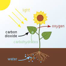
Photosynthesis is said to be the method by which the plants make their own food by use of sun, water and the carbon dioxide.

There is a concerned place called as stroma and is the place where light independent reaction in photosynthesis takes place. It has many enzymes that can work along with ATP and also NADPH to have carbon fixed.

Most of the life forms depend on the process of photosynthesis. This method is carried out by the plants, few type of bacteria, algae that help in capturing the energy from the rays to make oxygen and then store the chemical energy in the form of glucose which is a sugar. Herbivores tend to get the energy by consuming plants and the carnivores have it by eating the herbivores.

At the time of this, plants tend to take in carbon dioxide and also water from the soil and air. While at the inside of cells of the plant, there is oxidation of water which means that the electrons are lost and also at the same time the carbon dioxide are said to reduce themselves and then it gains the electrons. Due to this, the water is converted to oxygen.

light independent reaction in photosynthesis
Image credit-Light independent reactionWikipedia

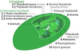
With having the conversion of water into oxygen the carbon dioxide is also transformed to glucose. The plant tends to release the oxygen again in the air and then stores the rest of the energy in the molecules of glucose. There are chloroplasts inside the plant cell and they are small organelles. It helps in storing of the energy from the sun. Inside the thylakoid there is membranes as well that help the chloroplast.

There is light independent reaction in photosynthesis that does not need light and thus the pigment called to be as the chlorophyll, is not responsible for it as it helps in absorbing the light. At this time, the chlorophyll tends to absorb the energy from the red and blue waves and then reflects the green waves as well that makes it look green in color. There are both light independent reaction in photosynthesis and also light dependent one. 

What is light independent reaction in photosynthesis?

Photosynthesis is a common process to all the plants and is also vital at the same time that helps in making of food and gives energy to it.

There are many steps at the back of this method and thus it can be broken down into two of the vital steps being light independent reaction in photosynthesis and light dependent phase in photosynthesis.

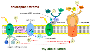
The reaction that is dependent on light takes place in the thylakoid and needs the rays of the sun thus is called to be light dependent reaction in photosynthesis. The chlorophyll tends to absorb the energy from the waves of sun as light and the transfers it into the chemical energy in the form of the NADHP and ATP molecules.

image 42
Image credit-PhotosynthesisWikipedia

On the other hand, light independent reaction in photosynthesis is called to be Calvin cycle and takes place in the stroma which is in between the membrane of thylakoids and the membrane of chloroplast and does not need light and thus called to be light independent reaction in photosynthesis. During this phase, energy from the NADPH and ATP molecules are used to assemble the molecules of carbohydrates like glucose from carbon dioxide.

Not all the forms of them are made to be equal for photosynthesis. There are several types of photosynthesis available that include the light independent reaction in photosynthesis and also the dependent one along with the C4 and C3 photosynthesis. Majority of the plants use up the method of C3. At the time of Calvin cycle they make a three carbon product called 3 phosphoglyceric acid that becomes glucose.

On the other part, C4 makes a four ring carbon product that is intermediate and then spits into the carbon dioxide and also again into a three form compound at the time of Calvin cycle. The best of C4 is that it gets to make a good level of carbon and then allows the plants to sustain in the surrounding with not much use of the water or light. C3 is concerned to be called as the Calvin cycle.

Where does light dependent reaction take place in photosynthesis?

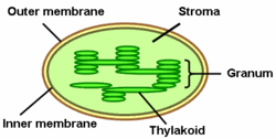
The chloroplast is surrounded by a membrane that is of type double and has an outer line and also an inner membrane. This is equal to that of mitochondria.

The light dependent reaction is seen in the thylakoids. The reaction takes place while the pigments of chlorophyll are seen inside the membranes of thylakoids that help in getting energy from sun.

The motif of this light dependent reaction is to gather a lot of energy from the dun and then break it down to molecules of water to make NADPH and ATP. The molecules tend to store energy and these are used up in the light independent reaction in photosynthesis. Chloroplasts are the pigments that are green and seen in the chlorophyll.

Chloroplast are the one that help in absorbing the light from sun. It is kept in the membranes of thylakoids and in the protein complex that is called the photosystem 1 and photosystem 2. The series of reaction that are light dependent starts when the sun hits the molecule of the chlorophyll and is seen in photosynthesis 2. This gets the electron excited and then leaves the molecule of chlorophyll.

image 115
Image credit-ThylakoidsWikipedia

After leaving the molecule they are seen to travel along the membrane of thylakoids through the series of carrier proteins which is called the electron transport chain. At this time, photosynthesis 2 gets the water molecules to be spitted up to have the lost electron gained back and then fills up the energy vacuum that is made by it. This method is used up by the humans but has failed in the labs. Each of the water molecules tend to break down into two molecules of hydrogen and only one for the oxygen.

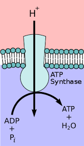
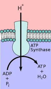
Oxygen is released as a product that is of no use with it being assembled to join again. The ions of hydrogen are made up in more concentration in the lumen of thylakoids. They tend to pass via the enzyme called as ATP synthase and then the move provides the energy that is needed to ass to the third of the ADP phosphate to make ATP. This energy is needed for make cell methods with also glucose being broken to make ATP later after respiration.

Where does light independent reaction take place?

The inner part of the chloroplast also has other membrane and is the thylakoid membrane that is folded in many forms linked to stacks of the discs.

The light independent reaction in photosynthesis takes place inside the stroma. It has many enzymes that help in the work of NADPH and also ATP and then has the carbon fixed from the carbon dioxide in the molecules that shall be built in the glucose.

The very goal of thelight independent reaction in photosynthesis is to gather the glucose molecules. This is a part of the food making process that needs carbon dioxide that is given by air to the plants. The plants get the carbon from CO2 and then makes the glucose blocks. An enzyme within the stroma is called as rubisco that bind with the five carbon molecule of the RubP linking itself to the carbon dioxide molecule.

This tends to make a molecule of six carbons that is broken again into the molecules of three carbons. This is the part of the light independent reaction in photosynthesis that is called to be carbon fixation. After this, the carriers of energy take up the light dependent reaction for its contribution. NADPH and ATP shares each of the 3 phosphoglycerate an atom of hydrogen and then makes two molecules of the simple sugar called the G3P. Finally, the molecules are used to make glucose.

image 43
Image credit-StromaWikipedia

This part of the light independent reaction in photosynthesis is sued up for carbon and also called as reduction. Calvin cycle uses up 6 molecules of carbon di oxide a single time and this shows that there are 12 molecules of the G3P that is made. Despite this, there are only two of the molecules that is sued to make glucose molecule and the rest of them are said to be recycled back to RubP so that this cycle can run again.

How is ATP produced in the light reactions?

The method is said to complete itself y sue of two other process in the field of the light reaction with one making ATP and other NADPH.

The proteins tend to diffuse outside the thylakoid via ATP synthase that gets to make ATP. One the electron reaches the PSI then it links the chlorophyll with a unique pair and then re excite it by absorbing light.

Electrons tend to transfer in a chain between the two type of photosynthesis with the photosynthesis 1 serving to make NADPH and then the second one making the ATP. In the light independent reaction in photosynthesis and also the other one, water is spitted to make a serve for the electrons and then give off oxygen in the form of byproduct ATP is made from ADP by adding of phosphate.

Protons diffuse out of the thylakoid lumen through the enzyme, ATP synthase, producing ATP in the process. Once the electron reaches PSI, it joins its chlorophyll a special pair and re-excited by the absorption of light. During the process of photosynthesis, light penetrates the cell and passes into the chloroplast. The light energy is intercepted by chlorophyll molecules on the granal stacks. Some of the light energy is converted to chemical energy.

image 116
Image credit-ATP SynthaseWikipedia

During this process, a phosphate is added to a molecule to cause the formation of ATP. In light reactions, water is split, providing a source of electrons and giving off O2 as a biproduct. H+ electrons from water reduce NADP+ to NADPH. Light reactions also generate ATP from ADP by adding a phosphate. The main function of light-dependent reactions of photosynthesis is to produce ATP molecules through oxidation-reduction reactions and chemiosmosis reactions in chloroplasts.

How are ATP and NADPH used in photosynthesis?

At the reactions, the sunlight is sued as energy and use the get the water oxidizes and then passes the electrons to NADP making NADPH.

In simple terms, both of these are made by the process called as electron transport chain. At the time of the light reactions, water is used and makes oxygen. This takes place at day as needs the sun.

Some of energy from light is used to have ADP converted to ATP. The ATP and NADPH are made subsequently and then sued as a power to have the sugar made at the time of Calvin cycle. Both of the ATP and NADPH are made together. Photosynthesis takes place in two stages that drives the sunlight to synthesize NADPH and ATP.

image 117
Image credit-ATPWikipedia

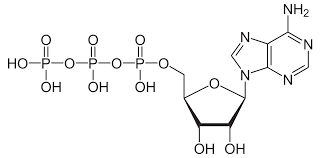
In a process called non-cyclic photophosphorylation (the “standard” form of the light-dependent reactions), electrons are removed from water and passed through PSII and PSI before ending up in NADPH. This process requires light to be absorbed twice, once in each photosystem, and it makes ATP. ATP can be used to store energy for future reactions or be withdrawn to pay for reactions when energy is required by the cell.

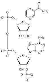
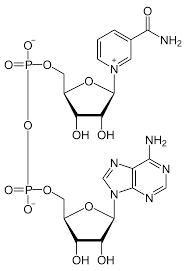
Animals store the energy obtained from the breakdown of food as ATP. Likewise, plants capture and store the energy they derive from light during photosynthesis in ATP molecules. NADPH is an electron carrier that accepts a pair of high-energy electrons and transfers them, along with most of their energy, to another molecule. NADPH plays a big role in the light-independent reaction when it is used, along with ATP, to produce high energy sugars.

Role of ATP and NADPH in the Calvin cycle

The very simple step function of these are to make electrons, generate energy and then also make hydrogen atoms in the Calvin cycle.

ATP is said to be the source of energy and while the NDPH gets to reduce the agent that adds to the light energy electrons t make sugar. The Calvin cycle makes three of the carbon molecules and the also G3P.

The two of the vital roles are to have the light dependent phase and the light independent reaction in photosynthesis to work together and also act as a energy source to have the energy taken away from the sun to have the light independent reaction in photosynthesis help itself to make food for itself. Some of the Calvin cycle needs ATP and other needs NADPH.

image 118
Image credit-NADPHWikipedia

This reaction is catalyzed by the enzyme rubisco. In the second stage, six ATP and six NADPH are used to convert the six 3-PGA molecules into six molecules of a three-carbon sugar (G3P). This reaction is considered a reduction because NADPH must donate its electrons to a three-carbon intermediate to make G3P. Both of these molecules carry energy; in the case of NADPH, it has reducing power that is used to fuel the process of making carbohydrate molecules in light-independent reactions.

Also Read:

Does The Krebs Cycle Produce Water, O2 and Co2: Why, How?

image 39

Krebs cycle is termed to be the chain of reactions in a cycle format that is catalyzed by the enzymes that includes pyruvate.

On regards to the question for does the Krebs cycle produce water, this cycle tends to eat up water and acetate with these two reduced to carbon di oxide. It uses no oxygen and tends to lose it.

Here there is a derivation of the pyruvate from the nutrients and is converted to Acetyl Coenzyme A which is completely said to be oxidized and then broken down to water and carbon dioxide to generate high energy compounds of phosphate which is also a source of the cell energy. There is no involvement of oxygen here.

Cell respiration is said to be a chain of the metabolic ways that harvest the biochemical energy from the substance that is organic mostly glucose and also then stores it in the form of energy called ATP that is used up for the activities that need up energy for the cell. The major steps or the processes for this needs three steps being glycolysis the Krebs cycle and then oxidative phosphorylation.

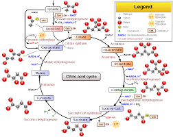
Mostly in being particular, with the part of does the Krebs cycle produce water, it contains the cyclic chain of all the reactions that include enzymes via which the pyruvate is converted to Acetyl CoA and then is oxidized to carbon di oxide. With this, the hydrogen ion is said to be removed from the molecules of carbon that helps transfer the atoms and the electrons making a good energy bond.

does the Krebs cycle produce water
Image credit-Krebs cycleWikipedia

The carbon di oxide that is made is taken from complete oxidation of the pyruvate and is made to detach itself from the cell in the blood. The hydrogen and the electron carriers, FADH2 and NADH are said to donate the electrons to the electron transport chain to make ATP via oxidative phosphorylation an and then the final metabolic thing is the cell respiration. In the eukaryotes, this cycle tends to occur in the mitochondria while prokaryotes have it is cytoplasm.

image 38
Image credit- Cell respirationWikipedia

Does the Krebs cycle produce water?

The very first step of this process involves Krebs cycle that helps in making of ATP from food via glycolysis and is taken by blood.

With considering the concern for does the Krebs cycle produce water there is no trace of water that is used up by Krebs cycle. It is the outcome finally of the last steps of respiration and the transport chain.

Glycolysis is the process of chemical reaction that is performed by the enzymes. They get onto having the glucose converted to 6 of the carbon sugar onto two of the pyruvate molecules that also have three molecules of carbons. In this method, the two of the molecules of ATP are also made as a couple of the molecules of NADH donating electrons.

image 97
Image credit-ATPWikipedia

At the moment, with does the Krebs cycle produce water, there are many cells that take up an add on step to glycolysis of the level of oxygen is not found. They need no oxygen. This method is quite old and ancient. After ATP tends to keep in energy two of the water molecules are made at this time. It is not till the late in this process that more of the water and ATP is made.

The very next or second step in the process of cell respiration is called to Krebs cycle. It is also termed as the TCA cycle or citric acid cycle. This is seen in mitochondria. Energy is converted into two carriers at this point, FADH2 and NADH along with the coenzyme that mostly plat a good role in making of energy. Some bodies find it hard to make ATP. The cells of the body also use up fatty acids here.

Specifically, the paragraph about does the Krebs cycle create water contains the cyclic chain of all the reactions that include enzymes that convert pyruvate to Acetyl CoA and subsequently oxidise it to carbon dioxide. The carbon di oxide produced comes from the oxidation of organic matter of pyruvate and is used to separate the cell in the blood.

How many water molecules are produced in Krebs cycle?

There is an involvement of one molecule of water while in the making of citrate and the other is made while making of malate from fumarate.

There is a total of 44 water molecules that is made from which there are 34 made from the synthesis of ATP. On addition to it 12 are made from cytochrome oxidase plus the 2 from the enolase and rest is taken by TCA.

There are three of the molecules of water that are used in this step with being 3 different ones. The very first is used while having oxaloacetate converted to citrate, then for having isicitrate transferred to cis aconite and then in the conversion of the fumarate from L malate. Later there is the citrate getting converted from the cis aconite.

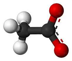
It is a balanced reaction and needs an input of two molecules of water. Water is used in many steps but the main point is to have the cycle in motion by balancing the outputs and the inputs. There is always an eye on the acetate. CH3COO- (acetate) + 2 H2O → 2 CO2.

image 39
Image credit-AcetateWikipedia

Acetyl-CoA + 3 NAD+ + Q + GDP + Pi + 2 H2O →CoA-SH + 3 NADH + 3 H+ + QH2 + GTP + 2 CO2. This cycle takes up two of the carbons in the level of the acetic acid and then release two molecules of carbon di oxide. This is said to be one of the vital goals in the cycle after having the final combustion done for the rest of the carbon skeletons and the carbohydrate of the amino acids.

The add on oxygen is taken from water and not from carbon di oxide. The rest of the process for combustion consists of the electron transfer from the FADH2 and NADH to the oxygen via the electron transport chain. This is the area where oxygen as two molecules of the gas come into play. It is then reduced to water so that of there is an end to the combustion reaction, fats, the skeletons and the carbs along with + O2 → CO2 + H2O

Does Krebs cycle produce oxygen?

This process can work without the presence of oxygen yet the very final step needs this gas to function and accept electrons.

On regards to the question for does the Krebs cycle produce water, giving out oxygen is not what this does. The process of electron chain transfer shall get jammed if the electrons are not done in the presence of oxygen.

The overall thing that is made by this after using of the acetyl CoA, it makes NADH, FADH2, GTP or ATP along with carbon di oxide. It does not make oxygen but is vital for the final stage of getting the electrons transferred. Oxygen are the final acceptors for electron. The Krebs cycle is not simple and thus is confusing. The processes that are anaerobic needs to oxygen but the rest does.

After the second turn through the Krebs cycle, the original glucose molecule has been broken down completely. All six of its carbon atoms have combined with oxygen to form carbon dioxide. The energy from its chemical bonds has been stored in a total of 16 energy-carrier molecules. These molecules are:

  • 4 ATP (including 2 from glycolysis)
  • 10 NADH (including 2 from glycolysis)
  • 2 FADH2

Oxidative decarboxylation of the pyruvate is the one vital method of the cell respiration that binds the Krebs cycle with glycolysis. It takes place in the mitochondria for the eukaryotes where the three carbon pyruvate is the end result of the method if glycolysis and is then carboxylate to make acetyl CoA being of two carbon molecules and this is done by pyruvate dehydrogenase. It increases cell turnover and helps eliminate accumulated dead skin cells on the epidermis — the top layer of the skin.

image 40
Image credit-GlycolysisWikipedia

Acetyl CoA enters this cycle and the carbon di oxide is then made to lose in the method. There is np involvement of the oxygen in direct basis and makes up some of the ATP and the reductants along with carbon di oxide, FADH and NADH. With no involvement of the gas oxygen, it ceases the view of FAD and NAD. The reaction can be kept in term of equation being- . 

image 95

Does the Krebs cycle produces Co2?

The mitochondria said to be the power house of the cell is the house for the Krebs cycle and also makes CO2 that we breathe out.

For this one method, there are two of the dioxide molecules, there of the NADH molecules and one of the GTP or ATP are made. While this makes carbon di oxide it does not have enough energy to make ATP.

The series of this reaction generates two molecules of carbon dioxide and also the reduced form of the FADH2 and NADH. It makes the use of two of the pyruvate molecules and then generates molecules of NADH for energy. There is only little quantity of ATP made after production of carbon dioxide. There is a total of 4 ATP containing two for glycolysis, 10 of NADH from the two including glycolysis and 2 FADH2.

image 98
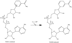
Image credit-NADHWikipedia

While the Krebs cycle does produce carbon dioxide, this cycle does not produce significant chemical energy in the form of adenosine triphosphate (ATP) directly, and this reaction sequence does not require any oxygen. There are two carbon dioxide molecules produced for every molecule of pyruvate in the Krebs cycle. They are produced during decarboxylation reactions. For one cycle, two molecules of carbon, three molecules of NADH one molecule of FADH2 and one molecule of ATP are made.

Prior to entering the Krebs cycle, the pyruvic acid molecules are altered. Each three-carbon pyruvic acid molecule undergoes conversion to a substance called acetyl-coenzyme A, or acetyl-CoA. During the process, the pyruvic acid molecule is broken down by an enzyme, one carbon atom is released in the form of carbon dioxide, and the remaining two carbon atoms are combined with a coenzyme called coenzyme A. This combination forms acetyl-CoA. In the process, electrons and a hydrogen ion are transferred to NAD to form high-energy NADH.

Does the Krebs cycle produce lactic acid?

The pyruvic acid is said to supply the cell that are alive with energy via the Krebs cycle in the presence of oxygen.

If there is no oxygen present, thus there is the fermentation for the making of lactic acid. With the question Does the Krebs cycle produce water, this process makes NADH, FADH and also ATP that are 38 in number.

Pyruvic acid supplies energy to living cells through the citric acid cycle (also known as the Krebs cycle) when oxygen is present (aerobic respiration); when oxygen is lacking, it ferments to produce lactic acid. Pyruvate is an important chemical compound in biochemistry. A turn of the Krebs cycle produces one ATP, three NADH, one FADH2, and two CO2Acetyl-CoA is not produced during Krebs cycle. It is produced from the decarboxylation of a pyruvate molecule, which occurs before the Krebs cycle can begin.

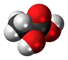
Lactic acid is mainly produced in muscle cells and red blood cells. It forms when the body breaks down carbohydrates to use for energy when oxygen levels are low. Times when your body’s oxygen level might drop include: during intense exercise Lactic acid is an organic acid. It has a molecular formula CH₃CHCOOH. Cellular respiration is a set of metabolic reactions and processes that take place in the cells of organisms to convert chemical energy from oxygen molecules or nutrients into adenosine triphosphate.

image 41
Image credit-Lactic acid in Krebs cycleWikipedia

Citric acid is the very first product that is made out of this cycle. It gets in pyruvic acid to carbon dioxide by breaking it down. This makes 2 molecules of ATP and then 6 of it is NADH. Oxygen is said to be the ultimate acceptor of electrons in the chain but without the use of oxygen this transport chain is jammed. Muscle cells add fermentation to its and thus pyruvate gets made to lactate.

Also Read:

Hypertonic vs Hypotonic: Comparative Analysis

image 25

Hypertonia means too much of the muscle tone. The newborns and the infants are mostly diagnosed with it.

With considering hypertonic vs hypotonic, hypertonic means more of muscle tone which also means less flexible. Hypotonic on the other hand is much opposite to that of Hypertonia.

  Hypertonic Hypotonic
Definition Too much of the muscle tone Muscle tone getting less or decreased
Cause Affects the central nervous system Affects the motor skills or brain.
Affect Affect both baby and adult damaging the central nervous   system, and is from birth. Affect adults as well but more is children and is seen early before the child is 6 months and time of birth.
Possibility It is less common than hypotonia in the baby and yet effects mostly the newborn with no rate discovered. It is most common in the new born and effects the motor skill. The exact reason is not known.
Symptoms No range of movement, loss of balance, pain, less movement. Baby feels limp, hard swallowing, muscle weak, crying, legs are weak.

With the sudden cause of hypertonia with less ability to be flexible, the children tend to have much poor way of mobility a loss stiff moves. They face good difficulty in reaching, walking, pushing and also feeding. The part of stress lies on the point of neck, arms and the legs.  In chemistry, solution is a special type of homogeneous mixture composed of two or more substances

Muscle tone means that the capacity of tension or resistance to move in the muscles. One is able to feel the tone in muscle of the pinch the bicep while it is in a relaxing mode. The resistance that is felt then is called muscle tone. The main motif of muscle toning is to allow a better posture maintaining ability while sitting that helps in the regulation of function of the organs in the build and controls the reflexes.

hypertonic vs hypotonic
Image credit-HypertonicWikipedia

If there is any muscle tone seen, the move seems shall be robotic as they are not able to relax the muscle a shall have minimum chance of being flexible. The children that have hypertonic have quite a lot of difficulties concerning the muscle movement. On the other hand, with hypertonic vs hypotonic, hypotonic means having a less muscle tone or a decreased one. It is its own situation. Muscle tone is defined as the continuous and passive-partial contraction of the muscle or the muscle’s resistance to passive stretch during the resting state.

In the case for hypotonic or so called hypotonia one faces the condition on its own and called the benign congenial hypotpnia. It can mean any other problems as well where there is a continuous loss of the tone in a muscle like that of cerebral palsy or also muscular dystrophy. This can be detected at the time of being an infant. One of the reasons for this can be damage of the brain at the time of fetal development. Appropriate muscle tone enables our bodies to quickly respond to a stretch.

For example, if someone took your arm and quickly straightened your elbow, your biceps muscle would automatically respond and contract in response to protect you from injury. Muscle tone can be seen when muscles are in a state of slight tension and they are ready for action. Regular training tones muscles and helps to create good posture . In addition, muscles will hypertrophy that is increase in size and develop better endurance .

Hypertonic vs hypotonic muscles

Hypertonic means to have increased in the level of muscle tone while hypotonic shall means a decrease in the level of muscle toning.

The very difference of both lies in the fact of its definition. Thus with one simple question with hypertonic vs hypotonic is one is an increase in its muscle toning and the other is decrease in toning of muscle. As directed by a doctor, stretching, body positioning may help improve muscle control.

There are two types of hypertonic muscles toning that is seen and they are the spastic hypertonia also called as spasticity. It is the reflex that has been exaggerated and then the muscle spasms lead to the increase in the movement. The other is rigidity called the Dystonic hypertonia which implies no change in movement.

Hypertonia is the phase that can attack or leaves its effect at both the time adults and babies that pay a glance to a damage in the central nervous system. The diagnosis of this injury can take place after any injury or any system if seen to undergo this condition that is present at the time of birth. It is generally found in babies after two years. It is mostly congenital.

image 27
Image credit-HypotoniaWikipedia

Hypertonia is said to be not so common or is less that hypotonic in the babies and is a more common scene that tends to affect the motor skills of the new born. The rate of it taking place is not so known and thus hypertonia can be a symptom to any other condition as well. Thus for this reason, hypertonia can be said to be any cause. Hypertonia is resistance to passive movement, it is not dependent on velocity, can be with or without spasticity.

Hypotonic is bad muscle toning. People that are diagnosed with so show less resistance while they move the joint for body mobility. Another term for thus can be the floppy infant syndrome. If one tends to relax the arm and pinch on the bicep the resistance is felt a there is a muscle tone. The toning of the muscle is a response for the body being forced and allows a god maintaining of posture.

image 28
Image credit- Floppy infant syndromeWikipedia

Hypotonia and muscle weakness are not the same. Muscle weakness is said to be a lack on the strength of the muscle which is often a good symbol for hypotonia. It is mostly seen at birth and the healthcare diagnose it mostly at the time of early birth and at the time of the child being 6 years. It is also affective in the adults.

Hypotonia is said to be much common for the motor skills of the newborn. It shares the same term with hypertonia stating no symptoms such available yet the rate is not known with it leading to any other disease. There is a dislink seen between the brain interaction with the muscle and the nerves that tell them to relax the muscles. The body also does not respond safe making it hard to move.

Symptoms of the hypertonia can be the result of the muscle becoming more stiff. It may vary from people to people that have been diagnosed with the same. The severe symptoms can depend on the injury location and spinal cord as well. Some of the symptoms can include less motion, loss of balance and more falling, muscle soreness, limited flexibility, myoclonus, pain.

Hypertonic vs hypotonic examples

Tonicity is said to be the amount of osmotic gradient that is effective on terms of pressure for the two of the solutions taken.

The very word hyper means more and thus hypo seems to have the solution that has less concentration on being compared to thence. Hypertonic is more concentrated. Thus with the hypertonic vs hypotonic comparison, it stands with the concentration.

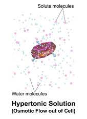
Hypertonic refers to the solution that has more osmotic pressure that the rest of the solution. In on the other definition, hypertonic is said to be one with the maximum number of solute particles with having less on the inside and more on the out. There is also more tension om the surrounding fluid under the comparison to make more of the outcome.

image 29
Image credit-Hypertonic solution-Wikipedia

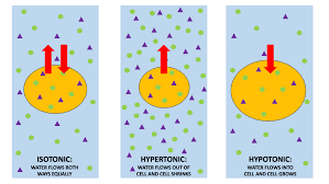
On considering biology, hypertonic vs hypotonic is quite of an easy term. Hypotonic means the solution in the cell has it all exposed eventually and it swells up for the molecules of water tend to go into the cell via the method of passive transport.  It means to have less of the osmotic pressure than the rest of the medium compared to the hypo organism.

An example of hypotonic can be the distilled water with having less solute than the cell and another example for the hypertonic one can be ocean water with having mote of the solute than the cell. Another example of hypertonic cell can be the red blood cell when compared to clear water. Hypotonic also means less of the muscle toning in the body. A hypertonic solution is a particular type of solution that has a greater concentration of solutes on the outside of a cell when compared with the inside of a cell.

Tonicity - Wikipedia
Image credit-Hypotonic solutionWikipedia

Some of the instances for the hypotonic solutions can be a thing that has more of water and then less solute on being compared to the cell. Some of the example being distilled water, 0.25% of saline water and 0.45% of the saline water. There are some cases that need the use of hypotonic solutions. At the time of diabetic ketoacidosis, the patient shall tend to be dehydrated and thus shall need the concentration of 0.45 of the solution that shall help get them rehydrated.

Some of the example of hypertonic solutions can be 5% of dextrose. 3% saline water also being the 5 in concertation of saline and then 5% dextrose in 0.9% salinity. Sea water is also said to be a hypertonic fluid and that means that is has more salt than the human blood intake. The within of the red blood cell when compared to the clear water can also be an example of it. This leads to water leaving the cell and flowing into the solution around it.

Seawater  has a high amount of salt particles compared to freshwater, making it a hypertonic solution. Freshwater fish can’t live in seawater because the water would rush from their cells into the surrounding saltwater. They would soon die from dehydration. Saltwater fish, on the other hand, have adapted to their hypertonic solutions and rely on saltwater to regulate their cells. The same works for saltwater plants; while typical plants need water to flow into their cells.

image 73
Image credit-Sea waterWikipedia

Hypertonic saline, particularly 7% or 23% hypertonic saline, is used to treat patients with traumatic brain injuries. It draws fluid out of cells to prevent cerebral edema. Typically, hypertonic saline over 3% is administered in a central line rather than in a standard IV drip. Water with a large amount of dextrose can be an effective way to replace fluids and calories in an IV drip. This hypertonic solution is helpful for babies who are at risk for hypoglycemia.

Hypertonic vs hypotonic dehydration

There can be three types pf dehydration possible being hypotonic, hypertonic and also isotonic. The most common seen is isotonic in humans.

If there is a greater loss of water with regards to the loss of sodium, then the concentration of the sodium serum tends to increase that results in the hypotonic dehydration with having less sodium concertation is hypotonic.

Dehydration occurs when the body loses more fluid than it takes in. This condition can result from illness; a hot, dry climate; prolonged exposure to sun or high temperatures; not drinking enough water; and overuse of diuretics or other medications that increase urination. Dehydration can upset the delicate fluid-salt balance needed to maintain healthy cells and tissues.

Hypertonic type of dehydration takes place when there is an imbalance seen in the level of salt and water in the body. Loosing too much of the water while having kept them in the fluid put of the cells shall result in the hypertonic dehydration. With hypertonic vs hypotonic dehydration, the most common drink type that shall help in the comeback is none. Increased thirst, dry mouth, light-headedness, fatigue, impaired mental focus, low urine output, dry skin, inability to produce tears, sunken eyes are the signs of dehydration.

image 30
Image credit-Dry skinWikipedia

Isotonic drink shall be used by more often with the hypotonic ones being a good solution for the ones looking for the replace of the fluids. Dehydration can have causes that aren’t due to underlying disease. Examples include heat, excessive activity, insufficient fluid consumption, excessive sweating or severe side effects. Hypotonic dehydration is loss of water associated with decreased effective osmolality of body fluids. Severe dehydration can cause more serious symptoms and may require good care.

Common symptoms of hypertonic dehydration include dry skin, dark urine, decreased urine production, and fatigue. However, as dehydration becomes more severe, one may experience additional symptoms, such as hypotension, muscle cramping, and dizziness. Causes include loss of isotonic fluids via the kidneys or gastrointestinal tract that has been partially compensated by drinking hypotonic fluids. Hypertonic dehydration occurs when water excretion from the body exceeds that of sodium excretion.

Also Read:
What Happens to a cell in a Hypotonic Solution: Detailed Insights

image 22

Any cell is said to be a mass of the cytoplasm that is guarded well by a cell membrane. They are all microscopic in size.

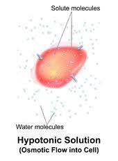
A cell in a hypotonic solution, has a net movement for water from the part of the solution in the body. A cell that is placed into a hypotonic solution shall seem to swell and then expand till it shall burst via a process called cytolysis.

A solution that is hypotonic or a cell in a hypotonic solution has a concentration that is lower than that of the solutes of the other solution. In the term of biology, the solution that is out of cell is said to be hypotonic only if it has less concentration for solutes considering to that of cytosol. There is a diffusion of water in the cell, for osmotic pressure and then the cell leads to often being turgid or also bloated.

In chemistry, a solution is a special type of homogeneous mixture composed of two or more substances. In such a mixture, a solute is a substance dissolved in another substance, known as a solvent. Some examples of solutions are salt water, rubbing alcohol, and sugar dissolved in water. When you look closely, upon mixing salt with water, you can’t see the salt particles anymore, making this a homogeneous mixture

 cell in a hypotonic solution
Image credit-Hypotonic solutionWikipedia

Unlike osmotic pressure, tonicity is influenced only by solutes that cannot cross the membrane, as only these exert an effective osmotic pressure. Solutes able to freely cross the membrane do not affect tonicity because they will always equilibrate with equal concentrations on both sides of the membrane without net solvent movement. It is also a factor affecting imbibition. A hypotonic solution has a lower concentration of solutes than another solution.

Solution are said to be a mixture of a compound made of a solvent and a solute. The substance that is the one that is present in more concentration is called as solvent and the one that is in less level is said to be solute. A salt water is a good example of hypotonic solution. Thus, cell in a hypotonic solution shall have less solute concentration than the cell. An iso-osmolar solution can be hypotonic if the solute is able to penetrate the cell membrane.

What happens to a red blood cell in a hypotonic solution?

The very simple definition of tension or more tone means to have a large osmotic pressure that the rest medium or fluid.

The time when cell in a hypotonic solution shall have lower concentration, the red blood cell kept in this solution shall have a free water net movement in the cell. This means less water than the cell.

On having a cell in a hypotonic solution or rather a red blood cell in it shall have it bloated up and might explode. If it is kept in a hypertonic mode, it shall shrivel that shall make the cytoplasm dense and the components shall remain concentrated and also can die. Thus, for this reason a plant cell seems to be ideal. To prevent the process of burst, the cell needs isotonic mode.

image 22
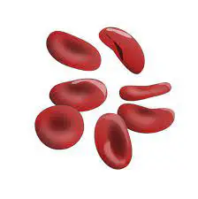
Image credit-Red blood cellWikipedia

After having the red blood cell in a hypotonic solution there shall be seen a net movement for free water inside the cell. This phase shall outcome in an increase volume in the cell with having less concertation of solute. The solution shall end up with finally high level of concentration. It would eventually lead to it swelling up and then bursting in the method called hemolysis.

To avoid situation like this, cell in a hypotonic solution can never be the red blood cell. Thus to level this up or avoid the process of hemolysis, on needs to place the cell in a solution called to be isotonic solution. This shall have 0.9% m/v of NaCl and glucose shall be in a concentration of 5% m/v. A solution is isotonic when there is equal amount if solutes on both side of membrane and no swelling or shrinking is seen.

Any example of isotonic solution can be the normal saline water that is 0.9% in concentration and the lactated ringers. These fluids are said to be useful at the time when there is a huge loss of body fluids from any trauma, dehydration, vomiting, diarrhea, blood loss or nausea. This term is used both in biology and in chemistry for transport across the semipermeable membrane.

What happens to an animal cell in a hypotonic solution?

Any cell in a hypotonic solution shall, have less amount if solute concentration and more solvent in it. Osmosis is the process involved in it.

The solutions that are hypotonic shall have less water than the cell. Thus with animal cell in a hypotonic solution shall be filled up with water completely and then shall burst. Pure water and tap water are hypotonic.

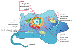
An animal cell is a eukaryotic cell type that do not have a cell all yet seems to have a nucleus that has a membrane and is true. It also has several other cellular organelles. The contents that it has are cytoskeleton, centrosome, lysosome, mitochondria, Golgi apparatus. Any typical cell that is of an animal shall have cytosol, a cell membrane, the organelles and cytoplasmic structure.

Wikipedia:Featured picture candidates/Typical animal cell - Wikipedia
Image credit-Animal cell-Wikipedia

The process of osmosis takes place at the time when animal cell in a hypotonic solution. Thus for this, the animal and plant cell seem to both appear a lot plump while placed in a hypotonic solution. After this has been seen under the microscope, the vacuoles seem to appear a lot larger considering only the plant cell. It has more of solute and net movement. It is all a result of the method called as osmosis.

The swelling of a cell in a hypotonic solution is for the less amount of solute in it and the net movement of water in the cell causes a breaking or swelling of the cell. There is a movement of water at a place where there is low outside fluid or osmolality to an area that has more of it. The cell shall also want to expand. Just like the animal cell, the plant cell shall mot burst. The increase in solute level leads to it being broken finally.

What happens to a plant cell when placed in a hypotonic solution?

Both the plants and animal ell are said to be different and thus perform and show different types of property when placed to conditions.

When a plant cell in a hypotonic solution is exposed it shows up osmosis that takes up water and then begins to swell up. There is a rigid wall in the plants that help prevent the cell from getting it busted making it turgid.

Osmosis is said to be a process having the spontaneous path or water diffusion system in rest of the solvents via a semipermeable membrane. It is the simple movement concerned to water from the place of high concentration for water to the place of low level water. It helps in getting the two of the solutions separated by having the solute concentrated.

image 23
Image credit-Plant cellWikipedia

If there is a link made between the two areas via a membrane, then the one that has more of the solute that the rest then the eater shall flow from the second point to the first. The ability of the solution to have the water move is said to be tonicity. The tonicity of any of the solution shall be called as osmolality. Concentration and solubility is directly related to each other.

Water seems to enter the cell for the plant and the also the plasma membrane that tends to swell up and the push up against the walls of cell. If the plant cell in a hypotonic solution has a concentration of solute less than the within if the cell the water shall tend to enter the cell via osmosis. The cell shall swell but with zero lyse as the cell wall gives the structure to expand. Thus, the cell membrane shall press up the wall and make a turgor force that shall give the support to plant.

The cell after attaining the osmolality in the hypotonic solution, shall not burst but expand. But the animal cell in a hypotonic solution shall explode. This is so as the plant cell have a wall for the cell that is rigid all the way via the plasma membrane. On getting to swell with the water they shall turn turgid. Hypotonic solutions also help in making the vegetables like bell pepper turn crisp.

What happens to a bacterial cell in a hypotonic solution?

Usually if there is a placement of cell in a hypotonic solution it is quite likely to get ruptured or swell up due to its tonicity.

If a bacterium cell is kept in a hypotonic solution, it shall seem to rupture by having the cell swell due to the gradient of osmotic pressure within the cell of the bacteria.

The bacteria are microorganism that have a cell boundary and is simple that that of the rest of the organism, they have their own center of control that have genetic data and is kept in a single loop for the DNA. Some of the bacteria do have an extra genetic circle of cell material and is called to be plasmid rather than being a nucleus. They are the prokaryotic cell type.

image 24
Image credit-Bacterial cellWikipedia

For the instance of osmotic pressure and the gradient that is made the relative presence of the hypertonic solution in the cell makes it swell and in the method it becomes much slow and some of them are finally resistance to each other in action by all means of the cell wall. If there is the control of water, the cell might lead to damage and burst. The plasma membrane helps in getting the pressure kept normal with bacteria having straight characters.

Most of the cell wall of the bacteria, fungi and algae have a cell wall that is rigid and are able to tolerate the osmolality and then enjoy the surrounding being hypotonic. If the solution seems to be hypertonic then the water that is in, then cell shall be able to leave it and then the bacteria will be shrinking. The cell movement outside the water is called to be osmosis. There is a prevention of the cell from expansion and thus have lysis.

Can a Hyperosmotic solution be hypotonic?

Osmosis is said to be the solute number that gets dissolve while tonicity leads to having no units. Osmolality is said to be comparison of two solutions.

Yes, it can be so. If the cell is placed in a hyperosmotic solution but the hypotonic part is like dextran of 10% concentration, there shall be a water movement. Thus, a hyperosmotic solution can be hypotonic.

With also have this question of can hyperosmotic solution can be hypotonic, it is not always needed to be so. Yet, the hyperosmotic solutions shall always remain hypotonic. There is a good response for the widespread presentation of tonicity and osmolality that had a good result. It also needs bit of an additional arrangement.

image 71
Image credit-OsmoticWikipedia

Hyperosmotic stress in most mammalian cells causes cell shrinkage due to osmotic efflux of water leading to increases in intracellular ionic strength. Hyperosmotic solution has been shown to disrupt mating pairs at two different time points. First, as described earlier, osmotic solution will erase the preparative changes brought about by “initiation”. The increase of intracellular ions and the accompanying influx of water cause RVI. 

A solution can be either isotonic or hyperosmotic and both. If a cell needs to be placed in a hypertonic solution, there will be a net flow of water out the cell and then the cell shall loose volume. A solution shall be hypertonic in a cell and thus its solute shall be having a high concentration. There is always a swelling due to influx and the cell shrinks.

Can a Hyperosmotic solution be isotonic?

A solution can be either be isotonic or also be hyperosmotic. Hyperosmotic refers to the capacity of achieving more than the normal osmosis.

The solutes that are hard to penetrate or actually have zero penetration via the cell membrane, thus the water movement across the membrane of the cell shall be able to take place to reach stability. Solutions of equal solute concentration are isotonic.

Tonicity is a measure of the effective osmotic pressure gradient; the water potential of two solutions separated by a partially permeable cell membrane. Isotonic is a term used to describe solutions and chemistry and, sometimes, muscles in human biology. In chemistry, a solution is said to be isotonic when it has the same concentration of solutes as another solution across a semipermeable membrane. The use of isotonic in human anatomy is used more rarely.

The very term hyperosmotic means the property of having a pressure with less osmosis. This means that the molecules in solute shall number in a single side of the membrane that shall allow only specific molecules to pass via the low side than the one that has on the rest side considering the number of solute. The water molecules shall be travelling fast and via the cell membrane that shall have the particles to solute in.

Hyperosmotic vs hypertonic

The solutions that tend to have less number of solute is called to be hypotonic. The word hypo means less and is same to hyperosmotic.

The solutions that are hyperosmotic are never always hypertonic. But the hypsometric ones are always said to be hypotonic. This depends on the tonicity and the osmolality of the solutions.

In the solutions that are hyperosmotic the solutes tend to move from the place where the surrounding has a high osmotic pressure that the rest part of the solution. On the other part, the hypotonic have the solutes that travel from the rea that have high part of concertation and move to the less area concentration or the surrounding.

In simple way, hyperosmotic refers to the character that has a pressure high is so osmolality while hypotonic refers to the feature of having an osmotic pressure that is less. Hyperosmotic also have pressure that is high in the rest of the area concerned to the cell while a cell in a hypotonic solution shall have less pressure out. Solution in hyperosmotic tend to move from the solution to rest while opposite in hypotonic.

image 72
Image credit-Cell shrinkageWikipedia

Hyperosmotic stress results from an extracellular osmolyte or solute concentration in the serum or medium that is higher than physiological, and high in comparison to the intracellular environment.  Hyperosmolality is classified as hypertonic or isotonic according to whether cell shrinkage occurs. A hypertonic medium contains solutes that are relatively membrane impermeable, such as peptides, metabolites, and small ions.

Also Read:
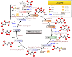
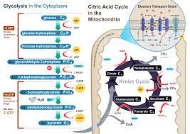
7 Krebs Cycle Steps and Process: Diagram, Explanation

Krebs cycle is also called to be citric acid cycle or the TCA cycle that helps in making two of the carbon dioxide molecules.

The Krebs cycle steps include a chain of reactions with reduced form of NADH and FADH2. The Krebs cycle steps including the phases like condensation, isomerization, oxidative decarboxylations and oxidative decarboxylation.

There are four of the carbon molecule that is involved, oxaloacetate that helps start the cycle and is regenerated after all the eight steps of the citric acid cycle is done. The Krebs cycle steps is a chain of hydration, decarboxylation, redox and dehydration reactions. All of the enzymes involved in the Krebs cycle steps are soluble.

Each of the turn in the cycle is made to form one ATP or one GTP as well with other three NADH molecules and one FADH2 molecules is further on used up in the Krebs cycle steps for the method of cellular respiration to make ATP for all the cells. It in simple term is a chain of reaction that is chemical by nature and done by aerobic organism.

krebs cycle steps
Image credit-Krebs cycleWikipedia

The Krebs Cycle can also be called the Citric Acid Cycle (CAC) or the Tricarboxylic Acid (TCA) Cycle. This cycle takes place in the Mitochondrial matrix and is the primary step of aerobic processing within a cell. The process oxidises glucose derivatives, fatty acids and amino acids to carbon dioxide (CO2) through a series of enzyme controlled steps.

The citric acid cycle keeps in the generation of energy along with having acetate oxidized and derived from fats, proteins, carbohydrates, proteins into the carbon dioxide. On the other hand, with being the same, Krebs cycle steps include the mechanism for oxidative analysis for acetyl units and then serves as the basic source for cellular energy.

There is a conversation of acetyl CoA, the Krebs cycle steps is seen in the matrix of mitochondria. In cellular biology, the mitochondria are an organelle that has a membrane which is often said to be the power house of the cell and also said to be cellular power plants as they are used up to make the maximum amount of ATP.

Cellular respiration Krebs cycle steps

There is only one enzyme for the Krebs cycle steps that is insoluble being the succinate dehydrogenase that is embedded inside the mitochondria.

The process of Krebs cycle steps includes 8 of the phases. Not like glycolysis, this cycle is not an open loop with the last stage of the path being regeneration of a compound that is used in the start step.

The eight of the steps are a complete chain of all the reactions that is possible and generates molecules of two carbon dioxide, NADH and FADH2, GTP or ATP. This pathway is said to be an aerobic one as the FADH2 and NADH is made and shall transfer the electrons to the next path in its system that makes the use of the gas oxygen.

image 18
Image credit-Cellular respirationWikipedia

Glycolysis produces pyruvate which, under aerobic conditions, gets moved into the mitochondria via a carrier protein within the membrane. There it is oxidatively decarboxylated by a huge enzyme complex called the pyruvate dehydrogenase complex. This reaction is irreversible and requires coenzyme A as well as producing 1 CO2 and picks up two electrons by NAD.  

The process of Krebs cycle steps includes-

Step 1

The very first phase of the Krebs cycle steps include the step for condensation that help combine the group of two-carbon acetyls.

This two carbon group is derived from the acetyl CoA that is linked with the oxaloacetate having four of the carbon molecule to make a molecule of citrate that has six carbon molecule.

CoA is said to link to a group called sulfhydryl and then makes it way to diffuse away that links eventually with the other group of acetyl. This process is said to be irreversible and the reason being is that it is too exergonic. The reaction rate for this step is observed by the not so positive feedback and the availability of the amount of ATP.

image 61
Image credit-Acetyl CoAWikipedia

There is an increase in the level of ATP with a decrease in the rate of the reaction. Along with working for many steps this reaction releases their molecules of NAD which are reduced to NADH. There is never a short of any reactants that shall be a hindrance to have a run. This cycle helps in making of energy that is used up by the cells.

Step 2

Citrate is said to be tricarboxylic acid and is derived from the group of three of the carboxyl group of the citric acid.

Citrate seems to lose one of the molecule of water and on the other hand also gains another as the citrate is converted to its isomeric form called isocitrate. Thus with the involvement of citrate, Krebs cycle steps completes its second phase.

In the second reaction of the Krebs cycle, the isomerisation of citrate to isocitrate takes place. As an intermediate, cis-aconitate is formed. Therefore, it is a two-step reaction sequence. The Krebs cycle is the second stage of cellular respiration. During the Krebs cycle, energy stored in pyruvate is transferred to NADH and FADH2, and some ATP is produced.

Once citrate has formed, the citrate then goes on to step 2 in which it is transformed into an isomer molecule called isocitrate. This step is carried out in two different reactions; a dehydrolysis reaction followed by a hydrolysis reaction. citrate is too stable for the reactions that follow. So, the Acontiase links to the Citrate to move one of its oxygen atoms to create a more unstable citrate isomer.

image 19
Image credit-Isocitrate-Wikipedia

The compound citrate has its role in the fundamental metabolisms. It is the anion citrate and the acid of tricarboxylic and also a conjugate base of the compound citrate and the conjugate acid of citrate. This compound citrate is used to make the urine in the blood less acidic or alkaline. This helps in the prevention of developing stones in kidney. 

Step 3 and 4

In this phase of Krebs cycle steps, there is an involvement of oxidization for the isocitrate that makes a carbon molecule.

The compound isocitrate when is oxidized forms a molecule of carbon that has five carbons in it. It also has an alpha ketoglutarate along with a molecule of carbon dioxide and two of the electrons.

The release of two of the electrons that is released are reduced from NAD to NADH. This steps are made to regulate via the negative feedback from the adenosine triphosphate and NADH and also effected by the positive outcome of the ADP. The third and fourth Krebs cycle steps consists of both the decarboxylation and oxidation steps.

This helps in the release of the electron that help in getting the NAD reduced to NADH and also helps release the group of carboxyl that makes molecules of carbon dioxide. The product of the third phase in the Krebs cycle steps is alpha ketoglutarate and a group of succinyl is made from the phase four of the Krebs cycle steps.

CoA seems to bind with the product of the step 4 which is the succinyl group to make succinyl CoA. The enzyme that this cycle uses is all independent of each other and available all the time and thus an enzyme is sued to work out the step 4 and thus is determined by a feedback which is inhibiter of ATP, NADH and also succinyl CoA.

Step 5

Any group of phosphate is said to be a substitute for the coenzyme A and has a good bond with high energy made for itself.

The energy that is made is used up at the level of substrate phosphorylation. It is at the time of having the succinyl group converted to succinate. This is done for either of the two reasons- to form either adenine triphosphate or guanine triphosphate.

The reaction of succinyl-CoA to succinate (see figure) proceeds via the intermediate succinyl phosphate. In the first step, the coenzyme A, which is located at the succinyl group, is substituted by a hydrogen phosphate ion Succinyl phosphate remains bound in the enzyme an dis therefore not released. In the second step, succinyl phosphate transfers its phosphoric acid residue on guanosine diphosphate.

There are enzymes seen and are typically of two types called the isoenzymes for this phase of Krebs cycle steps. This step depends on the type of tissue used mostly been animal tissue that is found. One of the form is seen in the tissue that have large amounts of adenosine triphosphate just like in the skeletal muscle and the heart. GTP and ATP can be converted into each other by the enzyme nucleosiddiphosphatkinase.

image 62
Image credit-ATPWikipedia

The form seen inside the tissue is said to make ATP. The form that is second for the enzymes is seen in the tissue that have mote number of pathways that are anabolic in its nature just like in the liver. This from is said to make GTP. ATP and GTP is basically said to be equal in terms of its energy with also its use being much restricted. The method of protein synthesis sues up GTP.

Step 6

This phase in the Krebs cycle step is said to the process of dehydration and helps in the conversion of a compound called succinate to fumarate.

It consists of the transfer of two types of hydrogen atoms into FAD that makes FADH2. There is energy stored in the electrons and this energy kept in the atoms is not enough to get NAD reduced but is more than enough to have FAD reduced.

With not quite likely for NADH, the career shall stay attached to the enzymes and help in the transfer of electrons to have the electrons chain directly transferred up. This method is seemingly easy for work out with by the enzyme that is localized in the step within the inner membrane of the matrix of the mitochondria.

image 20
Image credit-FAD-Wikipedia

With the carbons removed, the rearrangement process begins manipulating the hydrogen. When the Succinate dehydrogenase links to the substrate, it releases two hydrogen atoms attaching them to a carrier, ubiquinone (Q), or FAD Flavin adenine dinucleotide. With the additional 2 electrons ubiquinone forms ubiquinol being QH2 or FADH2 which is then transferred to power the electron transport chain.

Step 7

Water is said to be added to the fumarate at the 7th phase of the Krebs cycle steps along with the generation of malate.

The last and final stage of the Krebs cycle steps include the once again production of oxaloacetate by having malate oxidized. There is another molecule that is made called the NADH.

Two of the carbon atoms enter this cycle from the acetyl group that represents the four out of six carbons of the one molecule of glucose. There is a release of two carbon molecule with not needed for the carbo atoms to be carried. The atoms of acetyl carbon which are two in number shall be release later on with the original ones stays incorporated.

The overall is given by Acetyl CoA + 3NAD+ + FAD + GDP + Pi + H20
gives 2CO2 + 3NADH + FADH2 + GTP + CoA + 2H+

image 60
Image credit-NADHWikipedia

Finally, the Malate dehydrogenase recreates the Oxaloacetate substrate and moves electrons from the NAD+ to form NADH, the last energy produced by the Krebs cycle. Interestingly, this Malate–Oxaloacetate reaction is also used to move anaerobic energy from the cytoplasm into the mitochondria. While anaerobic reactions produce NADH, it cannot move from the cytoplasm to the mitochondria.

Krebs cycle vs fermentation

Both anaerobic respiration and fermentation are the two type of cell respiration methods that are use up for making of ATP for cell function.

Fermentation is the process that begins with the method of glycolysis but yet has no involvement of any steps of aerobic respiration unlike Krebs cycle and oxidative phosphorylation.

Fermentation is a metabolic process that produces chemical changes in organic substrates through the action of enzymes. In biochemistry, it is narrowly defined as the extraction of energy from carbohydrates in the absence of oxygen. In food production, it may more broadly refer to any process in which the activity of microorganisms brings about a desirable change to a foodstuff or beverage. The science of fermentation is known as zymology.

220px Fermenting
Image credit-FermentationWikipedia

Both of the methods are made to function in without the use of oxygen gas. They use the hexose sugar as a form of substrate. This hexose sugar is said to first undergo the method of glycolysis. The ultimate difference for the two processes is that fermentation seems to not use the Krebs cycle and the electron transport chain while the anaerobic process makes the use of citric cycle.  

Krebs cycle vs Ketosis

The time that the cell needs fuel, it shall pull up the ketone body from the blood and then turn in back into else.

After pulling up of ketone body and converting it to acetyl CoA it is transported via the method of Krebs cycle whet they make up energy in the form of ATP. The brain in quite fond of ketones that shall be increasing on along with keto diet.

Ketosis is the process that takes place when the body is not able to make carbohydrates enough to burn the energy down. Instead of it bringing out things called ketones and fats that shall help as aa fuel. Ketosis is the process that is mostly uses for Weight loss or the diabetic ones.

Ketosis happens when your carbohydrate intake is low. As your body breaks down fat, it produces an acid called ketones or ketone bodies, which becomes your body and brain’s main source of energy. Because ketosis shifts your metabolism and relies on fat for energy, your body can burn fat at a higher rate. In general, it should take you 2–4 days to enter ketosis. However, some people may find they need a week or longer. 

Also Read:

3+ Krebs Cycle Function: End Products, Equations and Facts

image 53

Krebs cycle is also called to be the TCA cycle having its full name as tricarboxylic acid and also called as citric acid cycle (CAC).

The Krebs cycle function in many ways. In simple, terms all of the pathways for the metabolism of energy are linked to this cycle. Some of the Krebs cycle function are-

It is defined as a series of chemical reaction that helps release the energy that is kept via the process of oxidation of the compound Acetyl-CoA and is derived from the proteins, carbohydrates and the fats. This cycle is also seen to be used by the organism that respire or the ones that ferment to produce energy either by the method of aerobic or anaerobic type of respiration.

Krebs cycle function
Image credit-Krebs cycleWikipedia

On addition, this method also helps the use of precursors for specific amino acids and also for the reducing agent called NADH which is used in any other reactions. It is of much vital central use for several biochemical paths that implies that it is one among the old methods or product for metabolism and shall develop abiogenically.

It is located in the matrix of mitochondria. One can be at comfort to help deduce each cell that possess the mitochondria and has quite a few physical conditions that is active in the TCA cycle. Despite all the cells need to perform this, there is one population of cell that has no mitochondria said to be the erythrocytes. Thus, there is no Krebs cycle or vitality of Krebs cycle function is this.

The Krebs cycle function makes the use of aerobic purpose for having a smooth path for it to work. The Krebs cycle that has the lack of the gas oxygen has quite of a limited velocity. There is a process of oxidation involved in this for the acetyl residues. This is the source for the reducing agents and yields FADH2 and NADH. It is said to be amphibolic pathway. Just like the method of electron transport chain (ETC), the process of transamination, then deamination if the amino acids and lipogenesis. 

Oxidation of the Acetyl residues

In the Krebs cycle function, the very first that comes up is having the residue of acetyl oxidized to carbon dioxide.

The method of Krebs cycle is an absolute for the source of the equivalents for reduction that are transferred for the cofactors said to be NAD and FAD. This results in FADH2 and NADH. This is called reduced coenzymes.

The reduced co-enzymes are made to enter the electron transport chain and the process of regeneration takes place. The method of regeneration is said to be the loss of electron or the loss of the reducing equivalents and thus this process is called the reoxidation.

The process of reoxidation is also called to be regeneration for the reduces coenzymes and is the one that links the TCA cycle with the main supply for the reduced coenzyme for the electron transfer chain and thus this cycle is very vital for the making and giving of ATP with having a turn over for making only one GTP.

image 53
Image credit-Electron transport chainWikipedia

Flow of many catabolic process

There are many of the catabolic ways that is a source to this. This is the second in the list for Krebs cycle function.

It can be a source to have the intermediates of TCA cycle, pyr, AcCoA. The fate for them can be having the carbon dioxide oxidizes and also to have the rest of the materials synthesized.

The oxidation number for C in the compound carbon dioxide is 4. This compound has a good and high TLV which is basically more and is quite inexpensive while compared to rest of the solvents. It is nonflammable and thus cannot be kore oxidized and thus it is an advantage.

The two of the carbon molecules of the Acetyl-CoA are made to release and thus make a molecule of carbon dioxide, Thus, for this reason they are made to enter the cycle in form of Acetyl-CoA with making a two of the molecules of carbon dioxide and oxidation takes place 4 times.

Providing precursor for the paths

The word precursors mean a substance from which any other alternative can be made mainly concerned with the metabolic path.

Some of the example for the Krebs cycle function as a precursor for many pathways are glycogenesis, having the amino acids synthesized, getting the tetrapyrroles biosynthesized and as a source of AcCoA for synthesizing of the fatty acids.

Glycogenesis is the method for having the glycogen formed and then the basic carbohydrates shall be kept in lover and the muscle cell for the animals and then makes glucose. Glycogenesis takes placed within the blood glucose while the level is sufficient more to have glucose and liver muscle.

Glycogenesis.png
Image credit- GlycogenesisWikipedia

Excretion of nitrogen

Alanine and the rest of the amino acids are seen to travel to the organ lover. It is the place of conversion.

In the liver, the conversion of the carbon to ketone body and the glucose is initiated and then nitrogen is converted to the urea which is then made to be extorted by the kidney. Urea takes place via nitrogen cycle.

Urea cycle is said to be the major path for having the nitrogen disposed from the humans. More than 90% of the protein that is ingested is made to metabolize to the urea and then is excreted via the urine. Ammonia is said to be derived from the various precursor of the protein sources.

425px The Nitrogen Cycle
Image credit-
Nitrogen cycleWikipedia

Krebs cycle Equation

Krebs cycle was named after Hans Krebs, who postulated the detailed cycle. He was awarded the Nobel prize in 1953 for his contribution.

The overall reaction for the citric acid cycle is as concerned- acetyl-CoA + 3 NAD+ + FAD + GDP + P + 2H2O = CoA-SH + 3NADH + FADH2 + 3H+ + GTP + 2CO2. Many molecules in the citric acid cycle serve as key precursors for other molecules needed by cells.

The citric acid cycle also produces 2 ATP by substrate phosphorylation. At the end of the Krebs cycle, the final product is oxaloacetic acid. This is identical to the oxaloacetic acid that begins the cycle. Now the molecule is ready to accept another acetyl-CoA molecule to begin another turn of the cycle. Reduced high energy compounds, NADH and FADH2 are also produced.

It is a series of eight-step processes, where the acetyl group of acetyl-CoA is oxidised to form two molecules of CO2 and in the process, one ATP is produced. Reduced high energy compounds, NADH and FADH2 are also produced. It is a series of eight-step processes, where the acetyl group of acetyl-CoA is oxidised to form two molecules of CO2 and in the process, one ATP is produced.

Where does Krebs cycle function?

The Krebs cycle is said to the main and vital source for the cells to have its energy and is vital for aerobic type.

The cycle is actually harmless and also has several chemical energies for acetyl coenzyme that is reduced to the power of nicotinamide adenine dinucleotide. The Krebs cycle function side the matric of the mitochondria.

It is actually a cycle for respiration that yields the making of ATP is large amounts and then consumes oxygen to work. It is actually for having the organism respire. Citrate is made in the Krebs cycle from oxaloacetate condensation and thus called the citric acid cycle.

The TCA cycle plays a central role in the breakdown, or catabolism, of organic fuel molecules that is glucose and some other sugars, fatty acids, and some amino acids. Energy is produced in a number of steps in this cycle of reactions. In step 5, one molecule of adenosine triphosphate also said to be ATP, the molecule that powers most cellular functions, is produced. 

Other functions include

What are the products of the Krebs cycle?

TCA cycle is the other name for Krebs cycle and is a portion for the large pathway where glucose is said to be oxidized.

At the start of this cycle, a molecule of glucose is first converted to the Acetyl-CoA. This method outcome in the yield of the 2 molecules of Acetyl-CoA to indulge into the cycle. Thus, the Krebs cycle function yields 7 in number of products-

Pyruvate is said to a molecule in biology and yield glucose as its product that reacts with ATP and also carbon dioxide that converts Acetyl-CoA a then ADP at the start of the Krebs cycle. It is generally included in the start or the first step of the cycle. It is derived from the method of glycolysis that dissociate quick.

NADH

It stands for the compound called nicotinamide adenine dinucleotide along with hydrogen and is seen in body.

It is a vital element in the chemical methods that is used to make energy. People also seem to use this element as a supplement for antibiotics with the base of NADH. Krebs cycle function yields 3 of it.

image 54
Image credit-NADHWikipedia

Nicotinamide adenine dinucleotide is said to be a coenzyme that is located at the central part and is vital for metabolism. It is seen in all the living cells and NADH is called to be a nucleotide as it has two of the nucleotides linked via the phosphate groups. One of the nucleotides have adenine and the other has nicotinamide.

NADH is a coenzyme found in all living cells; consists of two nucleotides joined through their 5′-phosphate groups, with one nucleotide containing an adenine base and the other containing nicotinamide. It has a role as a fundamental metabolite and a cofactor. NADH is necessary for cellular development and energy production: It is essential to produce energy from food and is the principal carrier of electrons in the energy-producing process in the cells

FADH2

This element stands for Flavin adenine dinucleotide. It has its use in the metabolite of E. coli and also for a mouse.

In terms of biochemistry Flavin adenine dinucleotide is a coenzyme related to redox activity and is linked with several proteins that is concerned with many other reactions. Krebs cycle function yields one of it. FADH, also known as 1,5-dihydro-fad or FADH2, belongs to the class of organic compounds known as flavin nucleotides.

The element of the Krebs cycle function is Flavin adenine dinucleotide and has many of the reactions that have enzymes included with it. A flavo protein is said to be protein that has the group of Flavin and may be in the form of Flavin mononucleotide or FAD. It has its chemical formula as C27H33P2N9O15 and is made by the reaction of reduction and oxidation.

A flavoprotein is a protein that contains a flavin group, which may be in the form of FAD or flavin mononucleotide (FMN). Many flavoproteins are known: components of the succinate dehydrogenase complex, α-ketoglutarate dehydrogenase, and a component of the pyruvate dehydrogenase complex. Both NADH and FADH2 are high energy/unstable compounds, like ATP.

CO2

In general carbon dioxide is a most common word in the product of Krebs cycle function and is a gas that has no color.

Krebs cycle function yields two of this element. There are two of the carbon atoms for the Acetyl-CoA that is released and each is made to form a molecule of carbon dioxide. Thus, for each acetyl that enters two of CO2 is made.

The energy that is made in the molecules of ATP, FADH2 and NADH is captured by them and is along with another compound that captures energy. CO2 is releases as a product that is a waste of the reactions. The final stage of the Krebs cycle function regenerates a compound called OAA which starts the Krebs cycle. Two carbon dioxide molecules are released on each turn of the cycle; however, these do not contain the same carbon atoms contributed by the acetyl group on that turn of the pathway.

GTP

It stands for guanine 5 triphosphate. It is a base of purine and is one of the building block that is needed to get the RNA synthesized.

The structure of GTP is said to be same as that of guanosine nucleoside with having only one difference being that the nucleotides in this like GTP that have the group of phosphates in the ribose sugar. Krebs cycle function yields one of this.

It is a molecule that is rich in the nucleotide analogue to adenosine triphosphate and is made up of guanine, three of the phosphate and ribose and is much vital during the time of protein synthesis. It is termed as guanosine triphosphate. It has its chemical formula as C10H16N5O14P3.

Guanosine triphosphate also called as GTP with chemical formula: C10H16N5O14P3 is a nucleoside phosphate comprised of a ribonucleotide and three phosphate groups. It means it has a ribose as its sugar and three phosphate groups attached. Heterotrimeric G-proteins are composed of three distinct subunits. There are many different α, β, and γ subunits, allowing a bewildering number of G-protein permutations.

Also Read:
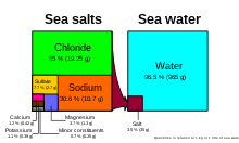
Parenchyma Cells in Plants: What, Structure, Types, Function, Origin

image 5

There are many terms lined with the word parenchyma and is mostly concerned with plants only being a tissue or a cell.

Based on anatomy, parenchyma cells in plants can be anatomically defined as the tissue that is functional in an organ and can be identified as the form of a connective tissue that also helps in supporting.

Not only based on anatomy but also considering the botany and zoologically, parenchyma cells in plants can be defined to be the cell tissue that is actually succinate as well as soft and is seen majorly in the soft areas of the lead and the pulp of the fruits, pith of stem and the bark as well.

The parenchyma cells in plants in terms of zoology can be said to be type cell tissue that stays between the organs of all the invertebrate organism and the body wall that lacks the part coelom for instance can be a flatworm.  It is actually a special tissue made up of all the cells that are alive and have a wall thin to the eye.

parenchyma cells in plants
Image credit-Parenchyma cellsWikipedia

Parenchyma cells in plants are the formation of the bulk of the plant in the ground tissue, that have a special way to work in for storage of food or energy or water or even the waste material, photosynthesis, transport and exchange of gases. They are the inner part of the vascular tissue and sends material inside the phloem and xylem.

As said already for the parenchyma cells in plants refers to the working portion of the plant body in an organ and is quite different from the one called to be interstitium or the stroma which means the structured tissue in the organs like that of the connective tissue. There are parenchyma organs that consist of the liver, kidneys, pancreas, spleen and the adrenal gland.

What is parenchyma tissue?

Parenchyma are found in plans and are tissue that are made of all the cells that are living and are thin walled.

They are not specific or special in its formation and thus are adaptable with easy to differentiate fir several functions they are called for. The parenchyma cells in plants are seen also in several areas.

The parenchyma cells are seen in the whole body of the plants and are much active in taking part for photosynthesis, having food stored and secretion. It also helps majorly in the method pf photosynthesis and rest of the plant life activities. Parenchyma is termed to be one of the main ground types for the three for them.

It combines with the dead tissue that support on the plant with the thick walls called the sclerenchyma and also with the support live tissue that have irregular wall and called the collenchyma. It fest to be part of the mesophyll that is called to be the inner later of the leaves and also forms the outer part of the plant called the cortex along with roost and stem.

The cells of this type are also seen in the fruits as soft tissue and also have themselves of the phloem and xylem to have the cells transferred and also for the bundle sheaths that sets around the vascular strands. The parenchyma tissue also shall be compact and will be extensive in space in the cells. The leaves also have parenchyma cells or tissue.

image 6
Image credit-Vascular strands-Wikipedia

The mesophyll in leaf is made of the parenchyma tissue. It can also be said that parenchyma cells in plants is seen in the mesophyll of the leaf. They are elongated and are palisade parenchyma that have large number of chloroplast per cell and is the ground area for photosynthesis in several plants. There is also an irregular sponge like parenchyma having chloroplast. 

Origin of parenchyma cells in plants

The parenchyma cells in plants are seen in the primary part of the plant body just like the cortex or the pith.

The origin of the parenchyma cells in plants is considered to be the ground meristem. Cambium and procambial tend to give rise to the phloem and xylem parenchyma of the secondary and the basic conducting tissue.

The parenchyma tissue cells are seen in the delicate portion of plants just like the roots and cortex, in the stems pf the ground tissue and also in the leaf mesophyll. Any of the organ is made up of the parenchymal type of cells and works for the parenchymal cells. They are said to be made up of many kinds of tissue with being a collection of all same kind.

Parenchyma is quite of a versatile cell that basically has the filler tissue in the sensitive part of the plants. It make up the another among the rest of the things like the outer are called the cortex, the central area called the pith  of the stems, the cortex area of the roost, the leaf mesophyll, the fruit pulp and the see endosperm. In simple words. They are a tissue collection that perform several work load.

image 7
Image credit-CortexWikipedia

In the leaves, the ground meristem and the protoderm give rise to the parenchymatous epidermis and also the mesophylls. On talking about the shape of the parenchyma cells in plants, they can be grown in the lab in the cultural media and are mostly sphere. In the cell gathering, there are polygonal as for their contact with the rest of the cells. The parenchymal cells with fourteen cells are common.

The parenchymal tissue that are compact tend to set without any of the intercellular gaps. One instance can be the seed having the endosperm tissue. They can be said to be loose in package yet that are well developed in the intercellular space and are seen in between them just like the leaves and the stem of the hydrophytes.

What is the parenchyma in plants?

Most of the tissue in the animals and plants are parenchymal. The very word parenchyma means content of vessel and is a Greek word.

In terms of biology, the parenchyma cells in plants are simple yet permanent type of tissue which is vital of the function of cells and perform in bulks. The chief way of understanding the parenchyma cells are getting to know its use.

The basic of the plants concerned with ground tissue are made up of their tope of cells called the parenchyma, sclerenchyma and the collenchyma. Parenchyma is the one having thin cell wall and is made of cell that are alive and work metabolically. Collenchyma is the one also made of living yet have thick cell wall and the last one is of dead cells and thick cell wall.

image 8
Image credit-Ground tissueWikipedia

The cells ofparenchyma are living type tissue that have the ability to process cell division at the adult stage. Thus, they offer the needed help in the regeneration of tissue and repairing of the tissues. The basic making of the reproductive cells like the gametes and spore is parenchymatous. Every cell that is parenchymatus being the part of zygote is totipotent that has the ability to grow in a whole plant.

They are a non-ending mass or cell in bulk that is found in the pith or the root cortex, the stem, the mesophyll in leaves, the flesh area of the succulents and the seed endosperm and these portions of the plant can be said to be parenchymatous. They also help in excretion, respiration, storage and transport in radial manner.

The cell wall of the plant for the parenchymal cells are thin and is made of the thin layer and is composed of hemicellulose and also cellulose. It is the primary layer made of only single part. The parenchyma cells are linked via the plasmodesmata. Plant cells have parenchyma accumulated in the storage place that have xyloglucan walls. There is a sugar concentration in the cells and also as a source of germination.

Parenchyma tissue in plants function

There are several work for the functioning of the parenchyma cells, and they are permanent simple tissue that divide.

They are used for storage of materials inside the plants, having the ability to gas exchanged, healing of wounds and regeneration of cells, secretion, photosynthesis. They parenchyma cells are the basic of the plant.

The cells of the parenchyma also help in mechanical support for the plants while they seem to be lignified and are thick walled. They have thin walls and stay activate and alive until mature. While this leads to make them being less useful in the application of structure, the cells need to move and have the water stored as they divide.

Types of parenchyma tissue in plants

Parenchyma cells in plants are simple and make a vital portion of the ground tissue and are embedded in vascular tissue.

The types of parenchyma cells in plants are-

Epidermal parenchyma

Epidermis is said to be the outer layer of the plant boy. In some of the older ones they are said to be a special type of parenchyma cells in plant.

With all of the consideration, in new terms epidermis is said to be dermal tissue where the parenchyma is said to a ground tissue. The epidermis serves quite of a function just like parenchyma cells.

image 9
Image credit-EpidermisWikipedia

Most of the leaves having the upper epidermis show the feature of dorsoventrally anatomy that means that the adaxial or the upper part and the abaxial or the lower part surface have some of the different form of construction and might as well function different.

Xylem parenchyma

The element of xylem being parenchyma cell is kin of complex and is also a tough tissue said to be called as xylem. The cells are used for storage.

Xylem and phloem both are said to be the conducting tissue of the vascular ones. The main role is to have the food transported for minerals, water and also food. Phloem is for food and xylem is for water.

image 10
Image credit-Palisade tissueWikipedia

Xylem are quite of a complex tissue that is concerned with having the water transported and also other type of nutrients in the plants. It involves dead cells while the parenchyma has cells that are alive also seen in the xylem. Xylem is said to be connected via the tyloses. They are store house for fats, food made in form of starch, crystals and also tannins.

Mesophyll parenchyma

Mesophyll is an inside tissue located in between the cell layers of epidermal in the leaf.

Mesophyll is made up of two kinds of tissue called the palisade parenchyma that is the upper later of the length chlorenchyma cells being large amount of the chloroplast. The other is the spongy parenchyma which is the lower area.

The palisade parenchyma is generally placed direct below the epidermis of the top leaf surface while the spongy one makes it fill the space between the palisade parenchyma. It helps in having gas engaged and also for photosynthesis through the pore called chloroplast.

Spongy mesophyll tissue is packed loosely for efficient gas exchange. The spongy mesophyll cells are covered by a thin layer of water. Gases dissolve in this water as they move into and out of the cells. he mesophyll is found between the upper and lower epidermis, it aids in gas exchange and photosynthesis via chloroplasts. The xylem transports water and minerals to the leaves.

Also Read:
Parenchyma Cells Function: In Plants, Xylem, Phloem, Roots
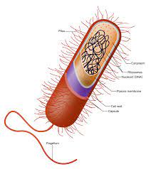
image 2

Parenchyma is a simple yet permanent tissue type that includes a major portion of the ground tissues for the plants with other having vascular ones.

The parenchyma cells are actually said to be alive and these parenchyma cells function is to have-

A parenchyma is a type of tissue that is non-vascular in form and is made of very simple and undifferentiated type of cell that is modifies to play a vital part in for several functions. The other two types of permanent simple tissues are sclerenchyma and collenchyma. The cells in it seem to have very less intercellular space or has no space at all being tightly packed.

The structure of parenchyma cells is that it is a living cell that has a good view of nucleus and the protoplast. The parenchyma cells functions to be isodiametric or is polyhedral in its shape. It can also be seen to be in the shape of oval round, sometimes elongated and polygonal. The cell wall of the parenchyma cells are composed of hemicellulose, cellulose.

parenchyma cells function
Image credit-Parenchyma cellsWikipedia

Plasmodesmata seen to connect the cells of all the tissue of parenchyma. They also have the feature of having many small and tiny vacuoles. In the parenchyma that are older small vacuoles get to link and form a large central vacuole that shall gather tannins or the anthocyanin. The vacuoles are not empty but have water.

Water is found in abundance inside the tiny vacuoles of the parenchyma cells and also serves to be a water reservoir. The parenchyma cells that are for storage might see to have thick walls of xyloglucan which are the endosperm of the date palm. The sugar is held to be used for the purpose of germination and making the wall thinner.

Along with parenchyma cells functions it also has fruits and flowers that have chromoplasts. The cells of parenchyma may have walls that are thick and lignified that helps in act hard to separate from the sclerenchyma. The mechanical strength for the parenchyma cells are taken from the cells property of hydraulic.

The following tend to show the parenchyma cells function-

Energy stored- The parenchyma cells are simple tissue that has cells that are non-specialized with having thin walls. They have cells that are not tight in packaging. It helps in getting the food storage and then provide plant support. The rest part of the part help in storing of nutrients and water. Parenchyma cells perform storage like starch and oils or secretory functions.

Store waste products- The cells of the parenchyma are actually bigger. They also have large vacuoles placed centrally that helps them to have the function of ion regulation and storage along with keeping in waste materials and water. The tissue that is specific for food keeping is made in the parenchyma cells. They keep in resins and gums and form the wood rays that are radially aligned tissues interspersed between the axial elements.

Help in photosynthesis– Some of the parenchyma cells are able to differentiate for phloem and then send special path for the sugars and the products for taking up photosynthesis to travel in the plant. These cells now have the product made from the leaves itself while they make its way via the whole root.

Gaseous exchange- Aerenchym are type of parenchyma cells that help in having the oxygen level maintained via the method of respiration. They are present in continuous form from the roots to shoots and thus help in having the air diffused from the leaves to the roots. They are the cells that are help through maturity.  These store various substances like water, starch, proteins etc. They act as a food and water reservoir. Parenchyma cells may be specialized as a water storage tissue in succulent plants such as Cactus or aloe Vera.

Secretion and transport-Parenchyma with chloroplast that helps in performing photosynthesis is called chlorenchyma. Sclerenchyma tissue, when mature, is composed of dead cells that have heavily thickened walls containing lignin and a high cellulose content with abut 60% to 80%, and serves the function of providing structural support in plants. Sclerenchyma cells possess two types of cell walls: primary and secondary walls.

Types of parenchyma

The parenchyma cells seem to have chloroplasts that are specific to photosynthesis.

The several types of parenchyma are-

Chlorenchyma

These are the parenchyma cells that are inherited with chloroplast and help in photosynthesis by performing the method.

It is seen in the green plants with situated mainly ate the region of sepal or stems. The mesophyll cells of the leaves that differentiate into spongy and palisade cells also have it.

The main criteria for this type of cell is that this type of parenchyma cells functions to repair. In the laves, they create the mesophyll and them are held active for generating photosynthesis and help in exchange of gases. These parenchyma cell types are in the leave mesophyll and are special type of parenchyma cells canned the chlorenchyma.

File:Herbaceous Dicot Stem Collenchyma, Sclerenchyma and Parenchyma in  Cucurbita (37411748021).jpg - Wikimedia Commons
Image credit-ChlorenchymaWikimedia

They are said to be a special type of tissue under parenchyma. It is said to be specific as it has chlorophyll that is green color pigment that helps in getting the process of photosynthesis done. All of the cells of this have same function as it is a permanent and simple tissue and thus all the chlorenchyma cells apart from the one having bulk area works for photosynthesis.

Chlorenchyma are the parenchyma cells which contain chloroplast. The tissue containing chlorenchyma is present in the leaf interiors as well out outer cortex of young stem. It is the tissue which carry out photosynthesis. Chlorenchyma tissues are the types of parenchymatic tissue having chlorophyll. It helps to perform the function of photosynthesis in plants.

Xylem parenchyma

They are the element having the type of complex tissue called to be xylem. The cells of parenchyma of the xylem have functions.

The parenchyma cells functions of the xylem parenchyma are to help in having the fats, the carbohydrates and the water stored for conduction. The plants cells are actually classified based on the functions and its strictures.

Xylem is seen to come from the work xylon that means wood. It was termed by Carl Nageli. It is a type of vascular tissue that is seen in the plants that helps in the primary support of water and also the nutrients from the stem and the leaves. They also provide with mechanical support to the plants. There can be two types of xylem based on its origin.

Xylem - Simple English Wikipedia, the free encyclopedia
Image credit-XylemWikipedia

The first can be primary xylem that generates from procambium and is further staged into metaxylem and the protoxylem. The secondary xylem has its vascular cambium as its origin. Xylem as a whole is made up of four types of elements that are different being vessels, xylem fiber, tracheid and xylem parenchyma.

In simple word, the parenchyma cells that are linked with xylem are called xylem parenchyma. There are two types of the parenchyma inside the secondary structure of xylem. They are the radial parenchyma cells and the axial parenchyma cells. The storage of the protein and fast vary with seasons. There is a migration of nucleus and cytoplasm.

The main attribute of parenchyma cells functions is-

  • Storing of the fats, crystals, starch and fats.
  • The parenchyma xylem cells are linked with the vessels or the tracheid via the outgrowth that are called the tyloses.
  • The parenchyma of xylem is connected with having to maintain the xylem transport tissue.
  • They are help actual for having the vessels restored and the help tracheids function when there is a cavity block as for the air bubble. Cavitation takes place due to the large water tension in the tissues of xylem
  • The parenchyma cells of xylem have the radial conduction going on inside it.

Arenchyma

They are said to be the artificial type of parenchyma Cells. It is made up of the sponge tissues that makes up space for the air.

The space is made in the roots, stems or the leaves of few plants that allot h passing of the gas exchange between the root and the shoot. The space can be said to be the channels and have air in them.

The channels seem to make low resistive for having the gases exchanged like ethylene or oxygen above water and then have the tissues submerged in it. This type of parenchyma cells is mostly common in the wetland and also the aquatic that shall grow in the hypoxic soils. The very word aerenchyma has been taken from a Latin word which means air infusion.

image 3
Image credit-AerenchymaWikipedia

While there is a flood in the soil, hypoxia takes place and the microbes eat up the oxygen fast that the method of diffusion. The presence of the hypoxic in soils is one of the greatest feature of the wetlands. There can be several other reasons for the hypoxia in the soils. In common terms, the lower level of oxygen helps in stimulating the plants and the trees like manganese and iron.

Prosenchyma

These are the type of parenchyma cells that have starch in them with walls made up of lignin mostly seen in Bougainvillea.

They are said to be a special type of cells of the parenchyma ad is also called the transfer cells which is linked in the short path of having the solutes moved or transfer by the cell.

The structure of parenchyma cells is that they are living cells with a clear view of the nucleus and protoplast. The parenchyma cells are either isodiametric or polyhedral in shape. It can also be seen to be oval, round, elongated, and polygonal in shape. The cell walls of parenchyma cells are made up of hemicellulose and cellulose.

A vessel element or vessel member that are also called to be trachea or xylem vessel is one of the cell types found in xylem, the water conducting tissue of plants. Vessel elements are typically found in angiosperms said to be the flowering plants but absent from most gymnosperms such as conifers. Parenchyma is living tissue, and it is a thin-walled and unspecialized structure, and it is adaptable.

They make a good amount of the plant tissue in the ground that helps them specialized in the process of transport, photosynthesis and storage. Parenchyma is an inside part of the vascular tissues that shows a path for having the materials exchanged inside and between the phloem and xylem. They are the ones which have elongated cell type and that interpenetrate the ends. 

Mesophyll parenchyma

Mesophyll is said to be a ground tissue that is seen in between the epidermal layer call of leaves and is made of two kinds if tissue.

The mesophyll is a parenchyma type of tissue that is simple as well as permanent. The palisade parenchyma is placed below the upper surface of the leaf and exactly lower to the epidermis and makes the spongy parenchyma fit the space.

Leaf mesophyll is made up of the parenchyma cells. The elongated parenchyma that is the palisade has several number of large chloroplasts per cycle and is the basic site for photosynthesis in many green plants. Mesophyll is found in both the upper and lower epidermis.

image 4
Image credit-Palisade cellWikipedia

Mesophyll conductance is a vital component of photosynthesis, whose importance for accurate characterization of photosynthetic limitations has increased during the last two decades. Carbon dioxide diffusion across the leaf mesophyll is a complex process implying both biochemical and anatomical factors. Among them, aquaporins, chloroplast distribution, and cell wall thickness are its principal determinants.

The mesophyll is used in having the exchange of gas and also supports in photosynthesis via the chloroplasts. The xylem helps in getting the water transported and also the minerals along the leaves while phloem helps in having the transfer of the products of photosynthesis to the rest part of the green plant. Mesophyll is the internal ground tissue located between the two epidermal cell layers of the leaf.

After the cells of the ground meristem tend to divide and the differentiate, they shall be able to distinguish in several numbers of tissue that consist of the cortex, pith rays and the pith. Inside the leaves, they give rise to many of the parenchyma cells for the mesophyll layers that get involved in for photosynthesis. The palisade layer of mesophyll is quite vital.

Also Read: