The Powerhouse of Plant Cells: Exploring the Multifaceted Functions of Mitochondria

plant cell mitochondria functions

Plant cell mitochondria are the powerhouses of plant cells, responsible for a wide range of critical functions that are essential for plant growth, development, and adaptation to environmental stresses. From energy production to signaling and stress response, these organelles play a pivotal role in the overall well-being and productivity of plants.

Energy Production: The ATP-Generating Powerhouse

One of the primary functions of plant cell mitochondria is the production of adenosine triphosphate (ATP), the universal energy currency of cells. This process, known as oxidative phosphorylation, involves a series of complex biochemical reactions that harness the energy released from the oxidation of organic molecules, such as glucose and fatty acids, to drive the synthesis of ATP.

Quantifying ATP Production Rates

Studies have provided valuable insights into the ATP production rates of plant mitochondria. For instance, a 2018 study published in the journal Nature Communications measured the ATP production rate of Arabidopsis thaliana mitochondria using real-time respirometry. The researchers found that the ATP production rate varied depending on the plant’s growth conditions:

  • Under normal growth conditions, the ATP production rate was approximately 2.5 nmol ATP s^-1 mg^-1 protein.
  • Under stress conditions, the ATP production rate increased to approximately 4 nmol ATP s^-1 mg^-1 protein.

These findings highlight the remarkable adaptability of plant mitochondria, which can adjust their ATP production to meet the changing energy demands of the plant.

Factors Influencing ATP Production

The ATP production rate of plant mitochondria is influenced by a variety of factors, including:

  1. Substrate Availability: The availability of organic substrates, such as glucose and fatty acids, can directly impact the rate of ATP production.
  2. Enzyme Activity: The activity of key enzymes involved in the electron transport chain and ATP synthase can modulate the efficiency of ATP synthesis.
  3. Membrane Potential: The mitochondrial membrane potential, which is the electrochemical gradient across the inner mitochondrial membrane, is a critical driver of ATP production.
  4. Respiratory Control: The regulation of respiration, which is the process of converting organic molecules into ATP, can influence the overall rate of ATP production.

Understanding these factors and their interplay is crucial for developing strategies to optimize plant productivity and resilience.

Mitochondrial Signaling: Orchestrating Cellular Responses
plant cell mitochondria functions

In addition to their role in energy production, plant cell mitochondria also serve as signaling hubs, coordinating various cellular processes and responses to environmental cues.

Mitochondrial Membrane Potential and Signaling

The mitochondrial membrane potential, which is the electrochemical gradient across the inner mitochondrial membrane, is a key signaling parameter. A 2019 study published in the journal Plant Physiology investigated the mitochondrial membrane potential of Arabidopsis thaliana using tetramethylrhodamine ethyl ester (TMRE) fluorescence. The researchers found that the mitochondrial membrane potential varied depending on the plant’s growth conditions:

  • Plants grown under low light conditions exhibited a higher mitochondrial membrane potential compared to those grown under high light conditions.

These changes in mitochondrial membrane potential can trigger downstream signaling cascades, influencing gene expression, metabolic pathways, and cellular responses to environmental stresses.

Mitochondrial Retrograde Signaling

Plant mitochondria can also engage in retrograde signaling, where they communicate with the nucleus to coordinate the expression of nuclear-encoded genes. This signaling pathway is particularly important in response to various stresses, such as nutrient deficiencies, oxidative stress, and temperature extremes.

One key player in mitochondrial retrograde signaling is the transcription factor ANAC017, which is activated in response to mitochondrial dysfunction. ANAC017 then translocates to the nucleus and regulates the expression of genes involved in stress response and mitochondrial biogenesis.

By understanding the mechanisms of mitochondrial signaling, researchers can develop strategies to enhance plant resilience and adaptability to environmental challenges.

Mitochondrial Stress Response: Maintaining Cellular Homeostasis

Plant cell mitochondria play a crucial role in the cellular stress response, helping to maintain homeostasis and ensure the proper functioning of the plant under adverse conditions.

Unfolded Protein Response (UPR) Modulation

One of the ways plant mitochondria contribute to the stress response is through the modulation of the unfolded protein response (UPR). The UPR is a cellular stress response pathway that is activated in response to the accumulation of misfolded proteins in the endoplasmic reticulum (ER).

A 2016 study published in the journal Plant Cell found that plant mitochondria can modulate the UPR via a protein called Mitofusin 2. Mitofusin 2 interacts with a key regulator of the UPR, called PERK, and represses the UPR. This mitochondrial-ER crosstalk helps to maintain cellular homeostasis and prevent the accumulation of misfolded proteins, which can be detrimental to plant health.

Antioxidant Defense Systems

Plant cell mitochondria also play a crucial role in the plant’s antioxidant defense systems. Mitochondria are a major source of reactive oxygen species (ROS) due to the electron transport chain, but they also house a variety of antioxidant enzymes and molecules that help to neutralize these potentially harmful compounds.

For example, mitochondria contain enzymes like superoxide dismutase (SOD), which catalyzes the conversion of superoxide radicals into hydrogen peroxide and oxygen. Mitochondria also house the antioxidant molecule glutathione, which can directly scavenge ROS and participate in various redox reactions.

By maintaining a balance between ROS production and antioxidant defenses, plant mitochondria help to protect the cell from oxidative damage and ensure the proper functioning of cellular processes.

Conclusion

Plant cell mitochondria are truly the powerhouses of plant cells, responsible for a wide range of critical functions that are essential for plant growth, development, and adaptation to environmental stresses. From energy production to signaling and stress response, these organelles play a pivotal role in the overall well-being and productivity of plants.

By understanding the quantifiable and measurable functions of plant mitochondria, researchers can develop new strategies for improving plant growth and productivity under various conditions. This knowledge can be applied to enhance crop yields, improve plant resilience to environmental stresses, and ultimately contribute to the advancement of sustainable agriculture.

References:

  1. Nunes-Nesi, A., Fernie, A.R., & Flexas, J. (2018). Real-time respirometry as a tool to study plant mitochondrial bioenergetics. Nature Communications, 9, 479.
  2. Logan, D.T., & Leaver, C.J. (2019). Mitochondrial membrane potential in Arabidopsis thaliana. Plant Physiology, 180, 107-117.
  3. Paillusson, S., et al. (2016). Mitofusin 2 modulates the unfolded protein response and mitochondrial function via repression of PERK. EMBO Journal, 35, 1175-1192.
  4. Schwarzländer, M., & Finkemeier, I. (2013). Mitochondrial energy and redox signaling in plants. Antioxidants & Redox Signaling, 18(16), 2122-2144.
  5. Ng, S., Giraud, E., Duncan, O., Law, S. R., Wang, Y., Xu, L., … & Whelan, J. (2013). Cyclin-dependent kinase E1 (CDKE1) provides a cellular switch in plants between growth and stress responses. Journal of Biological Chemistry, 288(5), 3449-3459.
Do Plant Cells Have Mitochondria?

do plant cells have mitochondria

Plant cells do contain mitochondria, which are essential organelles responsible for energy production and various cellular functions. Mitochondria play a crucial role in generating ATP (adenosine triphosphate) through cellular respiration and photosynthesis, providing the necessary energy for plant cells to thrive.

The Presence of Mitochondria in Plant Cells

Mitochondria are found in the cytosol of plant cells, distributed throughout the cell’s interior. These organelles are responsible for breaking down sugars, such as glucose, to produce ATP, which serves as the primary energy currency for the cell.

In plant cells, the process of energy production occurs through both photosynthesis and cellular respiration. During the day, plant cells utilize mitochondria to generate energy through photosynthesis, while at night, they rely on cellular respiration to produce energy.

Structural Differences in Plant and Animal Mitochondria

do plant cells have mitochondria

Interestingly, the structure of mitochondria in plant cells differs from those found in animal cells. In plant cells, mitochondria are typically fragmented, existing as individual organelles that interact through physical colocalization. This is in contrast to animal cells, where mitochondria form large networks and are often represented as edges and branch points as nodes.

In plant cells, mitochondria are represented as nodes, and the colocalization events are represented as edges, which is more analogous to the representation of social networks in other systems.

Quantifying Mitochondrial Characteristics in Plant Cells

The number, size, and shape of mitochondria in plant cells can be quantified using experimental characterization and mechanistic modeling. For instance, in Arabidopsis hypocotyl cells, researchers have been able to measure the basic size, shape, and number of mitochondria and use these parameters to parameterize the model.

Mitochondrial Size and Shape in Arabidopsis Hypocotyl Cells

  • Size: The average size of mitochondria in Arabidopsis hypocotyl cells is approximately 0.5-2 μm in diameter.
  • Shape: Mitochondria in Arabidopsis hypocotyl cells can exhibit a variety of shapes, including spherical, elliptical, and elongated forms.

Mitochondrial Number in Arabidopsis Hypocotyl Cells

  • Number: The number of mitochondria in Arabidopsis hypocotyl cells can vary, with an average of 50-100 mitochondria per cell.

By quantifying these mitochondrial characteristics, researchers can gain a deeper understanding of their function and behavior within the plant cell.

Mitochondrial Dynamics in Plant Cells

Mitochondria in plant cells are not static; they exhibit dynamic behavior, including fusion, fission, and movement within the cell. These dynamic processes are essential for maintaining the overall health and function of the mitochondrial network.

Mitochondrial Fusion and Fission

  • Fusion: Mitochondria can fuse with each other, allowing for the exchange of genetic material and the sharing of resources.
  • Fission: Mitochondria can also divide, or undergo fission, to create new mitochondria within the cell.

Mitochondrial Movement

Mitochondria in plant cells can move within the cytosol, often along the cytoskeleton. This movement is crucial for distributing mitochondria to areas of the cell that have high energy demands, such as actively growing regions or sites of cellular respiration.

Mitochondrial Genome and Inheritance

Plant mitochondria possess their own genome, which is distinct from the nuclear genome. The mitochondrial genome in plants is typically larger and more complex than the mitochondrial genomes found in animals.

Mitochondrial Genome Characteristics

  • Size: The mitochondrial genome in plants can range from 200 kilobases (kb) to over 11 megabases (Mb) in size, depending on the plant species.
  • Complexity: The plant mitochondrial genome can contain a variety of genetic elements, including protein-coding genes, ribosomal RNA genes, and a large number of introns.

Mitochondria in plant cells are typically inherited maternally, meaning that the mitochondrial genome is passed down from the female parent to the offspring.

Mitochondrial Functions in Plant Cells

In addition to their role in energy production, mitochondria in plant cells perform a variety of other essential functions:

  1. Cellular Respiration: Mitochondria are the primary site of cellular respiration, where they convert the energy stored in organic molecules (such as glucose) into ATP, the universal energy currency of the cell.

  2. Photosynthesis: During the day, plant cells utilize mitochondria to generate energy through photosynthesis, which is the process of converting light energy into chemical energy in the form of ATP and NADPH.

  3. Calcium Signaling: Mitochondria can act as calcium buffers, sequestering and releasing calcium ions, which are important for various signaling pathways within the plant cell.

  4. Apoptosis: Mitochondria play a role in the process of programmed cell death, or apoptosis, which is essential for plant development and response to environmental stresses.

  5. Thermogenesis: In some plant species, such as the skunk cabbage (Symplocarpus foetidus), mitochondria are involved in the production of heat, which helps the plant maintain a warm temperature and attract pollinators.

Conclusion

In summary, plant cells do contain mitochondria, which are essential organelles responsible for energy production and various cellular functions. Mitochondria in plant cells exhibit structural differences compared to animal cells, and their number, size, and shape can be quantified using experimental characterization and mechanistic modeling. Mitochondria in plant cells also exhibit dynamic behavior, including fusion, fission, and movement, which are crucial for maintaining the overall health and function of the mitochondrial network. Additionally, mitochondria in plant cells perform a variety of essential functions, including cellular respiration, photosynthesis, calcium signaling, apoptosis, and thermogenesis.

References:

Do Plants Absorb Light: A Comprehensive Guide

do plants absorb light

Plants are remarkable organisms that have evolved the ability to harness the energy from sunlight and convert it into chemical energy through the process of photosynthesis. This process is fundamental to the survival and growth of plants, and it is driven by the absorption of specific wavelengths of light. In this comprehensive guide, we will delve into the intricacies of how plants absorb light, the mechanisms involved, and the various applications of this knowledge.

The Spectrum of Light Absorption in Plants

Plants are capable of absorbing light across a wide range of the electromagnetic spectrum, but they are most efficient in the wavelength range of 400 nm to 700 nm, known as the photosynthetically active radiation (PAR) region. This region encompasses the visible light spectrum, which is the portion of the electromagnetic spectrum that is visible to the human eye.

Within the PAR region, plants exhibit a preference for the absorption of blue (400 nm to 500 nm) and red (600 nm to 700 nm) wavelengths. This is because the pigments responsible for photosynthesis, such as chlorophyll, have absorption peaks in these regions. In contrast, plants tend to reflect or transmit green light (500 nm to 600 nm), which is why they appear green to our eyes.

Wavelength Range Absorption Efficiency
Blue (400-500 nm) High
Green (500-600 nm) Moderate
Red (600-700 nm) High

It’s important to note that while plants primarily absorb blue and red light, they can also utilize green light, albeit less efficiently, for photosynthesis. This is a recent discovery that has challenged the traditional understanding of plant light absorption.

The Role of Photoreceptors in Light Absorption

do plants absorb light

Plants have evolved specialized photoreceptors that play a crucial role in the absorption and perception of light. These photoreceptors include:

  1. Phytochromes: Phytochromes are responsible for detecting red and far-red light, which are important for processes such as seed germination, stem elongation, and flowering.

  2. Cryptochromes: Cryptochromes are sensitive to blue and UV-A light, and they are involved in regulating the plant’s circadian rhythm, as well as various developmental processes.

  3. Phototropins: Phototropins are blue light-sensing receptors that control the plant’s growth and movement towards light, a process known as phototropism.

These photoreceptors absorb light at specific wavelengths, triggering a cascade of physiological and developmental responses in the plant. For example, the activation of phytochromes by red light can induce the expression of genes involved in photomorphogenesis, which is the light-dependent development of the plant’s structure and form.

Quantifying Light Absorption: Leaf Spectrometers

To better understand the light absorption properties of plants, scientists and researchers use advanced tools called leaf spectrometers. These devices are capable of measuring the spectral signatures of plant leaves, providing valuable information about the biochemical and physical properties of the plant.

Leaf spectrometers can measure the absorption, reflection, and transmission of light by the plant’s leaves across a wide range of the electromagnetic spectrum. This data can be used to:

  1. Pigment Profiling: Determine the concentrations and distribution of pigments, such as chlorophyll, carotenoids, and anthocyanins, within the leaf.

  2. Stress Detection: Identify early signs of stress, such as nutrient deficiencies or disease, by detecting changes in the leaf’s optical properties.

  3. Leaf Traits: Analyze the physical and structural properties of the leaf, such as leaf area, thickness, and water content.

  4. Photosynthetic Efficiency: Estimate the plant’s photosynthetic capacity and efficiency by measuring the absorption of light in the PAR region.

  5. Remote Sensing: Provide valuable data for remote sensing applications, such as monitoring plant health and productivity at the ecosystem level.

By using leaf spectrometers, researchers and growers can gain a deeper understanding of the complex mechanisms underlying plant light absorption and photosynthesis, which can inform the development of more efficient and sustainable agricultural practices.

Applications of Plant Light Absorption Knowledge

The knowledge of how plants absorb light has numerous applications in various fields, including:

  1. Horticulture and Agriculture: Understanding plant light absorption can help optimize lighting conditions in greenhouses and indoor farming systems, leading to improved crop yields and quality.

  2. Biofuel Production: Certain algae and cyanobacteria can be engineered to enhance their light absorption capabilities, improving their efficiency in converting light energy into biofuels.

  3. Architectural Design: Incorporating an understanding of plant light absorption into building design can help create more energy-efficient structures that maximize natural lighting and minimize the need for artificial lighting.

  4. Ecological Monitoring: Satellite-based remote sensing techniques that leverage plant light absorption data can be used to monitor ecosystem health, track deforestation, and assess the impact of climate change on vegetation.

  5. Biotechnology: Researchers are exploring ways to engineer plant photoreceptors and pigments to enhance their light absorption capabilities, potentially leading to the development of more efficient bioenergy crops or novel applications in the field of photonics.

By delving into the intricacies of plant light absorption, we can unlock a wealth of knowledge that can be applied across various disciplines, from agriculture and horticulture to biotechnology and environmental science.

Conclusion

Plants have evolved remarkable mechanisms to absorb light, which is the driving force behind their ability to convert light energy into chemical energy through photosynthesis. By understanding the spectrum of light absorption, the role of photoreceptors, and the applications of this knowledge, we can gain valuable insights that can inform the development of more sustainable and efficient agricultural practices, as well as innovative applications in various fields.

As we continue to explore the complexities of plant light absorption, we can expect to see further advancements in our understanding of these remarkable organisms and their interactions with the environment.

References

  1. Konica Minolta. (n.d.). Can Colored Lights Affect How Plants Grow? Retrieved from https://sensing.konicaminolta.us/us/blog/can-colored-lights-affect-how-plants-grow/
  2. 2HrAquarist. (2019). Light Wavelength In-Depth. Retrieved from https://www.2hraquarist.com/blogs/light-3pillars/light-wavelength-indepth
  3. ResearchGate. (n.d.). Why do plants absorb quite efficiently at wavelengths situated well beyond the end of the visible range (700 nm)? Retrieved from https://www.researchgate.net/post/Why-do-plants-absorb-quite-efficiently-at-wavelengths-situated-well-beyond-the-end-of-the-visible-range-700-nm
  4. Murchie, E. H., & Lawson, T. (2013). Chlorophyll fluorescence analysis: a guide to good practice and understanding some new applications. Journal of Experimental Botany, 64(13), 3983-3998. Retrieved from https://www.ncbi.nlm.nih.gov/pmc/articles/PMC7285096/
  5. CID Bio-Science. (2021). 6 Essential Applications of Leaf Spectrometers. Retrieved from https://cid-inc.com/blog/6-essential-applications-of-leaf-spectrometers/

Do White Blood Cells Have a Nucleus?

do white blood cells have a nucleus

White blood cells, also known as leukocytes, are a crucial component of the human immune system, responsible for defending the body against infections and diseases. One of the defining characteristics of white blood cells is the presence of a nucleus, which sets them apart from other blood cells, such as red blood cells (erythrocytes). Understanding the role and structure of the nucleus in white blood cells is essential for comprehending their function and the overall workings of the immune system.

The Nucleus in White Blood Cells

The nucleus is a vital organelle within white blood cells, as it contains the genetic material (DNA) that allows these cells to perform their essential functions. The presence of a nucleus in white blood cells is a key feature that distinguishes them from red blood cells, which lack a nucleus.

Nucleus Structure and Composition

The nucleus of a white blood cell is typically round or oval in shape and occupies a significant portion of the cell’s volume. The nucleus is surrounded by a double-layered membrane, known as the nuclear envelope, which regulates the movement of materials in and out of the nucleus.

Inside the nucleus, the genetic material is organized into chromosomes, which contain the genes that encode the proteins and enzymes necessary for the cell’s function. The chromosomes are made up of DNA, which is tightly coiled and packaged with histone proteins to form chromatin.

Nucleus Variation Among White Blood Cell Types

The shape and size of the nucleus can vary among the different types of white blood cells, reflecting their specialized functions:

  1. Neutrophils: Neutrophils have an irregularly shaped, multi-lobed nucleus, which is a characteristic feature that helps to identify them under a microscope.
  2. Eosinophils: Eosinophils have a distinctive bilobed nucleus, with the two lobes connected by a thin strand of nuclear material.
  3. Basophils: Basophils have a small, round or oval-shaped nucleus that occupies a relatively small portion of the cell.
  4. Lymphocytes: Lymphocytes, such as T cells and B cells, have a large, round nucleus that takes up a significant portion of the cell’s volume.
  5. Monocytes: Monocytes have a large, kidney-shaped or horseshoe-shaped nucleus.

The variation in nuclear structure among different white blood cell types reflects their specialized functions and the unique requirements of each cell type.

The Role of the Nucleus in White Blood Cell Function

do white blood cells have a nucleus

The presence of a nucleus in white blood cells is crucial for their ability to perform their immune functions effectively. The nucleus serves as the control center of the cell, housing the genetic material and providing the necessary instructions for the synthesis of proteins, enzymes, and other molecules required for the cell’s activities.

Protein and Enzyme Synthesis

The nucleus of a white blood cell contains the genes that encode the proteins and enzymes needed for the cell’s various functions. These include proteins involved in cell signaling, phagocytosis (the process of engulfing and digesting foreign particles or pathogens), and the production of inflammatory mediators.

Cell Division and Proliferation

The nucleus also plays a crucial role in the cell division and proliferation of white blood cells. When the body encounters an infection or an immune challenge, the nucleus of white blood cells can initiate the process of cell division, allowing for the rapid expansion of the immune response.

Immune Response Regulation

The nucleus of white blood cells is responsible for the expression of genes that regulate the immune response. This includes the production of cytokines, which are signaling molecules that coordinate the activities of different immune cells, and the expression of surface receptors that allow white blood cells to recognize and respond to specific pathogens.

Comparison to Red Blood Cells

The absence of a nucleus in red blood cells (erythrocytes) is a key difference that sets them apart from white blood cells. The lack of a nucleus in red blood cells allows them to be more efficient at carrying oxygen, as they can devote more of their volume to the hemoglobin molecules responsible for oxygen transport. In contrast, the presence of a nucleus in white blood cells enables them to perform their complex immune functions, such as protein synthesis, cell signaling, and proliferation.

White Blood Cell Counts and Abnormalities

The normal range for the total number of white blood cells in the human body is between 4,500 and 11,000 cells per cubic millimeter (mm³) of blood. However, this number can fluctuate in response to various physiological and pathological conditions.

Factors Affecting White Blood Cell Counts

Several factors can influence the number of white blood cells in the body, including:

  1. Infection or inflammation: Bacterial, viral, or fungal infections, as well as inflammatory conditions, can cause an increase in white blood cell production to combat the threat.
  2. Stress: Acute stress can trigger the release of hormones that stimulate the production of white blood cells.
  3. Medications: Certain medications, such as corticosteroids, can increase white blood cell counts, while others, such as chemotherapeutic agents, can decrease them.
  4. Chronic diseases: Conditions like autoimmune disorders, cancer, and chronic infections can lead to abnormal white blood cell counts.

Abnormal White Blood Cell Counts

Deviations from the normal white blood cell count range can indicate the presence of an underlying health condition:

  1. Leukocytosis: An abnormally high white blood cell count, often seen in response to infection, inflammation, or certain blood disorders.
  2. Leukopenia: An abnormally low white blood cell count, which can increase the risk of infection and is commonly associated with conditions like autoimmune disorders, cancer, or certain medications.
  3. Differential count abnormalities: Changes in the relative proportions of different white blood cell types, such as an increase in neutrophils (neutrophilia) or a decrease in lymphocytes (lymphopenia), can also indicate specific health issues.

Understanding the normal range and potential abnormalities in white blood cell counts is crucial for healthcare professionals in diagnosing and monitoring various medical conditions.

Conclusion

In summary, white blood cells, or leukocytes, are a vital component of the human immune system, and their defining characteristic is the presence of a nucleus. The nucleus of a white blood cell contains the genetic material that allows these cells to perform their essential functions, such as protein synthesis, cell signaling, and proliferation in response to infections or immune challenges.

The shape and size of the nucleus can vary among different types of white blood cells, reflecting their specialized roles. The nucleus is crucial for the proper functioning of white blood cells, and deviations from the normal white blood cell count range can indicate the presence of underlying health conditions.

By understanding the role of the nucleus in white blood cells and the factors that can influence their counts, healthcare professionals can better diagnose, monitor, and treat a wide range of medical conditions related to the immune system.

Reference:

  1. https://study.com/academy/lesson/do-blood-cells-have-a-nucleus.html
  2. https://www.britannica.com/science/blood-biochemistry/White-blood-cells-leukocytes
  3. https://socratic.org/questions/do-blood-cells-have-nuclei
  4. https://www.ncbi.nlm.nih.gov/books/NBK279395/
  5. https://www.merckmanuals.com/professional/hematology-and-oncology/approach-to-the-patient-with-abnormal-blood-cell-counts/overview-of-white-blood-cell-disorders
  6. https://www.ncbi.nlm.nih.gov/books/NBK482419/

11+ Commensal Examples: Detailed Facts

commensalism2 300x200 1

This article would highlight a series of commensal examples with detailed facts in order to gain an in-depth understanding about the symbiotic relationship commensalism. 

Commensalism is described as the symbiotic relationship between two species where one of them is benefitted while the other remains unharmed or unbothered. The species that benefited from the association is called commensal whereas the other unharmed species is termed as the host species.  

Commensal examples: Protozoa 

In the human intestinal tract, there are traces of nonpathogenic intestinal protozoa. These are single-celled parasites which do not associate themselves with any form of illness being caused to humans. These do not affect the human body even if they have a compromised or weak immune system. 

The species which are commonly found in the intestinal tract of the human beings without causing any harm are Chilomastix mesnili, Endolimax nana and more which serves as effective commensal examples. 

Another commensal example would be the amoeba Entamoeba coli, which is in endo-commensal with humans and feeds on the bacteria present in the lumen of the intestine. Then there is the presence ciliate protozoan Ephelota gemmipara, which is an ectocommensal on different types of marine invertebrates.

Commensal examples: Bacteria 

Commensal bacteria engage in supplying the host with beneficial nutrients and also defend the host against a series of opportunistic pathogens. These commensal bacteria are important in developing intestinal architecture along with immunomodulatory processes. The host on the other hand engages in providing the bacteria with essential nutrients to support the growth and ensure a favourable environment. 

Oral commensals

In the human body, there are oral commensals, namely Streptococcus oralis and Streptococcus salivarius, which involves in protecting the middle ear from inflammation, identified as the term otitis media which is caused by the pathogens namely S. pneumoniae and Haemophilus influenzae.

Skin Commensals

Another commensal example would be of Corynebacterium accolens, which is present on the skin of the human body and encodes lipase in order to catalyse the hydrolysis process of host triacylglycerols in order to produce free fatty acids which has antibacterial properties that has the ability to suppress the growth of S. pneumoniae. 

Commensal Examples: Flora and Fauna 

Few of the commensal examples in terms of flora and fauna within the ecosystem are as follows: 

Emperor Shrimp and sea cucumbers 

The emperor shrimp falls under the category crustacean and are found commonly in the Indo-pacific region. These are found attached to sea cucumbers in order to facilitate transportation as well as protection from various predators. 

This saves the emperor shrimp from spending unnecessary energy. Since the emperor shrimp is small in size, it does not affect the sea cucumbers in their movement. 

Caribou And Arctic Fox

The arctic fox helps to trail the caribou when the reindeers prowl for their food. The fox digs up soil which leads to the exposure of the lichen plants. This attracts the subnivean mammals to the site while the fox maintains distance to avoid being preyed on by the reindeers. 

Burdock Seeds On Animals

There are several plants with evolved dispersal features, which includes the curved spines. Burdock plants are situated mostly along the sides of the roads. The seeds of these plants have long and curved spines which is helpful in getting attached to the fur of the animals. 

This helps in further transportation of the seeds in various areas. The seeds are so light that the pressure is not recognised by the animals. 

Barnacles and whales 

Barnacles fall under the category of crustaceans and do not have the ability to move on their own. During the larval stage, barnacles tend to attach to other organisms like whales. Further growth occurs on the whales without affecting it negatively.

Barnacles further feed on various plankton along with other food materials as the whale engages in moving. Hence, it benefits barnacles in both nutrition and transportation without affecting the whale. 

Army Ants and birds 

The commensal relationship between army ants and birds has been identified to be unusual as both the species can prey on the other. 

The birds engage in trailing with the ants to feed on the various insects that would escape from the ants as they move through the forest. This helps the birds to catch easy prey while not affecting the ants. 

Pseudoscorpions and beetles

Pseudoscorpions are small insects that look like scorpions and lack stingers. It tends to hide on the surfaces which are exposed onto the host animals. The host animals are either fur-based animals or the bees or beetles with wings. 

Pseudoscorpions tend to gain transportation along with protection from various predators. Pseudoscorpions are so small that it does not cause any harm to the host insects like the beatles or bees. 

Sharks And Remora Fish

The remora fish are small in size and are the member of ray-finned fish. It has special suckers that can easily get attached to large organisms like sharks or whales. 

This helps the remora fish in transportation and even extends protection from various marine predators. The small size does not affect the large shark or whales. 

commensal examples
Commensal Example: Shark and Remora from Flickr

Commensal Symbiotic Relationship types with examples 

There are various types of commensal symbiotic relationships which are as follows with commensal examples: 

Inquilinism

In inquilinism, one of the species engages in using another species regarding permanent housing. 

Example: The birds that live by making holes in the trees like woodpeckers. 

Metabiosis 

Metabiosis is described as a commensalistic relationship where one of the species engages in forming habitat for the other species. 

Example: The hermit crab utilises the shells of dead gastropods in order to manage its own protection. 

Phoresy 

In phoresy, one of the species would engage in attaching to another species to facilitate transportation. 

Example: one is the attachment of anemone on the top of hermit crab shells. Another would be pseudoscorpions that live on mammals. 

Microbiota

Microbiota are identified as commensal organisms that engage in forming communities with the host organisms. 

Example: Bacterial flora that are found naturally on the top layer of the skin in humans.

Also Read:

4 Amensalism Examples: Detailed Facts

This article would define in detail about amensalism along with the different types and various amensalism examples to gain an in-depth understanding regarding the relationship. 

In the ecosystem, there are different types of relationships among various species and not all the relationships are positive. Amensalism would be an example of a negative relationship existing between two species within the ecosystem. 

What is amensalism? 

Amensalism is described as the “ecological interaction” existing between two species within the ecosystem, where one among the two would either be destroyed or eliminated from the ecosystem and the other species would remain completely unaffected.

This relationship is established as a form of interaction where a competitive behaviour is promoted or witnessed between two species. This interaction is one of the evident modes of evolution where the survival of the fittest generates the “natural selection”. 

In an amensalism interaction, the species that remains completely unaffected or are responsible for causing the harm to the other involved species usually engage in producing some kind of chemicals or any product that would have a negative impact on the species. 

Amensalism types

Amensalism can be broadly classified into two different types based on the species that are involved and the impact of the overall interaction. 

Competition

This is identified as a negative interaction in which there is involvement of two species, one large or strong and the other one is small. In this, mostly the stronger species deprives the smaller one from the food, living or other important survival aspects. 

In this interaction, the involved species belong from a common niche within the ecosystem which leads to the situation of scarcity for the weaker section of species in terms of living spaces and consuming nutrients. 

The stronger species involved in the interaction remains completely unaffected as the competition is the outcome of sharing a common ecological niche. This can occur between the same species as well. 

Antibiosis

This is the type of interaction where the weaker species is killed with a type of chemical secretion by another species, while itself remaining unaffected. The term antibiosis has been derived from the French word “antibiose” which defines an antagonistic relationship among species that colonise within similar ecological niches.

One of the species involved in the interaction secrete antibiotics against other species who are identified as predators as a defence mechanism. Some species even engage in secreting toxins.

This is identified as the interaction where one of the species interrupts the growth of the other species by inhibiting the development which allows them to have a greater access to more resources like food.  These interactions have been identified to be common in species of prokaryotes as well as eukaryotes.  

Amensalism examples

Few of the amensalism examples highlighted in terms of both the types are listed below:

Amensalism Examples of the type Competition

Example 1: Goats and insects

Herbivores for example goats engage in sharing the same ecological niche as that of the grasshoppers which leads to an interaction of competition between the two species for the resources like food.

The goats tend to consume the similar types of grass and shrubs that the insects feed on which leads to an increased scarcity of food for the insects. The goat on the other hand remains unharmed whereas the insects remain deprived from a considerable portion of food. 

Thus, this interaction highlights competition where the stronger species, which is the goat, has a negative impact on the growth of the weaker species, which are the insects due to their presence in the same ecological niche. 

Example 2: Larger and smaller fishes

Inside the aquatic habitats, the smaller fishes have to engage in competing with the larger fishes in terms of acquiring enough food as for both types, phytoplankton and zooplankton is the actual source of food. 

The larger fishes have an easier access to these food resources whereas the smaller fishes face a hard time to avail the food due to increasing scarcity. Thus, in this interspecies competition, the smaller fishes mostly get eliminated in the long-run. 

Amensalism Examples of the type Antibiosis

Example 1: Penicillium and bacteria

In the interaction between the fungal species Penicillium and other sets of bacterial species, it has been identified that Penicillium secrete penicillin as a secondary metabolite during the process of growth. 

Penicillin has been identified to have an antagonistic effect against different types of bacterial species. Thus, penicillin was identified as the first antibiotic which could kill or inhibit the growth of bacterial species. Thus, the bacteria sharing a common niche with Penicillium gets eliminated from the ecosystem. 

Example 2: Black walnut and herbaceous plants

Black walnut identified as Juglans nigra engages in producing the substance known as juglone that has been identified to have an antagonistic effect on different types of herbaceous plants that are situated in the root zone of the Black Walnut plant.

Amensalism examples
Black Walnut Tree inhibiting the growth of Herbaceous plants from Wikimedia

The walnut plant produces juglone as the self defence mechanism to remain protected from various pathogens and in the process any herbaceous plants that grow near the root zone of Black Walnut get eliminated from the ecosystem.

Also Read:

5+ Transferase Enzyme Examples: Detailed Facts

coa terferase 300x167 1

This article would focus on identifying the structure and functions of transferase enzymes along with the identification of various Transferase Enzyme examples. 

An enzyme is defined as a substance that works as a catalyst within living organisms in order to regulate the rate of various chemical reactions, but itself remains unaltered. Transferase is one among the various classes of enzymes in living beings. 

What is transferase enzyme?

The transferase group of enzymes engage in catalysing the transfer of a specific set of functional groups (e.g., glycosyl; or methyl group) from one molecule, known as the donor molecule to another molecule, known as the acceptor molecule. 

The transferase enzymes are associated with a numerous sets of reactions within the cells. These enzymes are identified to be an essential part in facilitating significant cycles in one’s life.   

The family of the transferase enzyme is highly diverse and seeks its usage in various types of biotechnological purposes and experimentations. 

Transferase enzyme Structure and classification 

Like all other enzymes, transferase enzymes are structurally built of a linear chain of amino acids. These enzymes are typically large and can range from containing 62 amino acid residues to 2500 amino acid residues. The active site of the enzyme consists of both the catalytic site and the binding site. In the case of transferase enzymes, transportation of only functional groups is facilitated. 

Based on the FC Number classification system, the Transferase group of enzymes can be divided into ten differentiating categories considering  the type of chemical group it transfers. Hydrogen does not get included in the functional group that is being targeted by transferase. 

EC number Examples Group(s) transferred
2.1 Methyltransferase
Formyltransferase
single-carbon groups
2.2 Transaldolase Transketolase  Aldehyde  Ketone groups
2.3 Acyltransferase acyl groups
2.4 Glycosyltransferase Hexosyltransferase Pentosyltransferase Glycosyl groups Hexoses  Pentoses
2.5 Riboflavin synthase Chlorophyll synthase alkyl/ aryl groups
2.6 Transaminase Oximinotransferase nitrogenous groups
2.7 Phosphotransferase Polymerase Kinase Groups containing phosphorus
2.8 Sulfurtransferase Sulfotransferase Groups containing sulphur
2.9 Selenotransferase Groups containing Selenium
2.10 Molybdenum transferase  Tungsten transferase Molybdenum Tungsten

Transferase enzyme Examples 

A few of the Transferase enzymes examples are as follows:

Peptidyl transferase

This particular enzyme engages in transferring an amino acid from the molecule of tRANA during the process of translation where protein biosynthesis is facilitated. This forms among the most important processes within living organisms. 

Coenzyme A transferase

This enzyme engages in the synthesis of fatty acids and facilitates oxidation of pyruvate in the C3 cycle. Basically it engages in moving thiol esters. 

transferase enzyme examples
Coenzyme A Transferase from Wikipedia

N-acetyltransferase

This enzyme has been identified to be essential for the pathways that would utilise tryptophan

Regulation of pyruvate dehydrogenase

This enzyme helps in converting pyruvate to acetyl CoA. The transferase enzymes are used in the process of interpretation. In terms of managing the situation, an amino corrosive chain is gathered which is moved by the peptidyl transferase enzyme. 

Transferase enzyme Examples in Biotechnology field 

Terminal transferases

These enzymes are the type of transferases which can serve to be essential in labelling DNA or even in producing various plasmid vectors. 

Both the activities are accomplished by the addition of deoxynucleotides through the form of a definite template which is attached to the downstreaming end of a DNA molecule, that is the 3′ end of DNA. 

Terminal transferase is recognised among the exceptional DNA polymerases that can effectively function without any RNA primer. 

Glutathione transferases

The family of this specific transferase enzyme is highly diverse and is used for a series of various biotechnological purposes. 

Plants engage in utilising glutathione transferases in order to segregate toxic seta of metals from all other parts of a cell. 

The set of glutathione transferases enzymes are also used in creating biosensors which could detect various contaminants associated with herbicides and insecticides.

In transgenic plants, Glutathione transferases are utilised in increasing the overall level of stress towards any biotic factor or abiotic factor of stress. 

There has been a potential of Glutathione transferases in the field of cancer biology where these enzymes are used as targets regarding anti-cancer medications in the role for drug resistance. 

Genes from glutathione transferase are being explored due to its ability in prevention of oxidative damage and its ability to improve the level of resistance in transgenic cultigens.

Rubber transferases

The Hevea plant (Hevea brasiliensis) is identified as the only available natural rubber used for commercial purposes. 

On a comparative analysis, natural rubber had been identified to be superior to synthetic rubber regarding commercial usage.  There has been exploration in the production of transgenic plants that would be capable of synthesizing natural rubber in addition with tobacco as well as sunflower. The exploration is based on sequencing the essential subunits of the rubber transferase enzyme complex with the aim of transfecting the genes to other sets of plants. 

Also Read:

Exploring the Intricate Ecosystem Structure: A Comprehensive Guide

ecosystem structure

Ecosystem structure is the foundation upon which the delicate balance of life in natural environments is maintained. It encompasses the physical and biological components of an ecosystem, including the arrangement and interactions of organisms and their abiotic environment. Understanding the complexities of ecosystem structure is crucial for preserving the health and resilience of our planet’s diverse habitats.

Quantifying Ecosystem Structure: Indicators and Assessment Methods

One of the primary ways to study ecosystem structure is through the use of ecosystem indicators. These are measurable characteristics that provide valuable insights into the condition and functioning of an ecosystem. According to a comprehensive study by Grima et al. (2023), there are a staggering 85 individual indicators that have been previously used to assess ecosystem services, each linked to various components and processes within an ecosystem.

Ecosystem Indicators: Remotely Sensed Data and Indirect Derivation

These indicators can be derived from remotely sensed (RS) data, which offers a powerful tool for gathering information about ecosystem structure and function. However, the study found that only a small fraction (6) of these indicators can be directly derived from RS data, while a larger portion (46) can be indirectly derived, and a significant number (33) are not derivable from RS data at all.

This highlights the importance of employing a diverse range of data sources and assessment methods to fully capture the complexity of ecosystem structure. Relying solely on remotely sensed data, while valuable, may not provide a comprehensive understanding of all the critical components that shape an ecosystem.

Ecological Integrity Assessment (EIA): A Holistic Approach

Another approach to quantifying ecosystem structure is through the use of ecosystem assessment methods, such as the Ecological Integrity Assessment (EIA) developed by NatureServe. The EIA method takes a holistic view, evaluating the structure, composition, and function of an ecosystem in comparison to reference ecosystems operating within the bounds of natural or historic disturbance regimes.

The EIA method employs a multi-metric approach, defining metrics as values derived from specific measures (e.g., basal area, stand structural class, species diversity) that inform the status of ecological factors or attributes of integrity. The primary rank factors and major ecological factors considered in this model include landscape context (landscape, buffer), size, and condition (vegetation, soils, and hydrology).

By assessing these key components, the EIA method provides a comprehensive understanding of the overall health and resilience of an ecosystem, which is essential for informed decision-making and effective conservation efforts.

Theoretical Frameworks for Ecosystem Structure

ecosystem structure

In addition to the empirical approaches mentioned above, there are also various theorems and theories that provide a theoretical framework for understanding ecosystem structure. One such theory is the theory of island biogeography, proposed by MacArthur and Wilson (1967).

The Theory of Island Biogeography

The theory of island biogeography offers a framework for understanding the distribution and abundance of species in fragmented habitats, such as islands or isolated natural areas. This theory suggests that the number of species in an isolated habitat is determined by a balance between immigration and extinction rates.

According to the theory, larger islands or habitats tend to have higher species richness due to their ability to support more diverse niches and resources, as well as lower extinction rates. Conversely, smaller islands or habitats are more susceptible to species loss due to their limited resources and higher vulnerability to environmental stressors.

This theoretical framework has important implications for understanding the impacts of habitat fragmentation and the design of effective conservation strategies, as it highlights the importance of maintaining large, well-connected natural areas to support diverse and resilient ecosystems.

Ecosystem Structure and Ecosystem Services

The structure of an ecosystem is not only a reflection of its physical and biological components but also a key determinant of the ecosystem services it provides. Ecosystem services are the benefits that humans and other organisms derive from the natural environment, such as food production, water purification, climate regulation, and recreational opportunities.

The Relationship between Ecosystem Structure and Ecosystem Services

The specific arrangement and composition of an ecosystem’s components, such as the diversity and abundance of plant and animal species, the distribution of resources, and the presence of key habitat features, can directly influence the type and quality of ecosystem services it can provide.

For example, a diverse and well-structured forest ecosystem may offer a wide range of services, including timber production, carbon sequestration, water filtration, and recreational opportunities. In contrast, a degraded or simplified ecosystem may have a reduced capacity to provide these valuable services.

Understanding the linkages between ecosystem structure and ecosystem services is crucial for developing effective management strategies that balance the needs of human communities with the long-term sustainability of natural environments.

Conclusion

Ecosystem structure is a complex and multifaceted concept that encompasses the physical and biological components of natural environments. By employing a range of quantitative and qualitative approaches, including ecosystem indicators, assessment methods, and theoretical frameworks, researchers and land managers can gain a deeper understanding of the intricate relationships and processes that shape the structure and function of ecosystems.

This knowledge is essential for developing informed conservation strategies, mitigating the impacts of environmental stressors, and ensuring the continued provision of vital ecosystem services that sustain human and natural communities alike. As we navigate the challenges of a rapidly changing world, a comprehensive understanding of ecosystem structure will be a critical tool in our efforts to protect and restore the delicate balance of life on our planet.

References:

  • Bourlière, F., & Hadley, M. (1973). Ecosystem Analysis: Combination of Qualitative and Quantitative Approaches. In D. E. Reichle (Ed.), Analysis of Temperate Forest Ecosystems (pp. 3-20). Springer, Berlin, Heidelberg.
  • Grima Nelson, J., Jutras-Perreault, M.-C., Gobakken, T., Ørka, H. V., & Vacik, H. (2023). Systematic review for a set of indicators supporting the Common International Classification of Ecosystem Services. Science of The Total Environment, 801, 149462.
  • MacArthur, R. H., & Wilson, E. O. (1967). The Theory of Island Biogeography. Princeton University Press.
  • NatureServe. (2021). Ecosystem Assessment | NatureServe. Retrieved from https://www.natureserve.org/ecosystem-assessment.

Closed Ecosystem Examples,Definition,Characteristics,Facts

Ecosphere Closed Ecological System

This article would define and highlight examples of closed ecosystems with a comparison between closed and open ecosystems. 

Ecosystems are of two broad types, open and closed based on the movement of the overall matter into or outside the entire system. Closed systems are extremely self-contained and engage in exchanging only energy and not matter.  

What is closed ecosystem?

Closed systems are described as the “self-replenishing” ecosystem which has the ability to maintain life without the aid of any external source. It ensures complete re-utilisation of all the available materials through the establishment of continuous cycles.

Here, exhaled carbon dioxide and other sets of waste materials are effectively converted, either chemically or through photosynthesis into products like food or water or oxygen. This is extremely self-contained. This is mostly used in describing the miniature form of man-made ecosystems and is scientifically interesting. These ecosystems tend to show a strong potential in establishing a life support system, especially during space flights or in any space stations or even in creating any space habitats. 

In a closed ecosystem, it is important to include at least one organism which is autotrophic in nature as almost all the closed ecosystem designed are based on the autotrophs, for example the green algae. It is also important that any form of waste product produced by one organism is utilised by the other within a closed ecosystem. 

The entire purpose remains to maintain a life, which can be a human or any other species, for which, waste products like faeces or urine or carbon-dioxide would need to be converted into food, water and oxygen. 

Closed ecosystem example

Biosphere 2: A large-scale artificial ecological system in Arizona, designed to explore the complex web of interactions within life systems. It includes various biomes like rainforest, ocean, and savannah.

Biosphere 2-Closed Ecosystem

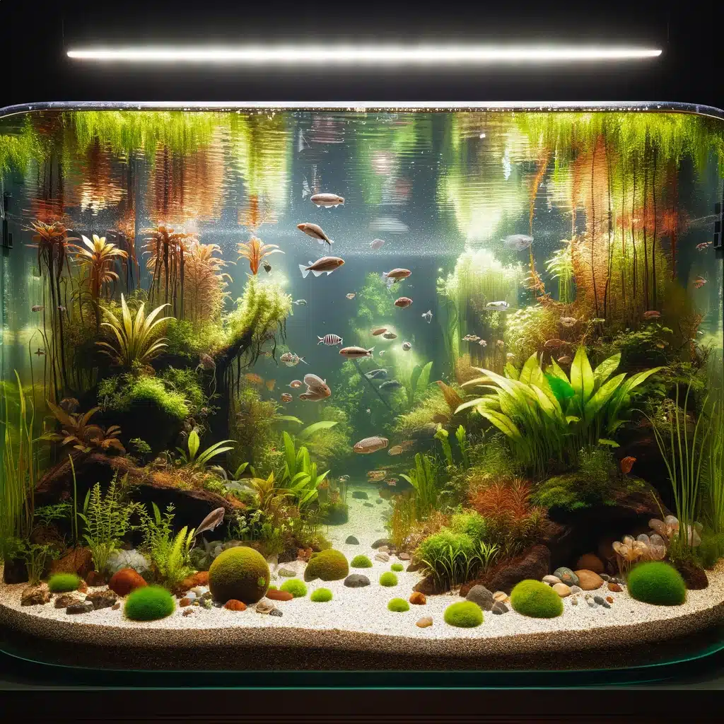
Aquariums: Closed aquatic ecosystems containing fish, plants, and microorganisms. These systems are carefully balanced to maintain a sustainable environment for aquatic life.

Aquariums-Closed Ecosystem

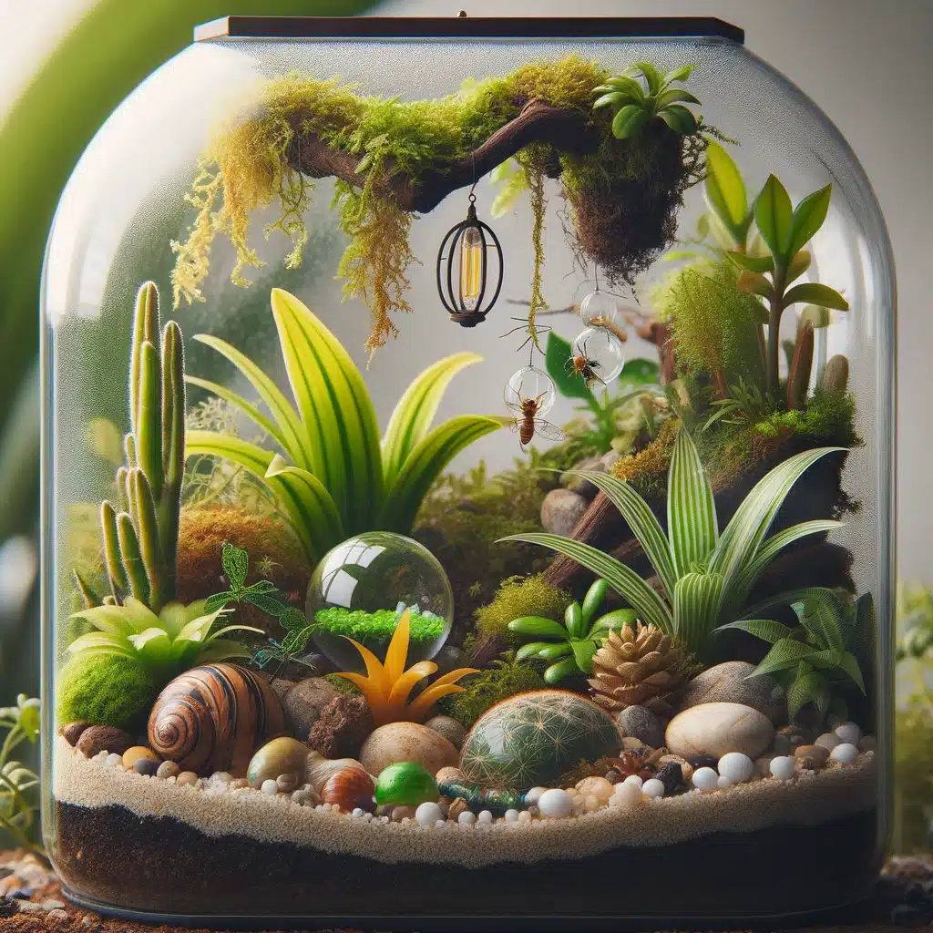
Terrariums: Enclosed environments for growing plants and sometimes small animals like insects. They simulate a mini ecosystem with controlled temperature, light, and moisture.

Terrariums-Closed Ecosystem

Bottle Gardens: Bottle gardens and small aquariums are either fully or partially closed glass containers which are self-sufficient in nature. Other man-made closed ecosystems can be MELiSSA, Biosphere 2, BIOS 1, BIOS 2 and also BIOS 3. These closed and artificial ecosystems were created with the aim of sustaining human life on earth. 

Bottle Gardens-Closed Ecosystem.webp
closed ecosystem
Closed ecosystem from Flickr

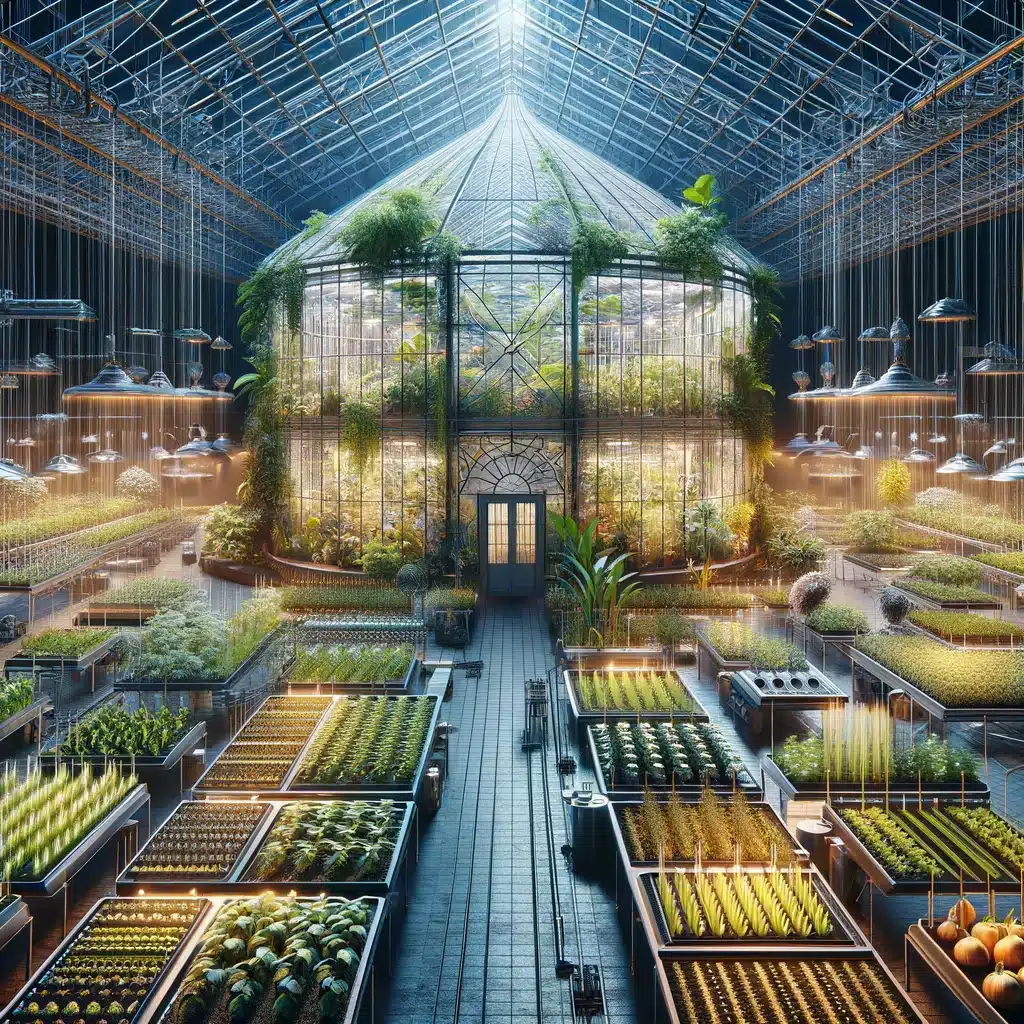
Greenhouses: Controlled environments used for growing plants. They manage temperature, humidity, and light to create an ideal growing condition, simulating a closed ecosystem.

Greenhouses Closed Ecosystem

Space Habitats: Conceptual or experimental habitats designed for life in outer space. These habitats aim to create a self-sustaining life-support system for long-duration space missions

Space Habitats

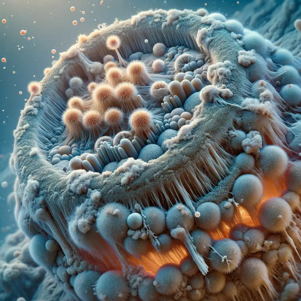
Microbial Mats: Natural formations of microorganisms, often found in extreme environments. They are self-contained ecosystems, performing all necessary biochemical processes within their structure.

Microbial Mats Closed Ecosystem

Closed water ecosystem

Closed aquatic ecosystems are identified as the sealed off ecosystem from the external world. These are self-sustained and are a miniature form of the aquatic world. Once this set-up is executed properly with adequate plants, sediments and animals, all the elements of the environment required to survive are generated naturally. These aquatic miniature ecosystems do not need any form of feeding or filtration or any external source other than the natural sunlight. 

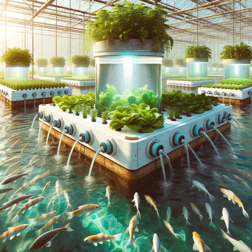
Aquaponics System: A sustainable ecosystem combining aquaculture (raising fish) and hydroponics (growing plants in water). In this system, the waste produced by farmed fish supplies nutrients for plants, which in turn purify the water.

Aquaponics System

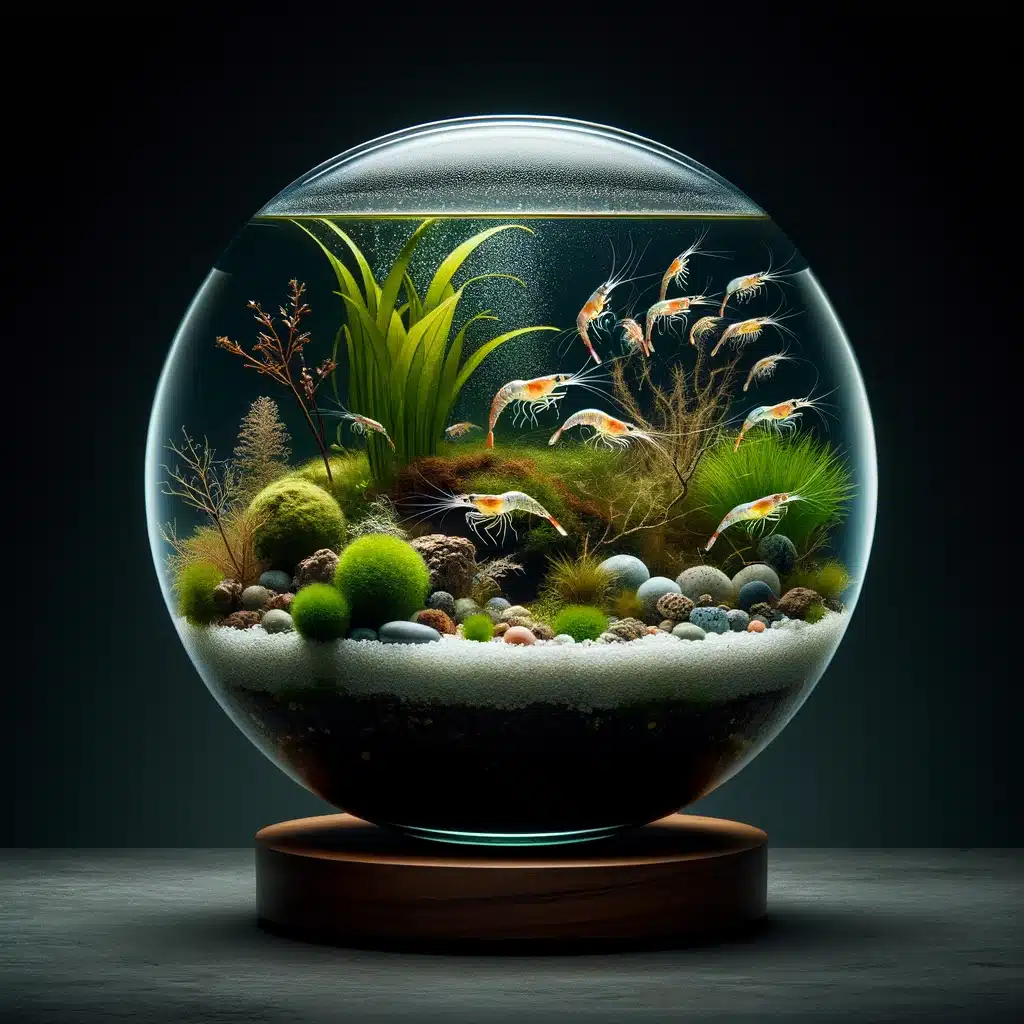
Ecosphere (Closed Ecological System): A sealed glass container holding an aquatic ecosystem that includes shrimp, algae, and microorganisms. The organisms and the environment in the ecosphere interact to maintain a balanced ecosystem.

Ecosphere (Closed Ecological System)

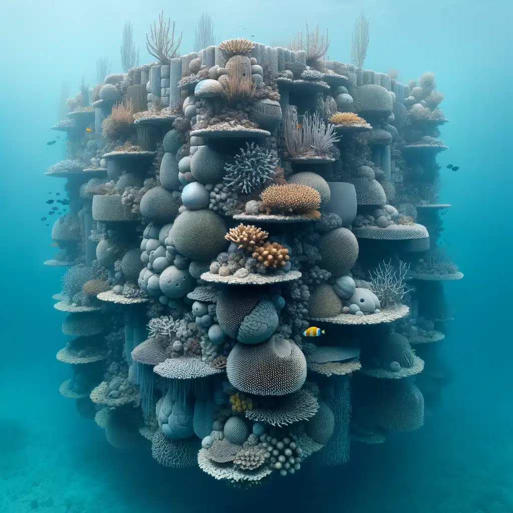
Artificial Coral Reef: These are man-made underwater structures designed to mimic the characteristics of natural coral reefs. They provide a habitat for marine life and help in balancing local marine ecosystems.

Artificial Coral Reef

How to make a closed ecosystem?

A self-sustaining closed environment, which can be an aquatic environment, would need three essential things, one is light, next is nutrients and the third is waste management. 

The source of light is mostly the sun which is utilised by the plants to create energy and the nutrients are further reused by the other elements of the environment. It is identified that all the organisms generate waste, but certain sets of bacteria have the ability to break down waste elements which helps in detoxifying the ecosystem. 

Thus, to make one, certain ingredients would include, a jar with a tight lid, a natural pond, a bucket, a shovel, a net and an optional magnifier. The simple series of steps to make the closed ecosystem with the ingredients are as follows:

Step 1: with the Shovel, it would be important to collect a few sediments or soil to be put in the bottom of the jar.  

Step 2: collect water from the nearby pond and leave for a few days for the water to settle. 

Step 3: add a set of few plants like water grass, duckweed, hornwort as algae would typically end up growing in the jar which would also work as an autotroph. 

Step 4: add a few sets of freshwater snails or any form of crustaceans. Please ensure to avoid adding large animals or fishes, as they tend to produce high bioload, as the waste products generated by these large animals cannot be processed in the small closed ecosystem. 

Step 5: keep it sealed for a few days and let the lives in the closed ecosystem survive and then maintain it in sunlight. 

Closed vs open ecosystem

The table would highlight the differences between the two broad types of ecosystems: 

Closed Ecosystem  Open ecosystem 
It engages in exchanging energy but not any form of matter with the surroundings  It engages in exchanging both, energy and any form of matter with the surroundings 
It is self-contained It is not self-contained. 
It does not require any external source to maintain life.  It requires external sources to maintain life. 
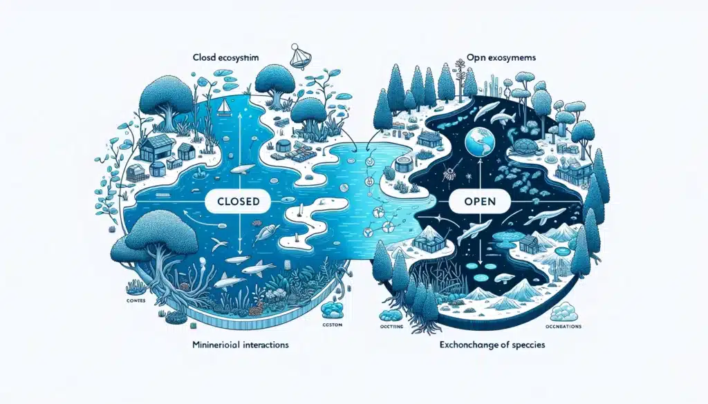
closed vs open ecosystem
  • The left side of the image shows a closed ecosystem, depicted as self-contained with minimal external interactions.
  • The right side represents an open ecosystem, interconnected with its surrounding environment, illustrating the exchange of resources and species.

Also Read:

5+ Hypotonic Solution Examples: Detailed Insights

hypotonic 1 220x300 1

This article would be presenting hypotonic solution examples with essential details. 

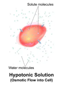
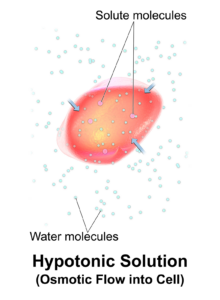
The solutions with lower concentration of solutes than the overall volume of water and which results in lower osmotic pressure are identified as hypotonic solutions. Cells that are placed in the hypotonic solution swell up as the water from the solution moves into the cells.   

Hypotonic solutions 

The solutions with lower osmotic pressure due to the result of lower concentration of solutes are identified as the hypotonic solutions. When a cell is being placed inside a hypotonic solution, the water from the solution tends to flow inside the cells which further swells it up. 

This is mostly because the concentration of water in the solution is greater than the concentration of water within the cell. In terms of solutes, the concentration is higher within the cells than the hypotonic solution.

hypotonic solution examples
Hypotonic solution from Wikimedia

Biological hypotonic solution examples 

Microbes 

Protists are organisms which lack a defined cytoskeleton or a cell wall, which would include examples like amoeba or paramecium but, due to the high ability of tonicity control the rigid structure is effectively maintained. 

Protists engage in living within a hypotonic solution which allows a constant input of water within the cells. These organisms have “contractile vacuoles” which are specialised organs used in collecting excess water from the cells to be discharged out and eliminate cell lysis.  

Plants and Fungi

Fungi and large plants engage in controlling the environment surrounding their cells in order to ensure an environment of hypotonic solution in comparison with the environment of the cells. This leads to the formation of turgid cells which leads to the cells pushing their cell walls outside which further pushes onto other cells to create a rigid structure. 

The turgor pressure is maintained with a continuous inflow of water within the cells of the plants under a hypotonic solution which is important for the plants to manage the stiffness and provide the structure. The turgor pressure helps the cells in plants to circulate water throughout the bodies, starting from roots till the top of the stem. 

In the Mangroves trees, a hypertonic cytosolic state within the root cells are developed to adapt with the marshy region which helps to absorb the water from the hypotonic environment that surrounds the roots of the plants. 

Animal cells

Animal cells lack cell walls and rely entirely on the cell membrane in separating the entry of any substance. The process involved in maintaining the overall solute concentration within an organism is termed as osmoregulation which occurs in all types of animals. 

Animals that are living in oceans tend to have salt glands which helps in expelling excess salt out of the body. The animals in the ocean drink gallons of water to maintain the water level in the body but the salt must be excreted to maintain a hypotonic environment for the cells.   

Freshwater fishes cannot thrive within seawater due to the differences in tonicity as well as osmotic gradient. Freshwater can be treated as a hypotonic solution which causes the fishes within freshwater to maintain a greater salt content within the cells than the environment surrounding them.  

Comparatively, freshwater fishes would have hypotonic cells when it would be exposed to salt water as the external environment would be hypertonic in nature. This would lead to the cells of the fishes to lose water causing extreme dehydration and finally death. 

Plasma membrane is identified as an important component for the cells as it is semi-permeable in nature and does not allow the passage of all solutes across the membranes. It helps in controlling the overall flow of the ions moving in and out of a cell. This leads to the establishment of variations in solute concentrations between the cytosol and the fluid of the cell. 

Hypotonic dehydration is a common condition where the sodium loss is higher compared to the water loss which decreases the serum osmolarity within the body. This is also termed as hyponatremia. Reduction in serum osmolarity leads to the water flowing from the extracellular to the intracellular region within the cells to cause cellular swelling and edema.

Sodium imbalance within the body can lead to a series of neurological symptoms like nausea, disorientation, headache, lethargy and weakening. In the worst cases it can lead people to coma and even death. Hypotonic dehydration leads to cellular swelling and edema due to excess water retention.

Click to read more on Hypotonic vs Isotonic.

Also Read: