Do Prokaryotic Cells Have a Cell Wall?

do prokaryotic cells have a cell wall

Prokaryotic cells, which include bacteria and archaea, do possess a cell wall, a rigid structure that lies outside the plasma membrane and provides additional protection to the cell. The cell wall is crucial for maintaining the cell’s shape and preventing osmotic lysis, which is the rupture of the cell due to the difference in osmotic pressure between the inside and outside of the cell.

The Composition of Prokaryotic Cell Walls

The chemical composition of the cell wall varies between different species of prokaryotes. Here’s a closer look at the cell wall composition of bacteria and archaea:

Bacterial Cell Walls

Bacterial cell walls contain peptidoglycan, a polymer made of sugars (N-acetylglucosamine and N-acetylmuramic acid) and amino acids (primarily L-alanine, D-glutamic acid, D-alanine, and diaminopimelic acid). Peptidoglycan is a unique component of bacterial cell walls and is not found in eukaryotic cells.

The peptidoglycan layer in bacterial cell walls can be thick or thin, depending on the species. Gram-positive bacteria have a thick peptidoglycan layer, while Gram-negative bacteria have a thinner peptidoglycan layer.

In addition to peptidoglycan, bacterial cell walls may also contain other molecules, such as:

  1. Teichoic acids: These are polymers of ribitol or glycerol phosphate found in the cell walls of Gram-positive bacteria.
  2. Lipoteichoic acids: These are amphiphilic molecules that anchor the teichoic acids to the cell membrane.
  3. Lipopolysaccharides (LPS): These are large molecules found in the outer membrane of Gram-negative bacteria, consisting of a lipid component (lipid A) and a polysaccharide component.

The composition and structure of the bacterial cell wall play a crucial role in determining the cell’s shape, rigidity, and resistance to environmental stresses.

Archaeal Cell Walls

Unlike bacterial cell walls, archaeal cell walls do not contain peptidoglycan. Instead, archaeal cell walls may contain a variety of other molecules, such as:

  1. Pseudopeptidoglycan: A peptidoglycan-like polymer found in some archaea, but with different chemical linkages.
  2. Glycoproteins: Proteins with covalently attached carbohydrate groups.
  3. Polysaccharides: Complex carbohydrates, such as glycans or heteropolysaccharides.
  4. Protein-based cell walls: Some archaea have cell walls composed primarily of proteins.

The diversity of archaeal cell wall compositions reflects the evolutionary adaptations of these organisms to various environmental conditions, such as extreme temperatures, pH, or salinity.

Gram Staining and Cell Wall Differences
do prokaryotic cells have a cell wall

Prokaryotic cells can be divided into two major groups based on their response to the Gram stain, a common laboratory technique used to differentiate bacteria:

  1. Gram-positive bacteria: These bacteria have a thick cell wall composed of multiple layers of peptidoglycan, along with teichoic acids and lipoteichoic acids. The thick peptidoglycan layer gives Gram-positive bacteria a purple or blue color when stained with the Gram stain.

  2. Gram-negative bacteria: These bacteria have a thinner peptidoglycan layer and an additional outer membrane containing lipopolysaccharides and lipoproteins. The thin peptidoglycan layer and the presence of the outer membrane give Gram-negative bacteria a red or pink color when stained with the Gram stain.

The differences in cell wall composition between Gram-positive and Gram-negative bacteria have important implications for their susceptibility to antibiotics, their interactions with the immune system, and their overall physiology.

The Importance of Prokaryotic Cell Walls

The cell wall is an essential structure for prokaryotic cells, serving several critical functions:

  1. Structural support: The cell wall provides structural integrity and maintains the shape of the cell, preventing it from bursting due to the high internal osmotic pressure.
  2. Protection: The cell wall acts as a barrier, protecting the cell from mechanical damage, osmotic stress, and environmental threats, such as antibiotics, toxins, and immune system components.
  3. Adhesion and interaction: The cell wall can facilitate the attachment of prokaryotic cells to surfaces, other cells, or host tissues, enabling them to colonize and interact with their environment.
  4. Cellular processes: The cell wall can play a role in various cellular processes, such as cell division, nutrient transport, and signal transduction.

The composition and properties of the prokaryotic cell wall can provide valuable information for identifying and classifying different species of bacteria and archaea, as well as for understanding their physiology, ecology, and interactions with their environment.

Conclusion

In summary, prokaryotic cells, including bacteria and archaea, do possess a cell wall, a crucial structural component that lies outside the plasma membrane. The cell wall composition varies between different species of prokaryotes, with bacteria typically having a peptidoglycan-based cell wall and archaea having a more diverse range of cell wall compositions. The differences in cell wall structure and composition have important implications for the physiology, ecology, and interactions of prokaryotic cells with their environment.

References:

  1. Lumen Learning. (n.d.). Structure of Prokaryotes. Retrieved from https://courses.lumenlearning.com/suny-biology2xmaster/chapter/structure-of-prokaryotes/
  2. Khan Academy. (n.d.). Prokaryote structure. Retrieved from https://www.khanacademy.org/science/ap-biology/gene-expression-and-regulation/dna-and-rna-structure/a/prokaryote-structure
  3. OpenStax. (n.d.). Unique Characteristics of Prokaryotic Cells. Retrieved from https://bio.libretexts.org/Bookshelves/Microbiology/Microbiology_%28OpenStax%29/03%3A_The_Cell/3.03%3A_Unique_Characteristics_of_Prokaryotic_Cells
Is Chromosome a Chromatin?

is chromosome a chromatin

Chromosomes and chromatin are closely related biological entities, with chromatin being the fundamental building block of chromosomes. Chromatin is a complex macromolecular structure composed of DNA, histone proteins, and non-histone proteins that collectively package the long DNA molecules within the nucleus of a cell. Understanding the relationship between chromosomes and chromatin is crucial for comprehending the organization and regulation of genetic information in living organisms.

The Structure and Composition of Chromatin

Chromatin is a highly organized and dynamic structure that serves to compact and regulate the DNA within the nucleus. The basic unit of chromatin is the nucleosome, which consists of approximately 147 base pairs of DNA wrapped around a histone octamer. These nucleosomes are further compacted into higher-order structures, such as the 30-nanometer chromatin fiber and ultimately the chromosome.

The histone proteins within the nucleosome play a crucial role in chromatin organization and function. There are five main types of histones: H1, H2A, H2B, H3, and H4. The histone octamer is composed of two copies each of H2A, H2B, H3, and H4, while the linker histone H1 helps to stabilize the higher-order chromatin structure.

In addition to the histone proteins, chromatin also contains various non-histone proteins, such as transcription factors, chromatin remodeling complexes, and architectural proteins. These non-histone proteins contribute to the dynamic regulation of chromatin structure and gene expression.

Levels of Chromatin Compaction

is chromosome a chromatin

Chromatin can exist in different states of compaction, ranging from the relatively loose and accessible euchromatin to the highly condensed and transcriptionally silent heterochromatin. The degree of chromatin compaction is regulated by various mechanisms, including histone modifications, chromatin remodeling, and the binding of architectural proteins.

  1. Euchromatin: Euchromatin is the less condensed form of chromatin, which is generally associated with active gene transcription. It is characterized by a more open and accessible chromatin structure, allowing for the binding of transcription factors and the recruitment of the transcriptional machinery.

  2. Heterochromatin: Heterochromatin is the highly condensed form of chromatin, which is typically associated with transcriptionally silent or repressed regions of the genome. It is characterized by a more compact and inaccessible chromatin structure, often marked by specific histone modifications and the binding of repressive chromatin proteins.

  3. Facultative Heterochromatin: Facultative heterochromatin is a form of chromatin that can transition between euchromatin and heterochromatin states, depending on the developmental or environmental cues. This dynamic regulation of chromatin structure is crucial for the control of gene expression during cellular differentiation and in response to various stimuli.

  4. Constitutive Heterochromatin: Constitutive heterochromatin is a more permanently condensed form of chromatin, typically found in regions of the genome that are consistently silenced, such as centromeres and telomeres. This highly compact chromatin structure helps to maintain genomic stability and prevent the expression of potentially harmful genetic elements.

The Relationship between Chromatin and Chromosomes

Chromosomes are the distinct, condensed structures that contain the genetic material during cell division. They are formed by the further compaction and organization of chromatin fibers, which are the basic building blocks of chromosomes.

During interphase, when the cell is not dividing, the chromatin is in a more decondensed state and is not visible as distinct chromosomes. However, as the cell enters mitosis or meiosis, the chromatin undergoes a series of structural changes, leading to the formation of the characteristic chromosome structures that can be observed under a microscope.

The process of chromosome formation involves the following steps:

  1. Chromatin Condensation: The chromatin fibers become increasingly compacted, with the nucleosomes and higher-order structures becoming more tightly packed.

  2. Chromosome Individualization: The condensed chromatin fibers organize into distinct, separate chromosomes, each containing a single, continuous DNA molecule.

  3. Chromosome Alignment: During cell division, the chromosomes align at the metaphase plate, allowing for the equal distribution of genetic material to the daughter cells.

The number, size, and shape of chromosomes vary among different species and can provide valuable information about the genetic makeup and evolutionary history of an organism.

Chromatin Dynamics and Epigenetic Regulation

Chromatin is a highly dynamic structure that undergoes various modifications and rearrangements to regulate gene expression, DNA repair, and other cellular processes. These changes in chromatin structure are often mediated by epigenetic mechanisms, such as histone modifications, DNA methylation, and the binding of chromatin-associated proteins.

  1. Histone Modifications: The histone proteins within the nucleosomes can undergo a variety of post-translational modifications, including acetylation, methylation, phosphorylation, and ubiquitination. These modifications can alter the interactions between the histones and the DNA, leading to changes in chromatin structure and accessibility.

  2. DNA Methylation: DNA methylation, the addition of methyl groups to the cytosine residues in the DNA, is another important epigenetic mechanism that can influence chromatin structure and gene expression.

  3. Chromatin Remodeling Complexes: Specialized chromatin remodeling complexes, such as the SWI/SNF and ISWI complexes, use energy from ATP hydrolysis to actively reposition nucleosomes, thereby altering chromatin accessibility and facilitating or repressing transcription.

  4. Architectural Proteins: Architectural proteins, such as CTCF and cohesin, play a crucial role in shaping the three-dimensional organization of chromatin within the nucleus, which can have significant implications for gene regulation and genome function.

These dynamic changes in chromatin structure and organization are essential for the precise regulation of gene expression, DNA repair, and other cellular processes, and they are crucial for the proper development and function of living organisms.

Conclusion

In summary, chromosomes and chromatin are closely related but distinct biological entities. Chromatin is the fundamental building block of chromosomes, and it is responsible for the compact packaging and regulation of the genetic material within the nucleus. The dynamic nature of chromatin structure and its epigenetic modifications play a crucial role in the control of gene expression and the maintenance of genomic integrity. Understanding the relationship between chromosomes and chromatin is essential for advancing our knowledge of cellular biology and the mechanisms underlying genetic inheritance and regulation.

References:

  • Luger, K., Mader, A. W., Richmond, R. K., Sargent, D. F., & Richmond, T. J. (1997). Crystal structure of the nucleosome core particle at 2.8 Å resolution. Nature, 389(6648), 251-260.
  • Kouzarides, T. (2007). Chromatin modifications and their function. Cell, 128(4), 693-705.
  • Strahl, B. D., & Allis, C. D. (2000). The language of covalent histone modifications. Nature, 403(6765), 41-45.
  • Dekker, J., & Mirny, L. A. (2016). Exploring the three-dimensional organization of genomes: interpreting chromosome conformation capture data. Nature reviews. Genetics, 17(12), 781-796.
  • Cremer, T., & Cremer, C. (2010). Chromosome territories, nuclear architecture, and gene regulation in mammalian cells. Cold Spring Harbor perspectives in biology, 2(10), a004975.
  • Dekker, J., & Heard, E. (2015). The spatial organization of chromatin in the nucleus. Nature reviews. Genetics, 16(11), 674-686.

Do Chloroplasts Have Ribosomes?

do chloroplasts have ribosomes

Chloroplasts, the organelles responsible for photosynthesis in plant cells, are known to possess their own genetic material and a complex internal structure. One of the key features of chloroplasts is the presence of ribosomes, which are essential for the synthesis of proteins required for their proper functioning. In this comprehensive blog post, we will delve into the details of chloroplast ribosomes, their structure, function, and the evidence supporting their existence.

The Presence of Ribosomes in Chloroplasts

Chloroplasts are semi-autonomous organelles, meaning they possess their own DNA and the machinery necessary for protein synthesis. This includes the presence of ribosomes, which are the cellular structures responsible for the translation of messenger RNA (mRNA) into functional proteins.

The presence of ribosomes in chloroplasts is well-established and supported by various lines of evidence:

  1. Structural Analysis: High-resolution structural studies, such as cryo-electron microscopy and X-ray crystallography, have directly visualized the ribosomes within chloroplasts. These studies have revealed that the chloroplast ribosomes are prokaryotic-type 70S ribosomes, composed of a small 30S subunit and a large 50S subunit, similar to those found in bacteria.

  2. Genome-wide Analyses: Genome-wide analyses of chloroplast translation have identified the presence of genes encoding ribosomal proteins and other translation-related components within the chloroplast genome. This provides strong evidence for the existence of a functional translation machinery within chloroplasts.

  3. Identification of Translation Factors: Researchers have identified specific factors that control protein synthesis in chloroplasts, such as translation initiation factors, elongation factors, and release factors. The presence of these translation-related components further supports the notion that chloroplasts have ribosomes and a functional translation machinery.

Structure and Composition of Chloroplast Ribosomes

do chloroplasts have ribosomes

The ribosomes found in chloroplasts are structurally and functionally similar to bacterial ribosomes, but with some notable differences:

Subunit Composition

  • Small Subunit (30S): The small subunit of the chloroplast ribosome, known as the 30S subunit, is composed of a 16S rRNA molecule and approximately 21 ribosomal proteins.
  • Large Subunit (50S): The large subunit of the chloroplast ribosome, known as the 50S subunit, is composed of a 23S rRNA molecule, a 5S rRNA molecule, and approximately 33 ribosomal proteins.

Ribosomal Proteins

  • The chloroplast ribosomes contain orthologs of most bacterial ribosomal proteins, indicating a close evolutionary relationship between chloroplasts and prokaryotes.
  • However, there are also some unique ribosomal proteins found in chloroplasts, which may have specialized functions or structural adaptations specific to the chloroplast environment.

Structural Deviations

  • While the overall structure of chloroplast ribosomes is similar to bacterial ribosomes, there are some structural deviations that may have functional consequences.
  • For example, the chloroplast 30S subunit has a unique structure in the region responsible for mRNA binding, which may influence the translation initiation process.

The Role of Chloroplast Ribosomes in Protein Synthesis

The ribosomes in chloroplasts play a crucial role in the synthesis of proteins required for the proper functioning of the organelle. This process, known as chloroplast translation, is essential for cellular viability and plant development.

Chloroplast Translation

  • Chloroplast translation is positioned at the intersection of organellar RNA and protein metabolism, making it a unique point for the regulation of gene expression in response to internal and external cues.
  • The chloroplast translation machinery is responsible for the synthesis of proteins encoded by the chloroplast genome, which include key components of the photosynthetic apparatus, as well as other essential chloroplast-specific proteins.

Regulation of Chloroplast Translation

  • The regulation of chloroplast translation is a complex process that involves various factors, such as translation initiation factors, elongation factors, and release factors.
  • These factors help to ensure the efficient and accurate translation of mRNAs into proteins, and they may also play a role in the coordination of chloroplast gene expression with the nuclear genome.

Importance for Cellular Viability and Plant Development

  • The proper functioning of the chloroplast translation machinery is crucial for the overall health and viability of the cell, as chloroplasts are essential for photosynthesis and other metabolic processes.
  • Disruptions in chloroplast translation can have severe consequences for plant development, as the chloroplast-encoded proteins are essential for the proper structure and function of the organelle.

Conclusion

In summary, chloroplasts do possess ribosomes, which are essential for the synthesis of proteins required for the proper functioning of the organelle. These ribosomes are prokaryotic-type 70S ribosomes, composed of a small 30S subunit and a large 50S subunit, and they contain orthologs of most bacterial ribosomal proteins.

The presence of ribosomes in chloroplasts is well-supported by various lines of evidence, including structural analyses, genome-wide analyses of chloroplast translation, and the identification of specific factors that control protein synthesis in plastids. The chloroplast ribosomes play a crucial role in the regulation of gene expression and the synthesis of proteins essential for cellular viability and plant development.

Understanding the structure and function of chloroplast ribosomes is an important aspect of plant biology, as it provides insights into the evolution and adaptation of these organelles, as well as their integration with the overall cellular metabolism and signaling pathways.

References:

  1. Tiller, N., & Bock, R. (2014). The translational apparatus of plastids and its role in plant development. Molecular plant, 7(7), 1105-1120.
  2. Yamaguchi, K., & Subramanian, A. R. (2000). The plastid ribosomal proteins: identification of all the proteins in the 50 S subunit of an organelle ribosome (chloroplast). Journal of Biological Chemistry, 275(37), 28466-28482.
  3. Schmitz-Linneweber, C., & Small, I. (2008). Pentatricopeptide repeat proteins: a socket set for organelle gene expression. Trends in plant science, 13(12), 663-670.
  4. Zoschke, R., Liere, K., & Börner, T. (2007). From seedling to mature plant: arabidopsis plastidial genome copy number, RNA accumulation and transcription are differentially regulated during leaf development. The Plant Journal, 50(4), 710-722.
  5. Manuell, A. L., Quispe, J., & Mayfield, S. P. (2007). Structure of the chloroplast ribosome: novel domains for translation regulation. PLoS biology, 5(8), e209.

Are Ribosomes Proteins? A Comprehensive Guide

are ribosomes proteins

Ribosomes are essential cellular organelles responsible for the synthesis of proteins, the fundamental building blocks of life. Contrary to the common misconception, ribosomes are not proteins themselves, but rather complex macromolecular structures composed of both ribonucleic acid (RNA) and proteins. Understanding the composition and function of ribosomes is crucial for comprehending the intricate mechanisms of protein synthesis and gene expression in both prokaryotic and eukaryotic cells.

The Composition of Ribosomes

Ribosomes are composed of two main subunits, the small subunit and the large subunit, which together form the complete ribosome. Each subunit is made up of a specific number of ribosomal RNA (rRNA) molecules and ribosomal proteins (r-proteins).

Prokaryotic Ribosomes

  • Prokaryotic ribosomes, found in bacteria and archaea, are smaller in size and have a sedimentation coefficient of 70S.
  • The small subunit (30S) contains a single 16S rRNA molecule and approximately 21 r-proteins.
  • The large subunit (50S) contains two rRNA molecules (5S and 23S) and approximately 34 r-proteins.

Eukaryotic Ribosomes

  • Eukaryotic ribosomes, found in the cytoplasm and organelles (such as mitochondria and chloroplasts), are larger and have a sedimentation coefficient of 80S.
  • The small subunit (40S) contains a single 18S rRNA molecule and approximately 33 r-proteins.
  • The large subunit (60S) contains three rRNA molecules (5S, 5.8S, and 28S) and approximately 47 r-proteins.

The rRNA molecules within the ribosomal subunits play a crucial role in the structural integrity and functional aspects of the ribosome, such as mRNA binding, tRNA positioning, and peptide bond formation. The r-proteins, on the other hand, contribute to the overall stability, assembly, and regulation of the ribosome.

The Function of Ribosomes

are ribosomes proteins

Ribosomes are the cellular factories responsible for the synthesis of proteins, a process known as translation. The primary function of ribosomes is to decode the genetic information stored in messenger RNA (mRNA) and use it as a template to assemble amino acids into polypeptide chains, which then fold into functional proteins.

The translation process can be divided into three main stages:

  1. Initiation: The small ribosomal subunit binds to the mRNA at the start codon, forming the initiation complex.
  2. Elongation: The large ribosomal subunit joins the initiation complex, and the ribosome moves along the mRNA, adding amino acids to the growing polypeptide chain.
  3. Termination: The ribosome reaches a stop codon on the mRNA, and the completed polypeptide chain is released.

During the elongation stage, the ribosome utilizes transfer RNA (tRNA) molecules, which carry specific amino acids, to assemble the polypeptide chain. The rRNA molecules within the ribosome play a crucial role in the positioning and binding of the tRNA molecules, as well as the formation of peptide bonds between the amino acids.

Ribosome Profiling: A Powerful Tool for Studying Translation

Ribosome profiling, also known as ribosome sequencing (Ribo-seq), is a powerful technique that allows researchers to study the dynamics of translation at the genome-wide level. This method involves the deep sequencing of ribosome-protected mRNA fragments, known as ribosome-protected fragments (RPFs), to obtain a snapshot of the actively translated regions of the transcriptome.

The key steps in ribosome profiling are:

  1. Ribosome Footprinting: Cells are treated with a translation inhibitor, such as cycloheximide, to freeze the ribosomes on the mRNA. The mRNA regions protected by the ribosomes are then digested with a nuclease, leaving behind the RPFs.
  2. RPF Isolation and Sequencing: The RPFs are isolated, and their sequences are determined using high-throughput sequencing technologies, such as Illumina or Ion Torrent platforms.
  3. Data Analysis: The sequencing data is analyzed to map the RPFs to the reference transcriptome, providing information on the positions of actively translating ribosomes and the relative abundance of translated mRNAs.

Ribosome profiling has several key applications:

  • Quantification of Translation Efficiency: By comparing the abundance of RPFs to the abundance of the corresponding mRNA transcripts, researchers can calculate the translation efficiency (TE) of individual genes, revealing post-transcriptional regulation mechanisms.
  • Identification of Translated Regions: Ribosome profiling can identify novel translated regions, including upstream open reading frames (uORFs), alternative start codons, and non-canonical translation events.
  • Characterization of Ribosome Dynamics: The precise positioning of RPFs along the mRNA provides insights into ribosome movement, pausing, and stalling, which can be used to study translation regulation and co-translational processes.
  • Linking Transcriptome to Proteome: By integrating ribosome profiling data with RNA-seq and proteomics data, researchers can gain a more comprehensive understanding of the relationship between mRNA abundance, translation, and protein levels.

Regulation of Ribosome Biogenesis and Abundance

Ribosome biogenesis and abundance are tightly regulated processes that are crucial for cellular homeostasis and adaptation to various environmental and physiological conditions.

Regulation of Ribosome Biogenesis

  • Ribosome biogenesis is a complex, multi-step process that involves the transcription of rRNA genes, the assembly of ribosomal subunits, and the transport of these subunits to the cytoplasm.
  • This process is regulated by various transcription factors, signaling pathways, and epigenetic mechanisms, which ensure the coordinated expression of the genes encoding rRNAs and r-proteins.
  • Disruptions in ribosome biogenesis can lead to a condition known as ribosomopathy, which is associated with various human diseases, including cancer, neurological disorders, and congenital syndromes.

Regulation of Ribosome Abundance

  • Cells can adjust the abundance of ribosomes in response to changes in nutrient availability, growth conditions, and cellular stress.
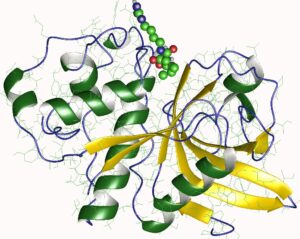
  • During nutrient starvation or stress, cells can rapidly downregulate ribosome abundance to conserve energy and resources, a process known as “ribosome hibernation.”
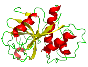
  • Conversely, rapidly growing or proliferating cells, such as cancer cells, often exhibit increased ribosome abundance to support their high protein synthesis demands.
  • The regulation of ribosome abundance involves the modulation of r-protein synthesis, ribosomal subunit assembly, and the selective degradation of ribosomes through processes like autophagy.

Understanding the complex mechanisms that govern ribosome biogenesis and abundance is crucial for elucidating the role of ribosomes in cellular homeostasis, disease pathogenesis, and therapeutic interventions.

Conclusion

In summary, ribosomes are not proteins, but rather complex macromolecular structures composed of both RNA and proteins. Ribosomes play a central role in the process of protein synthesis, decoding the genetic information stored in mRNA and assembling amino acids into functional polypeptide chains. The composition and function of ribosomes are tightly regulated to ensure cellular homeostasis and adaptation to various environmental and physiological conditions. Techniques like ribosome profiling have provided valuable insights into the dynamics of translation and the relationship between the transcriptome, translatome, and proteome. Continued research on ribosomes and their regulation will undoubtedly lead to a deeper understanding of the fundamental mechanisms of life and the development of novel therapeutic strategies.

References

  1. Biomarker Research. (2024). Ribosome profiling: A powerful tool for quantitative analysis of translation dynamics. https://biomarkerres.biomedcentral.com/articles/10.1186/s40364-024-00562-4
  2. NCBI. (2021). Ribosome profiling: a powerful tool for deciphering the translational landscape. https://www.ncbi.nlm.nih.gov/pmc/articles/PMC8274658/
  3. Nature Scitable. (n.d.). Ribosomes, Transcription, and Translation. https://www.nature.com/scitable/topicpage/ribosomes-transcription-and-translation-14120660/
  4. University of Manchester. (n.d.). Ribosome profiling: a powerful tool for studying translation. https://research.manchester.ac.uk/files/54535242/FULL_TEXT.PDF
  5. NCBI. (2020). Ribosome profiling: a powerful tool for the discovery of novel regulatory mechanisms. https://www.ncbi.nlm.nih.gov/pmc/articles/PMC7351614/

The Intricate Dance of Exocytosis Diffusion: A Comprehensive Exploration

is exocytosis diffusion

Exocytosis is a fundamental cellular process that enables the release of molecules from a cell’s interior to the extracellular space. At the heart of this mechanism lies the diffusion of these molecules from the vesicles that fuse with the plasma membrane. Understanding the intricacies of exocytosis diffusion is crucial for unraveling the complex dynamics of cellular communication, neurotransmitter release, and various physiological processes.

Measuring Exocytosis Diffusion: Experimental Approaches

Membrane Capacitance Measurements

One of the primary methods for quantifying exocytosis is by measuring the increase in cell membrane capacitance caused by the addition of secretory granule membranes. This technique allows researchers to determine the amount of vesicle membrane that is incorporated into the plasma membrane during the exocytosis process. By tracking the changes in membrane capacitance, scientists can gain valuable insights into the kinetics and extent of vesicle fusion with the cell surface.

For example, a study conducted by Neher and Marty in 1982 demonstrated that the capacitance of the cell membrane increases in a stepwise manner during exocytosis, corresponding to the fusion of individual secretory vesicles. This pioneering work laid the foundation for using membrane capacitance measurements as a reliable tool for studying exocytosis dynamics.

Electrochemical Techniques

Another powerful approach for investigating exocytosis diffusion is the use of electrochemical techniques, such as amperometry. These methods allow for the direct detection and quantification of neurotransmitters released from individual vesicles during exocytosis.

Amperometry involves the placement of a carbon fiber electrode near the cell surface, which can detect the oxidation of neurotransmitters as they are released into the extracellular space. This technique enables the measurement of the amount of neurotransmitter released, the kinetics of release, and the fraction of vesicular content that is actually released during exococytosis.

Interestingly, a study published in 2023 using amperometry revealed that exocytosis is predominantly a partial process, where only a fraction of the vesicular content is released. This finding has important implications for the regulation of neurotransmitter release and the plasticity of synaptic transmission, as it suggests that cells can fine-tune the amount of neurotransmitter released to modulate the strength of synaptic signaling.

Computational Modeling and Simulation

In addition to experimental approaches, computational modeling and simulation can also provide valuable insights into the diffusion of molecules during exocytosis. These techniques allow researchers to explore the complex interplay between various factors that influence the diffusion process, such as the cellular environment, vesicle properties, and the dynamics of vesicle fusion.
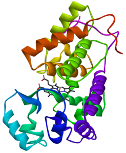
One such study, published in the journal Nature in 2023, utilized the STEPS software package to investigate vesicle diffusion in crowded cellular environments. The researchers found that vesicle diffusion is reduced in these crowded conditions, and that the effective diffusion rate of mobile vesicles is not constant over time. These findings highlight the importance of considering the cellular context when modeling exocytosis and diffusion processes.

Factors Influencing Exocytosis Diffusion

is exocytosis diffusion

Vesicle Size and Composition

The size and composition of the secretory vesicles play a crucial role in the diffusion of molecules during exocytosis. Larger vesicles, for example, may release their contents more slowly due to the increased distance the molecules must travel to reach the extracellular space. Additionally, the lipid and protein composition of the vesicle membrane can affect the rate and efficiency of diffusion.

Studies have shown that the size of secretory vesicles can vary significantly, ranging from small synaptic vesicles (diameter of ~40-50 nm) to large dense-core vesicles (diameter of ~100-300 nm). The larger vesicles typically contain a higher concentration of neurotransmitters or hormones, which can influence the amount and kinetics of their release during exocytosis.

Cellular Environment and Crowding

The cellular environment surrounding the secretory vesicles can also impact the diffusion of molecules during exocytosis. The cytoplasm is a highly crowded and complex environment, with a variety of macromolecules, organelles, and other cellular structures that can hinder the free diffusion of molecules.

As mentioned earlier, the computational modeling study using the STEPS software demonstrated that vesicle diffusion is reduced in crowded cellular environments. This finding suggests that the local microenvironment within the cell can play a significant role in the diffusion of molecules released during exocytosis, potentially affecting the spatiotemporal dynamics of neurotransmitter or hormone signaling.

Membrane Fusion and Pore Formation

The fusion of the secretory vesicle with the plasma membrane and the subsequent formation of a fusion pore are critical steps in the exocytosis process that can influence the diffusion of molecules. The size and dynamics of the fusion pore can determine the rate and extent of molecule release into the extracellular space.

Researchers have observed that the fusion pore can undergo various stages of expansion and contraction, which can modulate the diffusion of molecules. In some cases, the fusion pore may remain open for an extended period, allowing for a more sustained release of vesicular contents. In other cases, the pore may close rapidly, leading to a more transient and localized release of molecules.

Understanding the factors that govern the formation and dynamics of the fusion pore is an active area of research, as it can provide valuable insights into the regulation of exocytosis diffusion and its implications for cellular signaling and communication.

Implications and Applications of Exocytosis Diffusion

The study of exocytosis diffusion has far-reaching implications in various fields of biology and medicine. By elucidating the mechanisms and dynamics of molecule release during exocytosis, researchers can gain a deeper understanding of:

  1. Neurotransmitter Release and Synaptic Transmission: The diffusion of neurotransmitters from synaptic vesicles is a crucial aspect of synaptic communication. Insights into exocytosis diffusion can help elucidate the regulation of neurotransmitter release and the plasticity of synaptic transmission, which is essential for understanding neural function and the pathophysiology of neurological disorders.

  2. Hormone Secretion and Endocrine Signaling: Exocytosis is a key mechanism for the release of hormones from endocrine cells. Understanding the diffusion of hormones from secretory vesicles can provide insights into the regulation of endocrine signaling and its implications for metabolic, reproductive, and other physiological processes.

  3. Cellular Communication and Signaling: Exocytosis diffusion is not limited to the release of neurotransmitters and hormones; it also plays a role in the secretion of various other signaling molecules, such as growth factors, cytokines, and extracellular matrix components. Elucidating the diffusion dynamics of these molecules can shed light on the complex networks of cellular communication and their impact on tissue homeostasis, development, and disease.

  4. Drug Delivery and Therapeutic Targeting: The principles of exocytosis diffusion can be leveraged in the development of targeted drug delivery systems. By understanding the factors that influence the release and diffusion of molecules from secretory vesicles, researchers can design more efficient and targeted drug delivery strategies, potentially improving the efficacy and safety of therapeutic interventions.

  5. Biotechnological Applications: The quantitative data and insights gained from the study of exococytosis diffusion can also find applications in various biotechnological fields, such as the development of biosensors, the optimization of bioreactor systems, and the engineering of synthetic cellular systems for industrial or medical purposes.

In summary, the exploration of exocytosis diffusion is a multifaceted and dynamic field of study that continues to yield valuable insights into the fundamental mechanisms of cellular function, communication, and signaling. By integrating experimental, computational, and theoretical approaches, researchers can unravel the intricate dance of molecule release and diffusion, paving the way for advancements in biology, medicine, and beyond.

References:

  1. Neher, E., & Marty, A. (1982). Discrete changes of cell membrane capacitance observed under conditions of enhanced secretion in bovine adrenal chromaffin cells. Proceedings of the National Academy of Sciences, 79(21), 6712-6716.
  2. Amatore, C., Arbault, S., Bouret, Y., Guille, M., Lemaître, F., & Verchier, Y. (2006). Regulation of exocytosis in chromaffin cells by trans-insertion of lysophosphatidylcholine and arachidonic acid into the outer leaflet of the cell membrane. ChemBioChem, 7(12), 1998-2003.
  3. Shin, W., Ge, L., Arpino, G., Villarreal, S. A., Hamid, E., Liu, H., … & Wu, L. G. (2018). Visualization of membrane pore in live cells reveals a dynamic-pore theory governing fusion and endocytosis. Cell, 173(4), 934-945.
  4. Wen, P. J., Grenklo, S., Arpino, G., Tan, X., Liao, H. S., Heureaux, J., … & Wu, L. G. (2016). Actin dynamics provides membrane tension to merge fusing vesicles into the plasma membrane. Nature communications, 7(1), 1-13.
  5. Bao, H., Goldschen-Ohm, M. P., Jeggle, P., Chanda, B., Edwardson, J. M., & Chapman, E. R. (2016). Exocytotic fusion pores are composed of both lipids and proteins. Nature structural & molecular biology, 23(1), 67-73.
  6. Wen, P. J., Osborne, S. L., Zanin, M., Low, P. C., Wang, H. T., Schoenwaelder, S. M., … & Cousin, M. A. (2011). Phosphoinositides regulate vesicle fusion at the nerve terminal. The Journal of cell biology, 195(7), 1139-1153.
  7. Barg, S., Knowles, M. K., Chen, X., Midorikawa, M., & Almers, W. (2010). Syntaxin clusters assemble reversibly at sites of secretory granules in live cells. Proceedings of the National Academy of Sciences, 107(48), 20804-20809.
  8. Becherer, U., & Rettig, J. (2006). Vesicle pools, docking, priming, and release. Cell and tissue research, 326(2), 393-407.
  9. Neher, E. (1998). Vesicle pools and Ca2+ microdomains: new tools for understanding their roles in neurotransmitter release. Neuron, 20(3), 389-399.

19 Domain Bacteria Examples: Detailed Explanations And Images

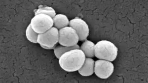
1200px Micrococcus luteus 9759 edited 300x169 1

Bacteria are prokaryotic cells that contain unique rRNA sequences different from Archaea and Eukarya. They are encapsulated with cell membranes composed of peptidoglycan and unbranched fatty acid chains attached to glycerol molecules by ester linkages.

The different domain bacteria examples are as follows

  1. Aquificae
  2. Thermotogae
  3. Thermodesulfobacteria
  4. Deinococcus
  5. Chrysiogenetes
  6. Chloroflexi
  7. Nitrospira
  8. Deferribacteres
  9. Cyanobacteria
  10. Chlorobi
  11. Proteobacteria
  12. Firmicutes
  13. Actinobacteria
  14. Planctomycetes
  15. Chlamydiae
  16. Spirochaetes
  17. Acidobacteria
  18. Bacteroidetes
  19. Fusobacteria
  20. Fibrobacterota

Aquificae

The phylum Aquificae is considered the oldest branch of bacteria.

Aquifex pyrophilus is a Gram-negative rod-shaped, microaerophilic bacterium. This thermophilic bacterium can thrive in temperatures ranging from 85°C and 95°C. Aquifex is a lithoautotroph that utilizes hydrogen, thiosulfate, and sulfur along with oxygen as terminal electron acceptors. 

Thermotogae

The second oldest branch is the phylum Thermotogae. This phylum includes Gram-negative, rod-shaped thermophiles enclosed in an envelope that is extended from both the ends as a balloon. They flourish actively in geothermal regions.

Contrastingly, Thermotogae is a chemoheterotroph that can survive anaerobically on carbohydrates and protein.

domain bacteria examples
Outline of a Thermotoga maritima From Wikipedia

Thermodesulfobacteria

These are sulfur reducing bacteria that utilizes sulfate as electron acceptor for anaerobic respiration. These bacteria can breathe sulfate. Example Thermodesulfobacterium hydrogeniphilum is thermophilic sulfate-reducing bacteria.

Deinococcus

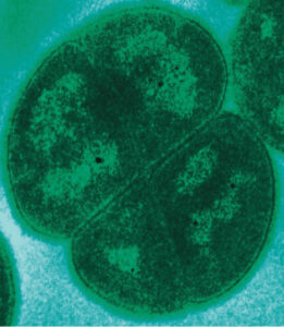
This group of bacteria is highly resistant to gamma rays and UV radiation. They have thick cell walls and can inhabit outer space for years. They have characteristic carotenoid pigment deinoxanthin which gives the bacterium pink color.

domain bacteria examples
Deinococcus radiodurans From Wikipedia

Deinococcus radiodurans is an important member of this phylum. It is a poly-extremophilic, non-endospore-forming bacterium that derives energy from organic compounds.

Chrysiogenetes

Chrysiogenes arsenatis is a member of this class that uses arsenate as a terminal electron acceptor. These anaerobic bacteria are usually found in arsenic-contaminated soils. They appear as rod-shaped motile cells with a single flagellum. They reduce arsenate to arsenite. It can utilize acetate, pyruvate, lactate, malate, and fumarate as its carbon source.

Chloroflexi

This phylum is now referred to as Chloroflexota which includes aerobic thermophilic bacteria, and anoxygenic phototrophs. They have a single membrane but appear as gram-negative after staining. These are green non-sulfur anoxygenic photosynthetic bacteria, that contain BChl a and c/d and carotenoids such as carotene, β—carotene, and myxocoxanthin as major photosynthetic pigments.

Nitrospira

It is a gram-negative bacterium that is capable of nitrite oxidation. It is helical in morphology and remains in aggregates. This chemolithoautotrophic bacterium is used in sewage water plants and reduces the emission of greenhouse gases. As it contains nitrite oxidoreductase genes thus it is mentioned as a nitrite oxidizer.

Deferribacteres

This group contains gram-negative anaerobic bacteria. They are rod-shaped and use iron, manganese, and nitrate for respiration. Geovibrio is a non-sporulating member of this class that uses sulfide, formate, and acetate as electron donors while sulfur, nitrate, and fumarate as electron acceptors.

Cyanobacteria

This group is the most diverse group of bacteria which contains about 2000 and more species. These gram-negative bacteria are also known as blue-green algae.

Cyanobacteria use chlorophyll a and photosystem I and II. The carbon reserve is glycogen and contains phycocyanin and phycoerythrin as pigments necessary for light reactions.  These pigments are stored in specialized organelles called phycobilisomes which are similar to chloroplasts of plants.

Some important cyanobacteria are as follows

Nostoc

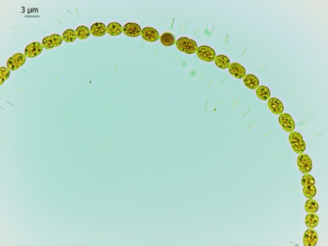
These are filamentous cyanobacteria distributed in both terrestrial and aquatic ecosystems. It is often involved in symbiotic interaction with plants and helps in nitrogen fixation. These bacteria have heterocyst, a specialized cell responsible for nitrogen assimilation. Nostoc is widely used as a biofertilizer.

Anabaena

Anabaena is another important genus of the cyanobacteria group that helps in nitrogen fixation. They contain heterocysts that convert nitrogen to organic ammonia and are often used as a biofertilizer in paddy fields.

domain bacteria examples
Anabaena From Wikipedia

Spirulina

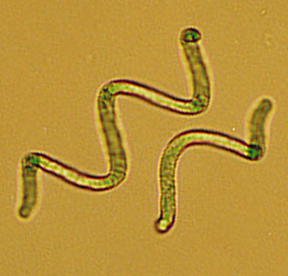
This genus is a worldwide famous group of cyanobacteria which is referred to as superfood. It doesn’t contain a heterocyst. However, Spirulina is very popular as a dietary supplement as it stores a high amount of protein (65-70%). It is rich in chlorophyll and carotenoids which act as antioxidants and contain vitamins such as folic acid, Vit B2, and Vit B12 along with other necessary minerals.

domain bacteria examples
Spirulina From Wikipedia

Chlorobi

Representative members of this group are Chlorobium tepidum and Chlorobium vibrioforme. They are anaerobic photoautotrophs, green sulfur bacteria that utilizes reduced sulfur compounds for electrons and performs non-cyclic photosynthesis. They contain BChl c, BChl d, and BChl e.

Proteobacteria

Proteobacteria is also known as Pseudomonadota. This phylum includes gram-negative bacteria that perform anoxygenic photosynthesis.

Alpha proteobacteria

The major photosynthetic pigments are BChl a/b and carotenoids such as lycopene, and rhodopsin. They utilize reduced hydrogen and sulfur compounds as electron donors and use photosystem II for photosynthesis.


Methylobacterium Can utilize C1 compounds, normally present in soil and water, facultative-aerobe
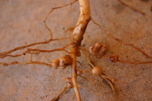
Rhizobium Nitrogen-fixing soil bacteria associated with Leguminosae plant, motile cells
Caulobacter Heterotrophs and oligotrophs with rod or vibrio shaped cells
Agrobacterium Plant pathogen causes tumors, chemoorganotroph, aerobic motile non-sporing rods
Rickettsia Obligate intracellular parasites, pleomorphic
Brucella Pathogenic, coccobacillus with flagella
Coxiella Obligate parasites causes Q fever
Rhodospirillum Photoheterotrophs either aerobic or anaerobic
domain bacteria examples
Rhizobia nodules on Vigna unguiculata From Wikipedia

Beta proteobacteria

These purple non-sulfur bacteria contain bacteriochlorophylls and carotenoids as major photosynthetic pigments. They can survive in a wide range of environments by using various metabolic strategies. They make use of reduced nitrogen and sulfur forms as electron donors.

Bordetella  Aerobic, nonmotile, coccobacillus parasites of mammals, need organic sulfur and nitrogen
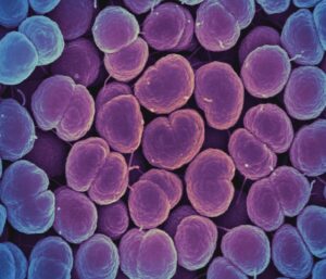
Neisseria Pathogenic causes gonorrhea and meningitis, oxidases-positive, resident of mucosal membranes of mammals
Burkholderia Aerobic, straight rods with a single flagellum, with Poly-β-hydroxybutyrate as reserve material, causes cystic fibrosis
Leptothrix Aerobic straight rods covered with a sheath containing iron and manganese oxides, present in clogged pipes
Nitrosomonas Ammonia-oxidizing, obligate chemolithoautotroph
Thiobacillus Autotrophs, produce sulfate by oxidizing H2S, beneficial for plants
domain bacteria examples
Neisseria gonorrhoeae From Wikipedia

Gamma proteobacteria

Azotobacter Aerobic bacterium, form cysts and helps in nitrogen fixation
Escherichia Facultatively anaerobic in nature, oxidase-negative, converts formic acid to H2 and CO2, common parasites for mammals
Methylococcus Methane-utilizing aerobic bacterium, usually form cysts
Haemophilus Facultative or aerobic, oxidase-positive, fermentative bacterium, frequently present as parasites of mammals
Pseudomonas Chemoheterotrophs with aerobic respiration, common pathogens for animals and plants
domain bacteria examples
Escherichia From Wikimedia

Delta proteobacteria

Bdellovibrio This is an aerobic pathogenic bacterium that grows in the periplasmic space of other gram-negative bacteria.  
Desulfovibrio Sulfur-reducing bacterium that inhabits polluted lakes and sewage lagoons
Desulfuromonas Sulfur-reducing bacterium mostly present in anoxic freshwater environments
Myxococcus Aerobic, motile bacterium usually present in soil and remain as dormant myxospores. These are micro predators and secret various digestive enzymes to lyse other bacterial cells.
domain bacteria examples
Desulfovibrio desulfuricans From Wikimedia

Epsilon proteobacteria

These are gram-negative rod-shaped microaerophilic proteobacteria.

Campylobacter This genus comprises both pathogenic and non-pathogenic members. These are fermentative oxidase-positive bacteria often found in the intestinal tract and oral cavity of animals.
Helicobacter These are both catalase and oxidase-positive, usually found in the gastrointestinal tract of humans.
domain bacteria examples
Campylobacter sp From Wikimedia

Zeta proteobacteria

This group is represented by Mariprofundus ferrooxydans. This bacterium is found in deep sea specially at hydrothermal vents as iron-oxidizing microorganism.

Firmicutes

This phylum contain gram-positive bacteria. Based on 16S rRNA analysis, this phylum contains bacteria with low G+C content.

Class : Mollicutes
Acholeplasma Mostly present in vertebrates, with optimum temp 30-37°C
Anaeroplasma Inhabit rumen of bovine and anaerobes
Entomoplasma Usually present in insects and plants with optimal growth at 30°C
Mycoplasma Present in humans and animals with 37°C optimal temperature, require sterol for its growth
Spiroplasma Helical filaments grow at 30-37°C in insects and plants
Ureaplasma Capable of hydrolysis of urea, present in human and animal
Class : Clostridia
Clostridium Anaerobic, chemoorganotrophs with oval or spherical endospores, rod-shaped pleomorphic, catalase negative 
Heliobacterium Anaerobic, photoheterotrophs with BChl g
Class : Bacilli
Bacillus Aerobic or facultative chemoorganotrophs with straight rod-shaped, have endospores, catalase-positive
Enterococcus Facultative spherical or ovoid cells, catalase-negative and undergoes fermentation, produce lactate with any gas, present in fecal matter
Lactobacillus Facultative or microaerophilic, non-sporing, fermentative, catalase-negative
Staphylococcus Chemoorganotrophs, catalase-positive, pathogenic, present in the skin and mucosal membrane of humans
Streptococcus Fermentative, produce lactate without any gas, catalase-negative, hemolysis-positive, parasites of animals

Actinobacteria

This is a fascinating group of bacteria that produces characteristic secondary metabolites with anticancer, and anti-helminthic properties. Their life cycle involves the development of filamentous hyphae that carry spores.

Arthrobacter Aerobic, present as rod-shaped cells (young) or small cocci (old), catalase-positive, mostly present in the soil
Corynebacterium Facultatively anaerobes, fermentative, oxidase-positive
Micrococcus Aerobic, present as tetrads or irregular cocci, catalase-positive, mammalian skin resident and also present in soil
Mycobacterium Aerobic, slightly curved rods, catalase-positive, present in soil and water, sometimes pathogenic in nature
Streptomyces Aerobic, branched vegetative mycelium with aerial mycelium containing spore, can utilize various organic compounds
Frankia Aerobic or microaerophilic, branched vegetative mycelium with no aerial mycelium, usually present as symbionts with plants
domain bacteria examples
Micrococcus luteus From Wikimedia

Planctomycetes

This group of bacteria is widely distributed in both aquatic and terrestrial environments. They play a significant role in carbon and nitrogen cycles and can survive under limited carbon and nitrogen sources. They lack the peptidoglycan layer and have separate internal compartments.

These anaerobes are capable of oxidizing ammonia to nitrogen inside a specialized organelle called anammoxosome which bears similarities with eukaryotic mitochondria.

Chlamydiae

This group comprises obligate intracellular parasites; some are pathogenic while others remain as symbionts of humans and animals. It also lacks a peptidoglycan layer. Example Chlamydia pneumoniae which causes pneumonia, and Chlamydia trachomatis that causes chlamydia.

domain bacteria examples
Chlamydia trachomatis From Shutterstock

Spirochaetes

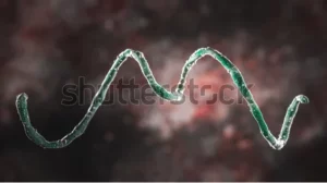
These are double membraned gram-negative bacteria with distinct corkscrew shapes. This group has distinct flagella known as endoflagella that is anchored at the end of the bacteria cell. It has a twisting motion. Most of the species are pathogenic in nature.

domain bacteria examples
Spirochete From Shutterstock

Acidobacteria

This phylum represents gram-negative, acidophilic soil bacteria. Mostly present in hot springs and metal contaminated soils. In soil, it represents 52% of the total soil bacteria community. It contains bacteriochlorophyll a and carotenoids such as Echinenone, lycopene, ɣ- and β- carotene. It utilizes organic compounds such as acetate and succinate as electron donors

domain bacteria examples
Acidobacterium From Wikipedia

Bacteroidetes

These can be either pathogenic or symbiotic bacteria. These are gram-negative, rod-shaped bacteria. Some species can degrade complex polysaccharides of plants such as starch, and cellulose. They exhibit protease and urease activity.

domain bacteria examples
Bacteroides biacutis From Wikipedia

Fusobacteria

These are obligate anaerobes with rod-shaped morphology. Species of this phylum colonizes the mucosal membrane of the humans and causes tonsilitis and peritonsillar abscess.

Fibrobacterota

This phylum includes bacteria that are capable of degrading cellulose and usually present in the rumen of bovine animals. This phylum is represented by two members Fibrobacter succinogenes and Fibrobacter intestinalis.

Summary

Domain bacteria comprises a diverse group of micro organisms that utilizes various metabolic strategies. Through such metabolic processes, these can thrive anywhere starting from deep sea hydrothermal vents to outer space. They can utilize organic and inorganic carbon sources. Some are pathogenic, commensal while others are beneficial to plants.

Also Read:

21 Protein Enzyme Examples: With Structures

Polyamine oxidase from Zea mays 194x300 1

Enzymes are “gnomes” of every cellular process. Enzymes are functional protein molecules. These biomolecules assist various metabolic reactions and participate in both anabolism and catabolism processes. They enhance the reaction rates and result in physiologically significant products.

  1. Dehydrogenase
  2. Oxidases
  3. Reductase
  4. Peroxidase
  5. Oxygenase
  6. Methyltransferase
  7. Acyltransferase
  8. Glycosyltransferase
  9. Transaminase
  10. Phosphotransferase
  11. Sulfur-transferase
  12. Nuclease
  13. Glycosylase
  14. Peptidase
  15. Lyase
  16. Isomerase
  17. Epimerase
  18. Racemase
  19. Mutase
  20. Ligase
  21. Kinase
  22. Decarboxylase

Dehydrogenase

Dehydrogenase is a type of oxidoreductase enzyme. It catalyzes the oxidation of the substrate by reducing an electron acceptor such as NAD+ or NADP++ or FAD or any flavin coenzyme. This enzyme either facilitates the removal of hydrogen to an electron acceptor along with the release of a proton or the transfer of two hydrogen atoms.

Examples- Aldehyde dehydrogenases, pyruvate dehydrogenase, succinate dehydrogenase

Oxidase

This enzyme prompts oxidation by transferring hydrogen from the substrate to the acceptor i.e., oxygen. It facilitates the oxidation of C-N and C-O bonds which are reduced to hydrogen peroxide. Examples – Xanthine oxidase, Cytochrome P450 oxidase, polyphenol oxidase.

protein enzyme example
Polyamine oxidase From Wikimedia

Reductase

This enzyme catalyzes the non-reversible reduction reaction. It also acts like a dehydrogenase enzyme. Example – Nitrate reductase. It is an important enzyme for nitrogen assimilation.  

protein enzyme example
Ribonucleotide reductase From Wikipedia

Peroxidase

This enzyme also belongs to the group of oxidoreductases. It facilitates oxidation of the substrate by the involvement of hydrogen peroxide and liberates water and oxygen molecules. Most of them contain a ferric heme protein at the catalytic site. These enzymes mostly act as antioxidants. Example – Manganese peroxidase, glutathione peroxidase.

protein enzyme example
Glutathione Peroxidase From Wikimedia

Oxygenase

This enzyme catalyzes the oxidation reaction. In this oxidation reaction, the substrate is oxidized by an oxygen atom which is obtained from molecular oxygen. Example – Tryptophan pyrrolase, tyrosinase.

Methyltransferase

This group involves enzymes that transfer methyl groups from S-adenosylmethionine (SAM) to the substrates. For example – DNA methyltransferases transfer methyl groups to cytosines.

protein enzyme example
DNA methylase From Wikipedia

Acyltransferase

This enzyme catalyzes the transfer of the acyl group. Example – Carnitine acyltransferases.

Glycosyltransferase

This enzyme facilitates the transfer of saccharide moieties to protein residues mostly tyrosine, serine, or threonine. These are transmembrane proteins adhered to the membranes of the Golgi apparatus.

Transaminase

This enzyme catalyzes the exchange reaction between an amino group and an alpha-keto group. It requires a coenzyme pyridoxal phosphate for its reaction.

Phosphotransferase

This enzyme is responsible for reversible phosphorylation reaction (addition of phosphate group). It transfers the phosphoryl group to hydroxyl, carboxy, and nitrogenase groups. The amino acids that are phosphorylated are serine, tyrosine and threonine.

The phosphorylase enzyme catalyzes the addition of the inorganic phosphate group to the substrate.

Sulfur transferase

These transferase enzymes are involved in the transfer of sulfur-containing groups. Example – Thiosulfate sulfurtransferase is a mitochondrial enzyme that converts cyanide to thiocyanate. The substrates of this enzyme are cyanide and thiosulfate and the products are sulfite and thiocyanate.

Nuclease

This enzyme is capable of cleaving the phosphodiester bonds present in nucleotides. It either creates a single cut or double cut. Depending upon its site of cleavage, it can be divided into endo and exonuclease. These enzymes are extensively used in biotechnology.

protein enzyme example
HindIII restriction enzyme From Wikipedia

Glycosylase

This is a type of hydrolase enzyme that is involved in the hydrolysis of glycosidic bonds present between glycosyl moieties. It can be of two types depending upon the site of action i.e., O- or S- glycosides or N-glycosides.

protein enzyme example
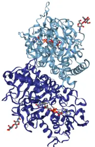
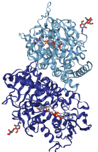
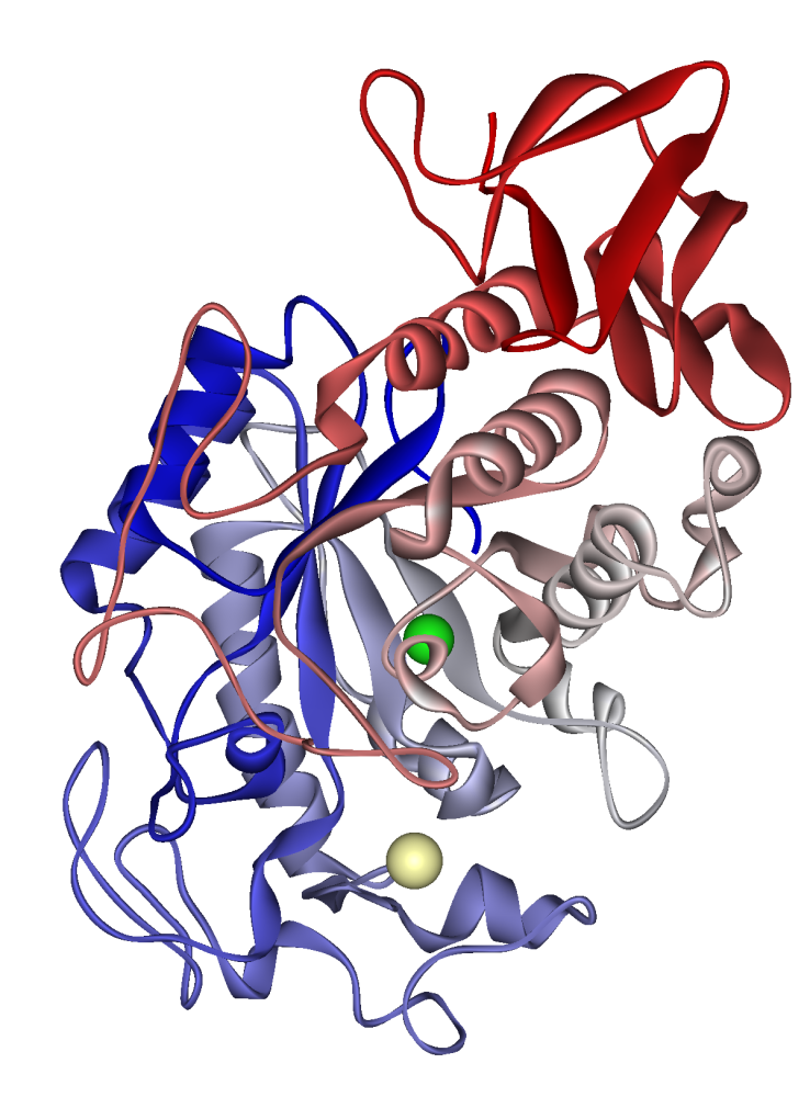
Pancreatic alpha-Amylase, A glycoside hydrolase From Wikimedia

Peptidase

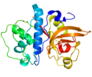
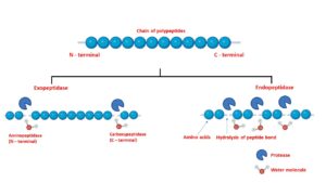
Peptidase or protease undergoes proteolysis, breaking down the peptide bonds present in polypeptide chains resulting in smaller polypeptides or amino acids. It can be seven types such as serine proteases, cysteine proteases, threonine proteases, aspartic proteases, glutamic proteases, metalloproteases, and asparagine peptide lyases.

protein enzyme example
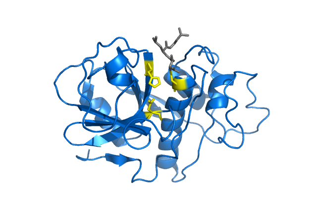
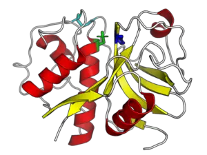
Cysteine protease From Wikipedia

Lyase

Lyases facilitate elimination or substitution reaction along with oxidation. It cleaves C-O, C-C, C-N, C-S, and P-O bonds. Mostly these enzymes are present either as peripheral membrane proteins or as transmembrane proteins.

protein enzyme example
Isocitrate lyase From Wikimedia

Isomerase

It facilitates the intramolecular structural or geometrical changes of one isomer to another isomer. The molecules are called structural isomers or stereoisomers.

protein enzyme example
Glucose-6-phosphate isomerase From Wikimedia

Epimerase

This enzyme is a type of isomerase enzyme. It catalyzes the stereochemical inversion around an asymmetric carbon in a substrate containing more than one center of asymmetry. Such molecules are called epimers. Epimer is a pair of diastereomers.  

protein enzyme example
Ribulose 5-Phosphate 4-Epimerase From Wikimedia

Racemase

Contrastingly, racemases facilitate the inversion around an asymmetric carbon in a substrate with one center of asymmetry. Example – methyl malonyl-CoA epimerase.

Mutase

This is a type of isomerase enzyme that accelerates the interconversion. In this reaction, the functional groups are shifted from one position to another. Example – bisphosphoglycerate mutase, phosphoglycerate mutase.

protein enzyme example
Phosphoglycerate mutase From Wikimedia

Ligase

This enzyme helps to join molecules or large moieties by creating new bonds. The most common example is DNA ligase. It ligates various bonds such as C-O, C-S, C-N, C-C, and also nitrogen metal bonds. These remain either as peripheral or transmembrane proteins.

protein enzyme example
DNA Ligase From Wikimedia

Kinase

This is a type of phosphotransferase enzyme where it transfers the phosphoryl group from ATP to a substrate.

protein enzyme example
Pyruvate kinase From Wikimedia

Decarboxylase

This enzyme is responsible for the addition or removal of the carboxyl group. Examples are glutamate decarboxylase, histidine decarboxylase, ornithine decarboxylase, phosphoenolpyruvate carboxylase, and pyruvate decarboxylase.

What is a protein enzyme?

Metabolic pathways are solely dependent upon these protein enzymes.

Enzymes are proteinaceous molecules. These biomolecules speed up the reaction rate by minimizing the intermediate activation energy. Most enzymes are present either as peripheral membrane proteins or as transmembrane proteins. Enzymes often require a coenzyme or cofactor for their catalytic activity.

Coenzyme is a small organic molecule facilitates the transfer of the atoms such as NAD, NADPH, FAD, FMN, flavin and ATP. These enzymes can be broadly classified into six groups. (i) Oxidoreductase (ii)Transferase (iii) Hydrolases (iv) Lyases (v) Isomerases and (vi) Ligases

Protein enzyme structure

Mostly these are globular proteins.

Enzymes contain linear chains of amino acids with disulfide bonds that give rise to a three-dimensional, globular structure. The enzyme size ranges from a few amino acid residues to more than 2500 residues. However, a smaller portion of this globular structure is involved in catalytic activity.

There are binding sites that are specific to a particular substrate, cofactor, or coenzyme. The catalytic and binding sites together form the active sites of an enzyme.

Conclusion

Enzymes are globular proteins present in cells either as peripheral membrane proteins or transmembrane proteins. Along with various coenzyme and cofactors that facilitate various biochemical reactions such as oxidation-reduction reactions, elimination, substitution, and inversion reactions.

Also Read:

17 Plant Enzyme Examples: Detailed Facts

1aeca 300x239 1

This article account the detailed facts about various plant enzyme examples.

Enzymes are also referred to as biological catalysts that accelerate cellular biochemical reactions by lowering the threshold energy. Plants also produce these proteinaceous biomolecules for their growth and development.

  1. Actidin
  2. Bromelain
  3. Papain
  4. Ficin
  5. Legumain-like proteases
  6. Horseradish peroxidase
  7. Peanut peroxidase
  8. Soybean peroxidase
  9. Invertase
  10. α-Amylase
  11. β- Amylase
  12. β- Glucanase
  13. Lipoxygenase
  14.  Phytase
  15. Lipase
  16. Hydroxynitrile lyase
  17. Nitrate reductase
  18. Urease

Actinidain

This is a type of cysteine protease commonly found in Kiwi fruit, pineapple, mango, banana and papaya. This is frequently used as a meat tenderizer and milk coagulant. The denaturation temperature is 60 °C.

plant enzyme example
Actinidin monomer From Wikimedia

Bromelain

It is obtained from the stems of the pineapple. This is a cocktail of various enzymes such as phosphatase, peroxidases, glycoproteins, cellulase and various other proteases. There are two types of bromelains. One is stem bromelain while the other one is fruit bromelain which shows greater proteolytic activity. It acts as a meat tenderizer with 50°C optimal temperature.

Papain

Papain is also a cysteine protease derived from papaya and is widely used in the food, textile, detergent, and leather industries. This enzyme performs several other activities such as endopeptidases, aminopeptidase, trans-esterase, and amidase. The optimal temperature ranges from 10-90°C with 5-7 pH.

plant enzyme example
Papain From Wikimedia

Ficin or ficain

This cysteine endopeptidase enzyme is present in the latex of Ficus (Ficus glabrata, F. elastica, F. carica). This enzyme exhibits anthelminthic activity. The optimal temperature is 45-55°C and pH is 5-8.

plant enzyme example
Ficain From Wikimedia

Legumain-like proteases

These are a group of proteases that reside in the vacuoles of plant cells and perform multifaced functions during plant growth.  These are functionally similar to animal caspases. They perform specific proteolytic activity after asparagine and aspartic acid residues. It was first isolated from the cotyledons of Vicia sativa.

Horseradish peroxidase

This is a heme-containing peroxidase enzyme obtained from the roots of horseradish (Amoracia rusticana) and is extensively used in biotechnology such as in the ELISA test, and immunoblotting. This enzyme is often conjugated with a fluorescent agent and can be easily detected when incubated with the proper substrate.

plant enzyme example
Horseradish peroxidase From Wikimedia

Peanut peroxidase 

This peroxidase is obtained from peanuts (Arachis hypogaea).

Soybean peroxidase

This peroxidase is obtained from the soybean (Glycine max) seed coat. This enzyme belongs to the family of class III plant peroxidases and exhibits excellent stability and catalytic properties. This enzyme is also useful in biotechnology as biosensor.

Invertase

This enzyme hydrolyzes the cleavage of disaccharides into hexose monosaccharides. In plants, there are three types of invertase which are present in apoplast, cytoplasm, and vacuole. These enzymes perform a pivotal role in various aspects of plant growth and development, carbohydrate partitioning, and other biotic and abiotic interactions.

α-Amylase

 This enzyme hydrolyses carbohydrates. In plants, it breaks down starch and usually plays an important role in seed germination. This enzyme is present in the aleurone layer of cereal seeds. There it hydrolyses the starchy endosperm and provides a constant source of soluble sugars necessary for root and shoot growth.

β- Amylase

This enzyme is mostly present in seeds and sweet potatoes which hydrolyses the glucose-glucose bonds and produces maltose. This enzyme is responsible for the sweetness of ripened fruit. The optimum pH is 4-5.5. It is present in the vacuole, cytoplasm, and the stroma of mesophyll cells.

β- Glucanase

β- Glucanase play a wide range of activities in plants. They play important role in cell division, seed maturation, translocating of materials through plasmodesmata, and specifically in plant defense. It is present in various crops such as barley, soybean, and wheat and is often expressed with other anti-fungal proteins.

Lipoxygenase

These are found in many plants. However, specifically in soybean, it shows enhanced activity. Soybean seeds contain three types of lipoxygenases. This enzyme catalyzes polyunsaturated fatty acids. It plays important role in the regulation of growth, antimicrobial activity and signaling molecules.

Phytase

This enzyme has been isolated from grains of wheat, rice, maize, and oilseeds of rapeseed, soybean, and rye. This is a type of phosphatase enzyme that hydrolyses phytic acid and releases inorganic phosphorus.

Plant Sources of phytase pH
Buttercup squash, canola seed, soybean seeds, tomato roots, rice, faba beans, hazel seed, sunflower peanut, rapeseed, barley, maize, wheat bran, oat, rye 4-6
Legume seed, mung beans, wheat bran 7-8

Lipase

This enzyme is obtained from pine nuts, lentils, coconut, oats, castor beans, and mungbean. It exchanges the position of fatty acid chains present in the glyceride. It also causes esterification and also transesterification along with hydrolysis of fatty acids.  This enzyme is used in the food, leather, textile, and paper industries.

Hydroxynitrile lyase

This enzyme catalyzes the enzymatic synthesis of a wide range of cyanohydrins. Cyanohydrins are of greater importance in pharmaceuticals and agrochemical industries. These are obtained from almonds, flaxseed, apple, apricot, peach, and rubber tree.

Nitrate reductase

This is the key enzyme necessary for nitrogen assimilation. It also regulates plant growth and promotes resistance against biotic and abiotic stresses. It catalyzes the rate-limiting step in the reduction of nitrate into nitrite. This enzyme is present in most crops.

What is plant enzyme?

Enzymes are highly specific biomolecules that catalyze various reactions.

Plant enzymes mainly focus on their own growth and development. Plant synthesizes an array of enzymes that performs multifarious functions. They can be classified broadly into peroxidase, protease, amylase, reductase, and invertase. These enzymes provide resistance against various biotic and abiotic stress by maintaining cellular homeostasis along with other regulatory functions.

Plant enzyme structure

These are proteinaceous biomolecules.

The precursor of these enzymes contains amino-acid residues arranged in polypeptide chains. Often these polypeptide chain contains disulfide bridges. Along with these, it contains a catalytic center with distinct structural domain.

Conclusion-

Plant enzymes are active proteinaceous biomolecules that perform a wide range of functions. The major groups of enzymes involves peroxidases, invertase, proteases, amylases. These enzymes perform various cellular function, act as defense mechanism against biotic and abiotic stress as well as these have industrial and biotechnological importance.

Also Read:

25+ Restriction Enzyme Examples: Detailed Facts

1QPS 300x225 1

With the advancement in biotechnology, restriction enzyme has become an indispensable tool for recombinant DNA technology.

A restriction enzyme also known as molecular scissors is a site-specific endonuclease encoded by bacteria and archaea. This article accounts for detailed facts about different restriction enzyme examples.

Different types of restriction enzymes:

Restriction enzyme examples and its recognition sites are listed below.

EcoRI

SourceEscherichia coli

Recognition sequence – 5’-GAATTC-3′ 3’-CTTAAG-5′ ; sticky ends

restriction enzyme example
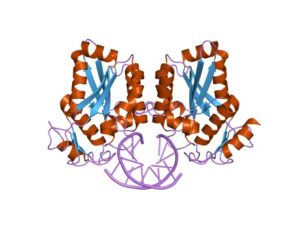
EcoRI From Wikipedia

EcoRII

Source Escherichia coli

Recognition sequence 5’-CCWGG-3′ 3’-GGWCC-5′; sticky ends

restriction enzyme example
EcoRII From Wikipedia

BamHI

Source-Bacillus amyloliquefaciens

Recognition sequence – 5’-GGATCC-3′ 3’-CCTAGG-5′: sticky ends

restriction enzyme example
BamHI From Wikipedia

TaqI

Source –Thermus aquaticus

Recognition sequence- 5’ TCGA-3′ 3’-AGCT-5′

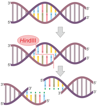
HindIII

Source – Haemophilus influenzae

Recognition sequence – 5’ AAGCTT-3′ 3’-TTCGAA -5′

restriction enzyme example
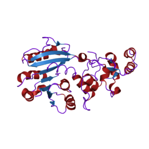
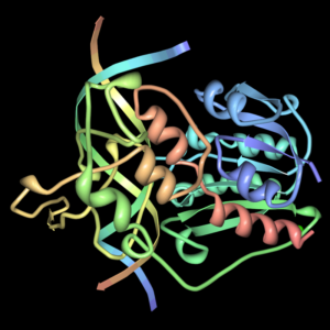
HindIII From Wikipedia

Sau3AI

Source – Staphylococcus aureus

Recognition sequence 5′ GATC-3′ 3′-CTAG-5′;

NotI

Source –Nocardia otitidis

Recognition sequence – 5′-GCGGCCGC-3′ 3′-CGCCGGCG-5′

PvuII

Source –Proteus vulgaris

Recognition sequence – 5′-CAGCTG-3′ 3′-GTCGAC-5′

HaeIII

Source – Haemophilus aegyptius

Recognition sequence- 5′ GGCC-3′ 3′-CCGG-5′

restriction enzyme example
HaeIII From Wikipedia

AluI

Source – Arthrobacter luteus

Recognition sequence- 5’-AGCT-3′ 3’-TCGA-5′

EcoRV

SourceEscherichia coli

Recognition sequence- 5′-GATATC- 3′ 3′-CTATAG- 5′

restriction enzyme example
EcoRV From Wikipedia

SalI

SourceStreptomyces albus

Recognition sequence – 5′ -GTCGAS-3′ 3′-CAGCTG -5′

ScaI

Streptomyces caespitosus

Recognition sequence- 5′-AGTACT-3′ 3′-TCATGA-5′

SmaI

Source Serratia marcescens

Recognition sequence – 5′-CCCGGG-3′ 3′-GGGCCC-5′

HinFI

Source Haemophilus influenzae

Recognition sequence- 5′-GANTC-3′ 3′-CTNAG-5′

HaeIII

Source Haemophilus aegyptius

Recognition sequence- 5′-GGCC-3′ 3′- CCGG-5′

HgaI

Source Haemophilus gallinarum

Recognition sequence- 5’GACGC-3′ 3′-CTGCG-5′

EcoP15I

Source Escherichia coli

Recognition sequence- 5′-CAGCAGN25NN-3′ 3′-GTCGTCN25NN-5′

KpnI

Source – Klebsiella pneumoniae

Recognition sequence– 5’GGTACC-3′ 3′-CCATGG-5′

PstI

Source – Providencia stuartii

Recognition sequence– 5′-CTGCAG-3′ 3′-GACGTC-5′

SacI

Source – Streptomyces achromogenes

Recognition sequence- 5′-GAGCTC-3′ 3′-CTCGAG-5′

SpeI

Sorurce –Sphaerotilus natans

Recognition sequence- 5′-ACTAGT-3′ 3′-TGATCA-5′

SphI

Source – Streptomyces phaeochromogenes

Recognition sequence- 5′-GCATGC-3′ 3′-CGTACG-5′

StuI

Source- Streptomyces tubercidicus

Recognition sequence- 5′-AGGCCT-3′ 3′-TCCGGA-5′

XbaI

Source –Xanthomonas badrii

Recognition sequence – 5′-TCTAGA-3′ 3′-AGATCT-5′

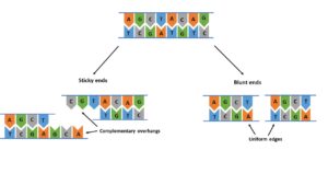
Restriction enzymes cut the DNA strands in two different ways.

Some restriction endonuclease cut the DNA at the same point. Such straight-cut within the recognition site creates blunt ends with no over-hanging ends. These ends are often ligated by DNA ligase enzymes. Examples: PvuII, Haelll, Alul.

Contrastingly, the second class of restriction endonuclease undergoes staggered cut resulting in complementary single-stranded over-hanging ends. Such ends are called sticky or cohesive ends. Examples EcoR1, BamH1, Taq1. These are frequently used for cloning purposes in biotechnology.

restriction enzyme example
Restriction enzyme cleaves DNA strand in two different ways

Types of restriction enzymes

Restriction enzymes can be classified into four groups depending upon the composition, type of cofactors requirements, the target site, and cleavage position.

Type I

This type of restriction enzyme are multifunctional proteins that cleave only one DNA strand at random as well as distant sites and also performs methylase activities. The target sequence is about 15 bp in length and cleaves non-specifically away from the recognition site. For catalytic activity, it requires Mg2+, ATP, and S-adenosyl – L- methionine. Examples – EcoK, EcoB.

Type II

This group comprises most orthodox restriction enzymes which are used in recombinant DNA technology. These enzymes are the most stable endonucleases which cleave DNA at specific sites. Thus, these generate desirable fragments of DNA. The recognition sequences are palindromic in nature of 4-8 bp and for catalytic activity, only Mg2+ ions are required. These enzymes can perform the nucleolytic activity only. Examples – Hinfl, EcoRI, PvuII, Alul, Haelll.

restriction enzyme example
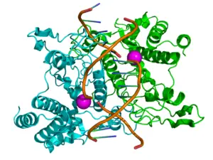
Type II restriction enzyme
(EcoRI in cyan and green color with two magnesium ions in magenta color) From Wikipedia

Type III

This group is an intermediate type between type I and type II. The length of the recognition sequence is 24-26 bp. They require both Mg2+ and ATP for their activity and cleave DNA sequences in close vicinity of target sites. Eg. Hinf III, EcoP1.

Type IV

The target DNA for this group is different from the rest of the types. It recognizes modified DNA sequences such as methylated, hydroxy-methylated, and also glycosylated bases. Eg., McrBC.

Restriction enzyme nomenclature

Restriction enzymes have unique nomenclature. Each enzyme is named after the bacterium from where it is isolated. The name contains the genus, species, and the strain of the bacterium.

The nomenclature of restriction enzymes follows a pattern

1. The first capital letter is the name of the genus from which bacterium is discovered.

2. The first two letters of the species name are written after the first initial.

3. Next is the strain identified which is written in subscript.

4. The number of enzymes produced by the bacterium.

5. Generally all restriction enzymes are prefixed with the general symbol R. This is used to distinguish from the methylases that are obtained from the same strains.

Example:  EcoRI name

Abbreviation                                Meaning

E                                                       Escherichia genus

co                                                     coli species

R                                                       RY13 strain

I                                                        First identified order of identification in the bacterium

What is a restriction enzyme?

The term restriction enzyme was derived from the studies of lambda bacteriophages where it was observed that these bacterial protein enzymes cleave the phage DNA and thus, restrict the activity. The own target sites of the bacterial cell are high methylated i.e., the addition of methyl groups to the adenosine and cytosine bases within the recognition sites. This methylation protects from cleavage.

A restriction enzyme is an endonuclease that enables site-specific cleavage of DNA sequence. These sites are called restriction sites or recognition sequences or target sites. These are usually synthesized by bacteria for defense mechanisms against invading bacteriophages. The mechanism comprising methylation along with restriction enzyme activity constitutes the restriction-modification system.

restriction enzyme example
Palindrome sequence From Wikipedia

DNA strand contains two strands. 5’ end-3’ end depicts the forward strand while the 3’ end – 5’ end is denoted as a reverse strand. At first, restriction enzymes recognize the specific DNA sequences and then make two incisions on each strand of DNA sequence. This specific sequence is called recognition sites. The sequences of recognition sites are palindrome sequences which reads the same on forward and reverse strands when read in the same orientation. The recognition sequences usually contain 4-8 nucleotides, mostly palindromic in nature.

Restriction enzyme structure

The most convenient restriction endonuclease enzymes belong to the type II enzyme.

The recognition sites are typically a short palindromic sequence of 4-8 bp and catalytic activity requires Mg2+ ions. It consists of two homodimers each 30kDa molecular mass, that recognize the palindromic sequences. The structural core of these enzymes consists of four β-strands and one α-helix. For this nucleolytic activity, it doesn’t require ATP hydrolysis.

As restriction enzymes are target-specific, 15-20 hydrogen bonds are formed between the dimers and bases of the target site.  In addition to hydrogen bonds, Van der Waals interaction also takes place. Due to these interactions, the enzyme undergoes conformational change leading to the activation of the catalytic center of the restriction enzyme. The catalytic center contains two carboxylates which are necessary for the binding of cofactor Mg2+. The resultants of the catalysis are DNA fragments with 5’-P and 3’-OH.

Summary

Restriction enzyme is a site- specific endonuclease that cleaves DNA strands at specific recognition sites. These are synthesized by bacteria as a part of their defense mechanism. For its activity some requires magnesium ions while others required ATP and S-adenosyl – L- methionine. Due to its nucleolytic activity these enzymes are extensively used in recombinant DNA technology.

Also Read:

23+ Protease Enzyme Example: Detailed Facts

papain 300x225 1

Protease enzymes are present ubiquitously and represent a pivot role in all physiological processes. Out of three industrial enzymes, protease is the major one. Proteases are also known as peptidases or proteolytic enzymes that are involved in protein catabolism. This article discusses some protease enzyme examples.

Some plant-derived protease enzyme examples

Protease enzymes present in animals

Microbial based protease enzyme examples

Papain

It is obtained from papaya. This enzyme is used as a meat tenderizer, denture cleaner.

protease enzyme example
Papain From Wikimedia

Caricain

It is also obtained from papaya. These enzymes are extensively used in gluten-free food processing.

Bromelain

It is obtained from pineapple. This enzyme play an important role as an anti-inflammatory and anti-cancer agent.

Ficin

This enzymes is isolated from fig. It is widely used in the pharmaceutical industry.

protease enzyme example
Ficin From Wikimedia

Actinidin

It is naturally present in various fruits such as kiwi, banana, mango, and pineapple. It is mostly used as dietary additives.

Zingipain

It is derived from ginger. These enzymes act as anti-proliferative agents.

Cucumisin

Mostly these enzymes are obtained from musk melon. These are involved in the hydrolysis of proteins.

Oryasin

This is obtained from rice and involved in milk clotting.

Phytepsin

This is obtained from barley. Similar to oryasin, it is also used in milk clotting.

Neprosin

This protease enzyme is derived from Nepenthes (the tropical pitcher plant). This plant-derived enzyme is widely used as a proteolytic agent in mass spectrometry, and histone mapping.

Trypsin

These are present in the small intestine, aids in protein digestion

protease enzyme example
Bovine trypsin From Wikimedia

Chymotrypsin

This is a proteolytic enzyme produced by the pancreas which is used in small intestine

protease enzyme example
Chymotrypsin From Wikimedia

Elastase

This is present in the pancreas which helps to breakdown proteins and fats.

protease enzyme example
Neutrophil elastase From Wikimedia

Collagenase

It helps to the breakdown of peptide bonds present in collagen.

Pepsin

This endopeptidase is present in the stomach and aids in digestion.

protease enzyme example
Pepsin From Wikimedia

Renin

It is secreted from juxtaglomerular kidney cells and helps to control blood pressure.

protease enzyme example
Renin From Wikimedia

Thrombin

It is a kind of serine protease that act as a procoagulant and anticoagulant.

Plasmin

This is another example of a serine protease that functions in a fibrinolytic mechanism.

Alcalase

Isolated from Bacillus licheniformis and widely used in the detergent industry.

Savinase

It is isolated from Alkalophilic Bacillus sp.. It is used in the detergent and textile industry.

Neutrase

It is obtained from Bacillus amyloliquefa-ciens.

Novozyme 3403

This is obtained from Bacillus licheniformis and substantially used as denture cleaner.

Novozyme 4551

This protease enzyme is isolated from Bacillus licheniformis and used largely in the leather industry.

Protease enzyme structure

Protease enzyme shows substrate and product specificity. Thus, these enzymes take part in a highly regulated cascade of cellular processes.

The protease enzyme consists of a single polypeptide chain. The polypeptide chain consists of 250 amino acid residues without any sulfhydryl groups.

Depending upon the pH range, protease enzyme can be of three types

Acidic

It functions at 3.8-5.6 pH; used as a seasoning agent in soy sauce, clearing agent in beer and fruit juices; mostly produced by fungal species Aspergillus niger, and Aspergillus oryzae secret extracellular acidic proteases known as Aspergilla opepsins. Other fungal members produce acidic proteases are Penicillium, Rhizopus, and Mucor.

Neutral

The pH of this protease enzyme ranges from 5-8; used in the brewing industry; obtained from Bacillus.

Alkaline

The optimal pH of alkaline protease ranges from 9 to 11; used in the detergent and leather industry; synthesized by Bacillus, mushrooms, and other bacterial species. Streptomyces sp. produces a type of alkaline protease that has keratinolytic activity.

Proteases can be further classified into two groups depending upon the site of cleavage.

Exopeptidases

It usually hydrolyses the ends of a polypeptide.  Based on the ends of a polypeptide i.e., C or N terminal is known as carboxy and aminopeptidases respectively.  Carboxypeptidases usually hydrolyze the C-terminal end of the polypeptide releasing a single amino acid or a dipeptide while aminopeptidases act on the N-terminal end resulting in dipeptide or tripeptide.

Endopeptidases

The active site of these enzymes are the internal regions of a polypeptide chain. Depending upon the catalytic site and the mechanism, they can be divided into serine protease, cysteine protease, aspartic protease, and metalloprotease.

protease
Types of protease enzymes

Serine proteases are denoted by the presence of a serine at the site of hydrolysis. These are low molecular masses and remain active at neutral to alkaline pH.  Example- trypsin, chymotrypsin

Aspartic acid protease is also known as acidic protease consists of aspartates at the active site. These are active in acidic pH. Pepsins, cathepsins, and renins are aspartic acid proteases present in eukaryotes.

Cysteine proteases consist of cysteine and histidine in a catalytic triad or dyad.  These are active in presence of reducing agents such as HCN or cysteine. Example – pepsinogen, papain.

Metalloprotease is a type of protease enzyme that requires divalent metal ions for its catalytic activity. Some require zinc while others use cobalt. 

What is a protease enzyme?

To maintain the growth and development of every animal an array of enzymes play significant roles, among which protease enzymes perform regulatory roles in all cellular processes.

The peptide bond that constitutes the polypeptide chain is hydrolyzed by these protease enzymes and results in the breakdown of protein into smaller polypeptides and amino acids. These are synthesized by animals, plants, fungi, and also bacteria. They cleave and result in smaller molecules or give rise to newer protein products.

Also Read: