What Is Conservative DNA Replication: What, Why, How, Several Facts

dna 508x1024 1

DNA is also referred to as deoxyribonucleic acid and is a molecule made up of two chains of polynucleotide which has coils around each of them making a double helix.

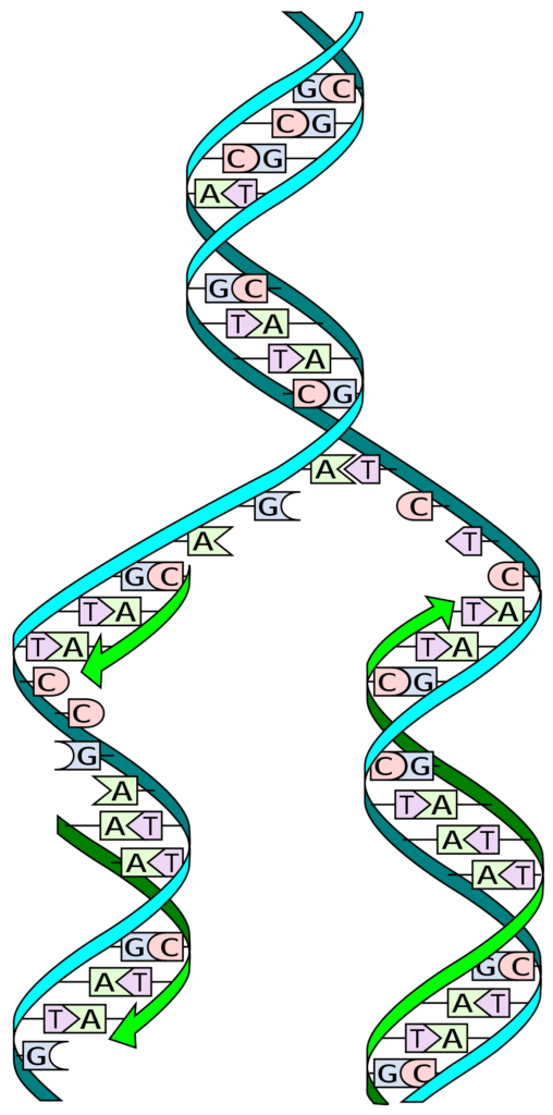
With concern to the question of what is conservative DNA replication, it is the whole original double helical structure of DNA that acts as a template for the new helix that is formed. It is made in such a way that each of the way of cell division makes one daughter cell with a whole new helix and another daughter cell.

The DNA is supposed to be that molecule which carries all the genetic data inside it so that the feature can pass via the parent to the new ones. It helps in growth, the function and the development of the organism along with also having the part in its reproduction. It is a type of nucleic acid. Nucleic acid is one of the vital types of macromolecules needed for life.

There are two strands in the DNA and is thus said to be double stranded. The two of the strands in DNA are called as polynucleotides and is made of tiny simple monomeric units known as the nucleotides. Each of the nucleotide is made up of one of the four bases of nitrogen having the nucleobases. DNA and RNA have the same kinds of nuclobases except one.

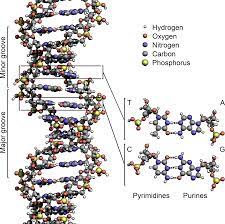
The nucleobases seen in the DNA are adenine, guanine, cytosine and thymine. It also has a sugar in it called the deoxyribose and a group of phosphate. They are joined to each other in a chain like format by the covalent bonding. This bond is seen in between the sugar and one of the nucleotide with the next being phosphate that results in a formation of an alternating phosphate-sugar backbone.

The conformation of the DNA can be in many possible ways which insist on B-DNA, A-DNA and also Z-DNA. The ones that have been seen now are the B and the Z DNAs. The adoption of the conformation of the DNA is based on the level of hydration, the sequence of DNA, the process of supercoiling and its way, the modification in the chemical composition of bases.
what is conservative dna replication
Image credit- DNA ReplicationWikipedia

The series for the steps in DNA replication for most of the organism are-

  1. Initiation
  2. Primer Synthesis
  3. Leading Strand Synthesis
  4. Lagging Strand Synthesis
  5. Primer Removal
  6. Ligation
  7. Termination

Is the process of replication called conservative DNA replication?

DNA replication is the method that takes place in all the living beings and is based on the concept of biological inheritance.

The process of DNA replication is called to be a semi conservative process and it is so cause of the one that is newly made DNA strand. One of them is the original and the other is new made from the original.

The method of getting the DNA replicated is actually semi conservative as each of the helix that is made have only single strand from the helical form that has been copied. It is also said to be conservative for the half of the portion of the each original helix is conserved in each of the new formed strand. They are ubiquitous enzymes in the cellular milieu functioning in diverse processes including DNA replication, DNA repair, RNA transcription and translation.

It has already been shown that the DNA that has been replicated by the process of semi-conservation method in the WT-4 cells has been grown at the 34 degrees or at the temperature of 38.5 degrees at the entire phase of log and into also the stationary phase. The initiation of DNA replication occurs in two steps. First, a so-called initiator protein unwinds a short stretch of the DNA double helix. 
hy
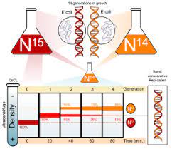
Image credit- Stahl and MeselsonWikipedia

Helicases are enzymes that bind and may even remodel nucleic acid or nucleic acid protein complexes. DNA helicases are essential during DNA replication because they separate double-stranded DNA into single strands allowing each strand to be copied. DNA replication occurs in all living organisms acting as the most essential part for biological inheritance. It  is a duplication process where exact copies of DNA within cells are replicated, with very low error rate.

There was provision for three of the points to be kept in mind during the method of DNA replication. It was explained by Stahl and Meselson who established a way to get to the fact of knowing the process of DNA replication to be either of the three. They conducted an experiment to prove it via-

  • There was suggestion for there of the model for DNA replication being dispersive, conservative and semi-conservative.
  • The method that was conservative suggested that the original DNA stays same and hooked up and the one that is made new links up with the original.
  • The method that was dispersive said that post the process of replication, the two of the daughter DNA that is formed have sugar segments in the alternate manner for both of the parents and the new one synthesized interspersed on two strands.
  • The process being semi conservative suggested that the two DNA that acted as parents served to be a template for the new formed DNA and post replication, each of the double strands of DNA had one parental and other to be the nee called the daughter strand.

Is prokaryotic DNA replication conservative?

Watson and Crick had discovered the DNA to be double helix and provided a hint about how the DNA had been replicated.

There needs to be a replication process to ensure the process of gene transfer and hereditary. Just unlike the eukaryotes, the pyrimidine is made to synthesize. The replication in it is concerned to be semi-conservative and not being conservative.

On regard to the question of what is conservative DNA replication, at the time of cell duplication each of the molecules of DNA is copied at its best to ensure that all the molecules move to each of the daughter cells. The double structure suggests that at its replication time it serves as a template that the two of the strands meet to get separate. DNA replication is the biological process of producing two identical replicas of DNA from one original DNA molecule.

In the process of conservative model of replication, the two of the pieces of DNA that is original is called the parent strands and appear to re base pair along with each other after being sued up as a template to get them synthesized and the two of the new ones are called as daughter strands acting as base pair. The double helix separates so that each old strand serves as a template for a new strand. Two new double helices result, each containing one new strand and one old strand.

One of the strands that are referred is called the old one and the other is referred to as the new one. In the process of semi-conservative one each of the two original strands act as a template for the new ones and after getting replicated each of the DNA strands make base pair with the complementary new one including the old one. The parental molecule directs synthesis of an entirely new double-stranded molecule.

Stahl and Meselson were quite interested in getting to know about the replication of DNA. They tested in the bacteria called E.coli for multiple generations in the medium that had heavy isotopes of nitrogen being 15N and 14N. N14 is said to be the light one and with the other being heavy form of isotope and thus the cell grew. In bacteria, the initiation of replication occurs at the origin of replication, where supercoiled DNA is unwound by DNA gyrate.

Is eukaryotic DNA replication conservative?

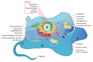
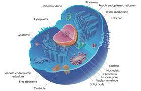
Eukaryotes are the organism that has nucleus covered inside a nuclear envelope. They have organelles having a membrane.

Within the eukaryotes, the majority of the synthesis in DNA takes place during the S phase of the cell cycle and the whole genome is said to untangled and copied to make the daughter cells. This process of DNA replication is semi conservative in eukaryotes.

The process of DNA replication in the eukaryotes is quote a conserved method that gets the restriction of replication per cycle. The central for replication is the chromosome and is vital for having the genome of the eukaryotes maintained. It is a process being semi conservative. A laboratory technique used to replicate, and thus amplify, a specific DNA segment.

The action of polymerase having to synthesize the strand of DNA complementing the original strand of DNA is said to be DNA replication. The cells of eukaryotes have within the organelles that have membrane like that of Golgi apparatus and mitochondria. Plants and the algae have chloroplasts in them. It contains one newly synthesized strand and one newly synthesized strand. The model of semi-conservative DNA was anticipated by Nikolai koltsov.
mnmn
Image credit-
EukaryotesWikimedia

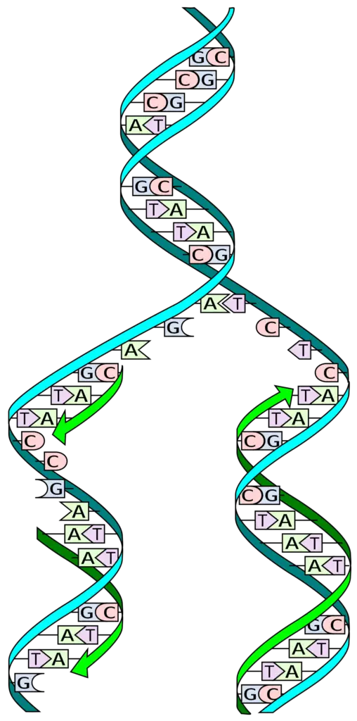
DNA helicase helps in getting the DNA strands to unwound making a form of fork called the replication fork and have two of the template being single stranded. The process of replication entertains the copy of the single DNA into two helixes. The major use of enzyme is carried out by the replication fork and is much conserved from eukaryotes to prokaryotes.  

One of the threads mentioned is called old and the other thread is called new. In semi-conservative analysis, each of the two original strands serves as a template for a new strand, and after replication, each DNA strand base-pairs with a new, complementary strand, including the old one. Both the eukaryotes and prokaryotic cells have replication process to be semi conservative.

Is bacterial DNA replication conservative?

Bacteria are the organism that have single cell and are microscopic and are found in millions of its forms and in every surrounding in and out of organism.

The method of DNA replication is anyhow seem conservation in any type of organism be it prokaryotes or eukaryotes. Thus bacteria have the process of replication being semi-conservative where one behaves as parents and the other is new.

While there was an experiment that said the process of replication to be semi conservative in bacteria, it has been said that it is just not in E.coli that the method is semi conservative but applies to other forms of bacteria as well. There has been no proof that says the process to be dispersive or conservative form of replication. DNA replication is the process of making copies of DNA. DNA replicates by semi-conservative replication.

this means which means that one strand of the parent double helix is conserved in each new DNA molecule. As for the question of what conservative DNA replication is, during cellular replication, each DNA molecule is copied in the best possible way to ensure that all molecules migrate to their respective daughter cells. The double structure suggests that the two strands meet and serve as a template for separation during replication.

The method used to replicate DNA is actually semi-conservative. This is because only a single strand of the spiral is copied to each generated spiral. This is also called conservative, as half of each original helix is ​​preserved in each of the newly formed strands. The parental strands f DNA re always separated to make a new one. For most eukaryotes and prokaryotes, the process of DNA synthesis is done semi conservatively. Semiconservative replication for E. Coli is initiated by way of a replicon structure which behaves as an autonomous unit of DNA replication.

DNA replication is said to be semiconservative because each newly made DNA molecule has one original and one new strand of DNA. A laboratory technique used to replicate, and thus amplify, a specific DNA segment. This process is known as semi-conservative replication because two copies of the original DNA molecule are produced, each copy conserving (replicating) the information from one half of the original DNA molecule. The conservative method of replication suggests that parental DNA remains together and newly-formed daughter strands are also together.

Also Read:
The Semiconservative Replication of DNA: A Fundamental Process in Molecular Biology

is dna replication semiconservative

The semiconservative replication of DNA is a fundamental process in molecular biology, which was first demonstrated through the classic density labeling experiments of Matthew Meselson and Franklin W. Stahl. This process ensures that the genetic information encoded in the DNA of a parent cell is faithfully replicated and passed on to its daughter cells, maintaining the integrity of the genetic material across generations.

Understanding the Semiconservative Replication of DNA

The semiconservative replication of DNA is a highly coordinated and intricate process that involves the unwinding of the double-stranded DNA molecule, the synthesis of new complementary strands, and the precise distribution of the parental and newly synthesized strands into the daughter molecules.

The Meselson-Stahl Experiment

The Meselson-Stahl experiment, conducted in 1958, provided the definitive evidence for the semiconservative replication of DNA. In this experiment, Meselson and Stahl grew Escherichia coli (E. coli) bacteria in a medium containing the heavy isotope of nitrogen, 15N, until the DNA of the cells was completely labeled with 15N. They then transferred the bacteria to a medium containing the lighter isotope of nitrogen, 14N, and allowed the cells to undergo several rounds of cell division.

At various time points, the researchers extracted the DNA from the cells and analyzed its density using equilibrium sedimentation in a cesium chloride (CsCl) density gradient. The results of this experiment were as follows:

  1. After the first round of replication, the DNA extracted from the cells formed a single, intermediate-density band, indicating that each new DNA molecule contained one strand of 15N-labeled parental DNA and one strand of 14N-labeled newly synthesized DNA.

  2. After the second round of replication, the DNA extracted from the cells formed two distinct bands: one at the intermediate density (hybrid DNA) and one at the lighter density (completely new DNA).

  3. Subsequent rounds of replication resulted in a gradual decrease in the proportion of hybrid DNA and an increase in the proportion of completely new DNA.

These results clearly demonstrated that DNA replication is a semiconservative process, where the parental DNA strands are conserved and equally distributed into the daughter molecules.

Autoradiographic Studies

The semiconservative replication of DNA has been further supported by autoradiographic studies, which have visualized the replication of the bacterial chromosome. These studies have shown that the replication fork, the site where new DNA strands are synthesized, moves in a unidirectional manner, with one strand being synthesized continuously (the leading strand) and the other strand being synthesized discontinuously (the lagging strand).

The autoradiographic studies have also revealed that each daughter molecule contains one old and one newly synthesized strand, consistent with the semiconservative model of DNA replication.

Theoretical Calculations

In addition to the experimental evidence, the semiconservative replication of DNA has also been supported by theoretical calculations. It has been shown that the concentration distribution of a single macromolecular species, such as DNA, in a constant density gradient should be Gaussian, and that the standard deviation of the band is inversely proportional to the square root of the macromolecular weight.

This model has been tested with homogeneous DNA samples from bacteriophage T4, and the results have been found to be remarkably consistent with the theoretical predictions, further validating the semiconservative replication of DNA.

The Significance of Semiconservative DNA Replication

is dna replication semiconservative

The semiconservative replication of DNA is a fundamental process that is essential for the proliferation and survival of all living cells. It ensures that each daughter cell receives essentially the same genetic information that was encoded in the DNA of the parent cell, maintaining the genetic integrity of the organism.

This process is crucial for the accurate transmission of genetic information from one generation to the next, as it allows for the faithful replication of the entire genome, including the coding regions that contain the instructions for the synthesis of proteins and other essential cellular components.

Furthermore, the semiconservative replication of DNA is a highly regulated and coordinated process, involving a complex network of enzymes, regulatory proteins, and other molecular machinery. Understanding the mechanisms and dynamics of this process has been a central focus of research in molecular biology and has led to numerous advancements in our understanding of cellular function, genetic inheritance, and the development of various biotechnological applications.

Conclusion

The semiconservative replication of DNA is a well-established and fundamental process in molecular biology, supported by a wealth of experimental and theoretical evidence. This process ensures the accurate transmission of genetic information from parent to daughter cells, maintaining the genetic integrity of living organisms. The Meselson-Stahl experiment, autoradiographic studies, and theoretical calculations have all contributed to our understanding of this crucial biological process, which continues to be a subject of ongoing research and exploration.

References:

  1. Cairns, J. (1963). The bacterial chromosome and its manner of replication as seen by autoradiography. Journal of Molecular Biology, 6, 208–213.
  2. Hanawalt, P. C. (2004). Density matters: The semiconservative replication of DNA. Proceedings of the National Academy of Sciences, 101(52), 17889-17894.
  3. Meselson, M., & Stahl, F. W. (1958). The replication of DNA in Escherichia coli. Proceedings of the National Academy of Sciences, 44(7), 671–682.
  4. Meselson, M., Stahl, F. W., & Vinograd, J. (1957). Equilibrium sedimentation of DNA from bacteriophage T4. Proceedings of the National Academy of Sciences, 43(11), 976–983.
  5. Watson, J. D., & Crick, F. H. C. (1953). A structure for deoxyribose nucleic acid. Nature, 171, 737–738.

RNA Replication Enzymes: Detailed Facts

mm 300x146 1

RNA are referred to as ribonucleic acid and are the molecule same to that of DNA. Just unlike them RNA has a single strand.

RNA polymerase is a RNA dependent enzyme used up for the RNA replication. It is also called as RNA replicase and is RNA replication enzymes. It helps in catalyzing the process of getting the RNA replicated from a template of RNA.

Mostly it helps in getting the process of synthesis of the single strand of DNA fast and complementary to that of a given template of RNA. Thus it is totally different from that of the RNA polymerase that is dependent on DNA. This enzyme helps an organism to make the use of catalyst for getting the transcription of RNA done from a DNA template.

RNA is strand which has a backbone and is made up of ribose which is a sugar and is placed alternately and also has phosphate groups attached with it. It also has itself linked with sugar along with the four bases of nitrogen called the adenine, cytosine, guanine and uracil. It can also be referred to be a molecule which is polymeric.

RNA is a nucleic acid and so is DNA. Just like the DNA, RNA also has been joined as a chain with its constituents being nucleotides but RNA is single stranded and seen in nature to fold onto itself not being double. Cellular living beings use messenger RNA to get itself pass the genetic code and also helps in directing the synthesis of few proteins.

Some of the molecules of RNA play a good role inside the cell by getting the biological process catalyzed with having the gene expression under control. It also helps in communicating several responses and sensing the responses to have the signals of the cell activated. One of the processes it is involved in is protein synthesis and also getting the ribosomes protein synthesized.

Some RNA molecules play a good role in the cell by catalyzing biological processes by controlling gene expression. It is also useful for communicating multiple responses, detecting the response and activating the cell’s signal. One of the processes involved is protein synthesis and ribosomal protein synthesis.

RNA replication

The replication of the genome is essential for the continuity of life. The molecular mechanism is very similar in all groups of organisms.

Many scientists consider RNA to be the first replicated macromolecule. Like DNA, RNA can, in principle, create a negative blueprint by spontaneous base pairing. Unlike double-stranded DNA, RNA can take a variety of spatial forms and therefore also functions as an enzyme that catalyzes its own replication.

Many bacteria divide once every thirty minutes, others replicate even faster. Eukaryotic cells only replicate their genome when new cells have to be created. This happens as a result of external signals, for example tissue loss or inflammation. The life cycle of eukaryhttps://lambdageeks.com/eukaryotic-cells-vs-bacterial-cells/otic cells underlies an accurately defined sequence of activities that can be divided into different phases.

However, the situation is different at the end of the chromosome, where the lack of base prevents the replication fork from proceeding. At the point on the lagging strand where the DNA primase places the last RNA primer, the DNA polymerase will not be able to continue replication. The terminal DNA segment cannot replicate. DNA strands become shorter and shorter as the number of cell divisions increases.

To protect themselves against the rapid shortening of the DNA, eukaryotic chromosomes possess sequence repeats (telomeres) at their extremities, which do not code for proteins. Since the telomeres do not contain any vita;l information, the key parts of the DNA are protected. The telomeres get shorter each time a cell divides. The length of these so-called telomere caps defines the number of possible divisions and hence the lifespan of a cell. 

rna replication enzymes
Image credit-
RNA replication Wikipedia

RNA replication enzymes in eukaryotes

Eukaryotes are the organism that has a cell and the cell is enclosed inside a nuclear envelope. They also have other organelles with membrane.

At the level of the eukaryotes, the replication fork there is a polymerase seen for replication which is three in number and is a complex that helps the DNA get itself replicated. The main RNA replication enzyme is said to be RNA polymerase.

The enzyme of RNA polymerase helps in getting the RNA synthesized by pointing a strand of DNA. This enzyme is also the one to play a role in copying of the DNA and its sequence into a sequence of RNA at the time of transcription. The process of RNA replication in the eukaryotes takes place in the nucleus.

ss
Image credit-
EukaryotesWikipedia

The strand of DNA is made to undergo the process of replication and then transcription within the nucleus and the proteins are then made inside the cytoplasm. RNA thus is made to travel across the membrane of the nucleus which also surrounds it before the time it goes into translation. On addition to RNA polymerase there can also be the presence of other small materials for it.

On addition to the RNA polymerase, there is α-primase and two of the DNA polymerases called the δ and ε that are needed for the process of the strand of DNA to get replicated. The δ polymerase is the major or basic polymerase inside the leading path for synthesis and the rest being δ and ε are the basic polymerase for the lagging synthesis of strand.

Mostly the enzyme that are present inside the cell for the help of getting the smooth functioning of the RNA is mostly for the purpose off good function of all the chemical reactions. RNA polymerase enzyme helps to synthesize RNA by pointing a strand of DNA. This enzyme is also the enzyme that plays the role of copying DNA and its sequence into an RNA strand at the time of transcription.

RNA replication in eukaryotes takes place in the nucleus. Some RNA molecules play important roles in catalyzing biological processes within cells and regulating gene expression. It also helps to activate cellular signals by transmitting multiple responses and recognizing them. One of the techniques its miles concerned in is protein synthesis and additionally getting the ribosomes protein synthesized.

RNA replication enzymes in prokaryotes

Prokaryotes are the organism that have only one cell and cannot be seen without microscope. They do not have a nucleus unlike the eukaryotes.

Inside the prokaryotes, the origin for the point of replication to take place is only one in number and takes place in two of the opposing directions at the similar tome and is seen to occur inside the cytoplasm o the cell.

On the other if the eukaryotes are taken into consideration, they have many site for getting the process of replication conducted and with not being concerned to two paths. The process is made to be useful in several ways and is unidirectional at the part of replication inside the cell within the nucleus.

Inside the prokaryotes there are several ways to get done with the process of the DNA to get it replicated. They are dispersive, semi-conservative and conservative. The very common one to be described is the conservative one that says that the original DNA stays linked and the new made strands of it called the daughter strands are also made to connect.

pp
Image credit-
ProkaryotesWikipedia

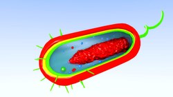
The prokaryotes are divided only into two domains. One is said to be the bacteria and the other is archaea. The bacteria are said to be common and the one known widely for the cells of prokaryotes. They lack the entire inside membrane and have flagella for moving. The oldest known prokaryote is 3.5 billion years old.

Both of the type of organism found being eukaryotes and the prokaryotes use an enzyme that is common to both and that is the RNA polymerase which is used to transcribe the RNA from DNA. The prokaryotes use up only one RNA polymerase for all the types of RNA or transcription. They do have only one site for replication but has several sequences for getting it repeated in form of array.

RNA replication enzymes in bacteria

Bacteria are said to be the organism that live freely and are single cell also regard to as the prokaryotes. They are not much longer and can be seen at any place

The microbes of RNA have themselves replicated via either of the two ways. The first being the process of the RNA being dependent on the synthesis of RNA and the other being the process of reverse transcription that is the RNA dependent on synthesis of DNA and is followed by transcription.

Inside the bacteria, all the transcription that Is seen is by a single and special type of RNA polymerase. This polymerase has with it four of its varieties and catalytic units with only one regulatory unit called as the sigma. They only have one copy site but many strings to make it repeat in a loop. The manner is made to be beneficial in numerous methods and is unidirectional on the a part of replication within the mobileular in the nucleus.

Some bacteria are harmful, but most serve a useful purpose. They support many forms of life, both plant and animal, and they are used in industrial and medicinal processes. Bacteria are like eukaryotic cells in that they have cytoplasm, ribosomes, and a plasma membrane. Features that distinguish a bacterial cell from a eukaryotic cell include the circular DNA of the nucleoid, the lack of membrane-bound organelles.

RNA-dependent RNA polymerase or RNA replicase is an enzyme that catalyzes the replication of RNA from an RNA template. DNA replication initiates at specific points, called the origins, where the DNA double helix is unwound. A short segment of RNA, called a primer, is then synthesized and acts as a starting point for new DNA synthesis. An enzyme called DNA polymerase next begins replicating the DNA by matching bases to the original strand.

Also Read:

Is Cytosine A Pyrimidine: Why And Detailed facts

nn

Cytosine is said to be one of the four bases in both of the nucleic acid being DNA and RNA and is denoted with the alphabet C.

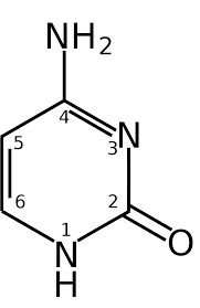
With regards to the question is cytosine a pyrimidine, the answer is a yes. The base of cytosine is derived from the pyrimidine that is found in the nucleic acids and is a component that helps in getting the genes controlled for all of the living cells and in also a few of the coenzymes.

The structure of this base cytosine along with the rest is usually preferred to be taken as a ring of nine and double membrane structure adenine and the guanine as the base for purine and the structure of thymine for the six membrane ring line being single and uracil and cytosine are said to be pyrimidine.

The principal pyrimidine are said to be thymine, uracil and cytosine that consists of the uridine.  Along with thymidine and the cytidine ribonucleosides and all other corresponding deoxynulceotides. The building blocks of DNA are said to be both thymine and cytosine while uracil and cytosine are seen in only RNA.

Cytosine being one of the four bases of building blocks for RNA and DNA is also one among the four nucleotides that are seen in both of the strands’ and each of the cytosine base makes up a portion of a code. This base also has the ability to hold a unique feature that helps in binding of the double helix and is seen opposite to guanine which is also another base. The base of guanine seems to pair up with cytosine and thymine pairs with adenine in DNA.

Cytosine can be found as part of DNA, as part of RNA, or as a part of a nucleotide. As cytidine triphosphate (CTP), it can act as a cofactor to enzymes, and can transfer a phosphate to convert adenosine diphosphate (ADP) to adenosine triphosphate (ATP). In DNA and RNA, cytosine is paired with guanine. Cytosine and thymine are pyrimidines which are structures composed of a single six-sided ring.

Why is cytosine a pyrimidine?

Cytosine is a derivative of pyrimidine and has an aromatic ring that is heterocyclic and have two of the substituents linked and is seen in both DNA and RBNA.  

With is cytosine a pyrimidine, it is a yes. The structure of this general cytosine with the remainder is generally preferred as a nine-ring structure, and the adenine and guanine double membrane is the basis for the purine and thymine structures for the six-loop membrane, and uracil and cytosine are given is being pyrimidine.

Any base which is same t that if the look of benzene which is a structure of six member ring ad contains the bases of thymine, uracil and cytosine as the bases acted for RNA and DNA can be said to be a pyrimidine. It helps in getting the genes to control the materials in the living cells.

is cytosine a pyrimidine
Image credit-
CytosineWikipedia

This base also has the ability to hold a unique feature that helps in binding of the double helix and is seen opposite to guanine which is also another base. The base of guanine seems to pair up with cytosine and thymine pairs with adenine in DNA.  The paring of the bases are done with the help of hydrogen bonds that are said to be interstrand.

Adenine is said to make hydrogen bond that is two in number along with uracil and cytosine makes up three bonds of hydrogen with only the base guanine. Thus we can all say that the bases of guanine, cytosine and adenine are found in both RNA and DNA. So these are the common bases of nitrogen in both of the nucleic acids.

Cytidine is said to be a nucleoside of cytosine and in the base pairing model of Watson and Crick, it seem to make a hydrogen bond with guanine being three in number. The chemical formula for this is C4H5N3O and has molar weight of 111.1 g with melting point being 320 degree Celsius and decomposes at 593 K. Cytosine can be said to be a part of either DNA or a part of RNA or a portion of nucleotide.

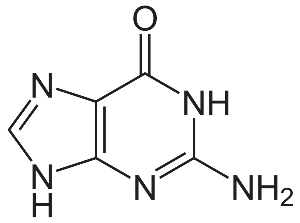
The bases of nitrogen that are purine are feature but the presence of a single group of amino with adenine being at the sixth place of carbon and guanine sitting at the second carbon place. Just like this, the cytosine, thymine and the uracil have a simple structure of ring which is made from pyrimidine and thus these are the derivative of pyrimidine bases. Guanine, an organic compound belonging to the purine group, a class of compounds with a characteristic two-ringed structure

Why cytosine bonds with guanine?

The four of the bass in DNA and RNA are supposed to pair uo with each other according to the system of complimentary bond pairing and thus cytosine pairs up with the base of guanine in both.

Cytosine and guanine link up together and make a base pair as the free bond of hydrogen donor and the acceptor of hydrogen bond link up together with each other within space. Cytosine and guanine are said to be complementary to each.

In the basic pairing formula the thymine makes sup with adenine and the rest guanine binds with cytosine. There is a shift in the place of the nucleotides that makes a wobble I between the general guanine and normal thymine. Thus they are paired together. Within the DNA molecule, guanine bases located on one strand form chemical bonds with cytosine bases on the opposite strand. The sequence of four DNA bases encodes the cell’s genetic instructions.

gg 2
Image credit-
GuanineWikipedia

The paring of both the bases is done by lining them up with three bonds of hydrogen. This makes a difference in the power in between the sets of Watson and Crick bases. Cytosine and guanine links up and thus has strong base pair than the adenine and thymine bond in DNA. Cytosine and guanine form three hydrogen bonds between each other, while tyrosine and adenine form two hydrogen bonds.

The guanine paired with the cytosine has three hydrogen bonds. This makes the difference in strength between the two sets of Watson and Crick basses. Base pairs associated with guanine and cytosine are stronger than those associated with thymine and adenine in DNA. Within the DNA molecule, guanine bases located on one strand form chemical bonds with cytosine bases on the opposite strand.

Also Read:

Types of RNA Splicing: Detailed Facts

gg 1

RNA splicing is the method in the field of molecular biology where the new made precursor messenger RNA transcription is converted to the mature mRNA.

There are several ways by which RNA splicing takes place sin nature and it is based on the splicing type on the base of the intron and the used catalyst that shall help it to take place. The types of RNA splicing are

Self-Splicing

It is the type of RNA splicing that takes place in few of the rare introns that are able to help promote the phosphodiester bond gaps.

This type or gap helps in making of the phosphodiester bond without the use of other proteins or rest spliceosomes. The introns that are involved in it are unique as they shall be able to mediate the excision from the precursor RNA and the ligation of the subsequent of the flanking of the exons in the very simple salt buffer.

This reaction is self-splicing and is worked upon by the tertiary part of the intron that shall help in making of the base for intron and helps it with the ability to recognize the sites of splicing for the precursor RNA and help it perform the ligation and the cutting part in the precise way possible.

The pattern that is there in these introns is able to perform being a ribozyme that shall help in the regulation of the whole process. There are there groups done for this part of RNA splicing and are termed to be Group I, Group II and Group III. The functions of each of the group are same but also different at the same time.

The Group I and Group II introns help in making of the process same to spliceosomes, t can be said that the introns are evolutionary related to spliceosomes. At this time, the 5’ is identified by a small element in the introns and is called the internal guide sequence. It consists of two reactions of Tran’s esterification an involved removal of the introns and the exon ligation.

gg 1
Image credit-
Self SplicingWikipedia

Alternate splicing

It is an alternate process for having the RNA spliced at the time of gene expression that shall helps in a single gene to code itself for the other proteins.

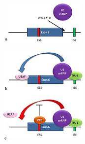
Here in this type of RNA splicing the exons of the gene shall be taken into play from the resulted mRNA that is made from the gene. It states that the exons that are linked in several combinations lead to varying strands of messenger RNA.

It occurs in a good state in the eukaryotes. It has the ability to increase its diversity of proteins here which also can be encoded by the genome. In us, 95% of the multi existing genes are made to splice alternate. There are the presences of many nodes for this process that can be seen under which the most general form is the skipping of exon.

The process of this is based in the system if acting up the Trans proteins that shall link to the cis pint on the primary transcript to itself. Just like it, the proteins slicked activators also use it to promote the use of the particular site for the slice and the restores reside the use of this site. The method for this process is much variable and some of the examples for this is also been taken into eye mostly for the use of the process throughout.

kk
Image credit-Alternate splicingWikipedia

tRNA splicing

There are many of the introns that interrupt the work of many genes in the eukaryotes and thus are removed by splicing and it is the vital step in expression of the genes.

In the eukaryotes there are four types of method that are used. The messenger RNA takes place in the phosphotransfer reaction on the complex and the dynamic machine. This function is related to the mechanism for two of the self-splicing way concerned with the introns of Group I and Group II.

The ones in the Group II are linked up with the identical way of splicing that helps in getting the common space of ancestor diverged. tRNA genes are also allowed to catalyze by all the three enzymes and the proteins and them are intrinsic of all the need for the hydrolysis of the ATP.

oo
Image credit-
tRNA splicingWikipedia

Mostly in bacteria that are self-spliced. It was seen that the process of this in the eukaryote and the archaea are not related buy its seen now that the enzyme first used here is tRNA endonuclease is seen in the evolution of the structures if the archaea and the eukaryote. There are many upright being surprised have been seen out of the boom and thus the stricture with way comparison of the enzyme from these of the two are in much diverge line.

Recursive splicing types of RNA splicing

Recursive splicing (RS) is an evolutionarily conserved process of removing long introns via multiple steps of splicing. It was first discovered in Drosophila and recently proven to occur also in humans.

RNA splicing is an eukaryotic gene expression process in which this genetic information is modified as RNA. During splicing, specific regions of the RNA copy are cleaved and adjacent sequences glued together. The parts of gene sequences that are expressed in proteins are called exons, because they are expressed.

RNA splicing is a eukaryotic gene expression process in which this genetic information is modified as RNA. The key difference between RNA splicing and substitution splicing is that RNA splicing is the process of joining exons together of the main copy of mRNA whereas alternative splicing is the process of creating distinct combinations of exons of the same one gene.

Trans-splicing

It is a special type of getting the RNA process. Here the exons are of two types being the primary transcript of RNA that are link to the end of the final part and the other is the ligated.

Normal type of it is called the cis type and consists of a single molecule. The Trans splicing makes a single RNA transcript from the several separate pre messenger RNA. This way can be used for molecular where they help with the mutate product of genes.

When there is a view of any fusion taking place in the transcripts during the normal process in human cell, his mechanism acts behind having the oncogenic transcript of fusion.  On the other hand, the translated proteins are made to slice and have many different types of amino acids sequence and gave different biological use.

Also this process get into the help allowing the human genome to have direct synthesis for many of the proteins that shall be an outcome expected from the 20,000 of the genes coding proteins. The transfer RNA are made to interrupt the introns but the process of splicing is actually different as it helps in getting them catalyzed and all takes place in Archaea, bacteria and the eukaryote.

What is RNA splicing?

RNA splicing is the way that makes the use of both precursor and mature RNA. It helps by getting the introns removed.

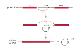
The very process of RNA splicing is started with getting then spliceosomes or the ribonucleoproteins linked with the introns that are seen on the site of RNA splicing. This linking of the ribonucleoproteins outcomes in the biochemical way and is called the Trans esterification in between the nucleotides.

At the section where the 3’OH is there of a concerned nucleotide on top of the intron that is defined with the assembly of spliceosome and outcomes to a attack of nucleophile on the very first nucleotide of the intron at the site of 5’ splice.  The spliceosomes are said to be a different part of protein group.

types of rna splicing
Image credit-
RNA splicing-Wikipedia

This shall result in getting the ends of 5’ and 3’ folded up making it look like a loop. On the other hand, the adjacent parts of the exons are also liked up together. At the end, finally the intron loop is kept detached from the sequence of the spliceosomes. The spliceosomes are also called to be ribozymes and are in control and regulation process.

After all the 1st phase, the second transesterification reaction takes place at the time of ligation of the adjacent segment of the exon. Here, the 3’OH part of the 5’exon that is released and performs in an attack of electrophilic on the very start present nucleotide and exactly at the site of the last present nucleotide at the intron on the 3’ splice site.

This causes the bonding of the two parts of the exon with the intron part being removed. Earlier, the very intron is kept outside at the time of splicing and is thought to be a unit of junk. Thus, yet it is still observed now that these are a part of the method where the proteins are kept to be removed.

Also Read:

Types of RNA Polymerase: Purpose, Various Elements, Detailed Facts

hh

Polymerase is any enzyme that helps in synthesizing the large chains of nucleic acids or the polymers. RNA polymerase helps in copying of the DNA sequence.

There are different types of RNA polymerase in both for prokaryotes and eukaryotes. All of the eukaryotes have several polymerases that can help in getting the genes transferred. There is RNA polymerase I, RNA polymer II and also the RNA polymerase III. Thus the types that are found are-

  1. RNA polymerase I
  2. RNA polymerase II
  3. RNA polymerase III
  4. RNA polymerase IV
  5. RNA polymerase V

RNA polymerase I

This complex is used up for the production of ribosomes which is vital for the synthesis machine of cellular proteins that helps in its growth along with getting to underline may of the other functions. The accurate transcription of human rRNA genes by RNA polymerase I requires two transcription factors, upstream binding factor. Pol I is a 590 kDa enzyme that consists of 14 protein subunits.

RNA polymerase II

It is responsible for transcription of nuclear genes encoding messenger RNA and some small nuclear RNAs. Twelve of its subunits have identical or related counterparts in RNA polymerase II (Pol II) and RNA polymerase III (Pol III). The other two subunits are related to Pol II initiation factors and have structural homologues in Pol III. RNA polymerase allows in copying of the DNA sequence.

RNA polymerase III

RNA polymerase III (Pol III) transcribes a variety of small, stable RNAs that are essential for multiple cell signaling pathways, including premRNA splicing (U6 snRNA) and protein synthesis. The last for the eukaryotes is the polymerase III that gets the 5S ribosomal RNA gens and the tRNA transcribe.

RNA polymerase IV

RNA polymerase IV is an enzyme that synthesizes small interfering RNA that suppresses gene expression in plants. The RNA polymerase I gets the genes of ribosomal RNA transcribed, RNA polymerase II helps in getting the snRNA, the miRNA and the messenger RNA gets itself transcribed. RNA polymerase is seen in all types and kinds of organism and thus yet the composition and the number of the DNA.

RNA polymerase V

Determining the composition and diversity of the Pol IV and Pol V RNA subunits is an important step in understanding their function. The ones that are being used in two different types of species are a bit different from each other. The data is kept inside the molecule of the DNA and is made to copy in a new molecule for the messenger RNA. Just for taking an example the entire organism needs methods by which they can carry on transcription.

RNA polymerase is said to be an enzyme that makes to take part in copying of the sequence in DNA and help in get converted to a sequence of RNA at the time of a method called transcription. It is same as that of the molecule that is made of the protein subunits and is much complex. The enzyme called as RNA polymerase helps in controlling its acting to get flowed with transcription at the time where the information is kept stored.

Considering an example for just one type of bacteria has only a small type of RNA polymerase while in the yeast and the multicellular organism often said to be the eukaryotes do have three types of it and are all different. Despite these not so similar features there are also many other characters that make them alike by which they can come hands on with the method of transcription.

Types of RNA polymerase in prokaryotes

Prokaryotes are the organism that have only one cell and cannot be seen under naked eye. They are single called along with being unicellular and having either flagella or cilia for their movement. Bacteria and archaea can be its example.

Within the bacteria or the prokaryotes, the polymerase is seen in two phases. One of them being called the core enzyme that helps in getting the RNA synthesized but is not able to link the DNA with the targeted promoter.

The second part of it is the holoenzyme which helps in two ways one being the synthesis of RNA and the other gets to recognize the promoter. Along with this, the prokaryotes use a single type of RNA polymerase for its use. The organism being prokaryotes helps themselves by suing the same polymerase of type RNA to get the genes transcribed.

Four if the units in them are denoted by α, α, β, and β′ and these are a part of the core enzyme. These units kink all the time into a genes and then is transcribed and disassemble it after transcription is done. While the eukaryotes have three types of RNA polymerase in use it also uses up there of the DNA polymerase.

types of rna polymerase
Image credit-
RNA polymeraseWikipedia

How many types of RNA polymerase in eukaryotes

The cells of the eukaryotes are one of the complex materials found in several ways that also consists of the term of the process transcription. These are the organism that have a nucleus and is covered inside the nuclear envelope.

Thus out of all the types of RNA polymerases eukaryotes take the use of three types of them. These types are typical in view to carry other types of organelles that have membranes just like the Golgi body and the mitochondria and also the chloroplasts that can be found in the algae and plants.

They are different in their type of unit they carry and also vary in number. There is also a variation seen in the type of class the three of each belong to. The three of them used are RNA polymerase I, RNA polymerase II and RNA polymerase III. The RNA polymerase I helps with the ribosomal RNA, the RNA polymerase II help with the transcription of messenger and the RNA polymerase III goes with the transfer RNA.

The RNA polymerase II helps with the encoding of the gens in proteins it is of more importance to the researchers that spy on the gene expression of the eukaryotes and its use. Taking into consideration, it is said that RNA polymerase II gets to bind the DNA with the promoter genes called as the TATA box that helps in the initiation of transcription.

All together having same motif for getting a small sequence of DNA these are the ones that have the core promoter. Despite these the changes in polymerase II gets to influence the genes that surround the DNA while recruiting the factors for transcription. Although they method of transcription is quite vital, they are contracted in the area of just effectiveness.

jj
Image credit- RNA polymerase in eukaryotesWikipedia

Types of RNA polymerase in bacteria

Bacteria are said to be organism having single cell and are microscopic that can be seen in millions of places in millions of forms covering in and out of a species.

Inside the bacteria despite being so many types of RNA polymerase there is the similar enzyme that helps in getting the messenger RNA synthesize and also the RNA that is not coded like that of ncRNA. There is a large size molecule that is called the RNAP.

The base of the enzyme contains of five units in it. In the bacteria they are just in one form of the rest for they are the prokaryotes. Bacteria are the organism that are actually ubiquitous and are almost free living ones. They have only one cell and have a large domain in its space.

mm 1
Image credit-ncRNAWikipedia

In the bacteria, one polymerase stays function. It has four subunits for catalyst and only one regulatory unit called the sigma. With the curious fact, there are many factors of sigma that have been found and each of them looks at transcription fir a unique set of genes. Thus the factor for sigma are said to be discriminatory with each of them binding to a specific sequence of promoter.  .

Types of RNA polymerase in enzyme

In defining this enzyme in much similar terms it helps in synthesizing the RNA from any DNA template with using the help of helicase.

It is much vital for sustaining life and is seen in all the living beings and many other microbes. Based on the species, a polymerase of RNA can be a complex of protein or also a small unit or subunit that by each shows a lineage that is not dependent.

One is located in the archaea, bacteria and the eukaryotes and also shares a same base structure and method. Each of the eukaryotes has multiple polymerase of RNA that functions on its own style in synthesizing. RNA polymerase is said to be an enzyme that helps copy a DNA sequence and convert it into an RNA sequence by a method called transcription.

It is composed of protein subunits and is the same as a very complex molecule. An enzyme called RNA polymerase helps control the action of information stored in a DNA molecule so that it flows with transcription when it is copied into a new molecule of messenger RNA. One is found in archaea, bacteria and eukaryotes and shares the same basic structure and methods.

Also Read:

Nitrogenous Bases in RNA: A Comprehensive Guide

nitrogenous bases in rna

Nitrogenous bases are the fundamental building blocks of RNA (Ribonucleic Acid), playing a crucial role in various biological processes. These bases, including adenine (A), guanine (G), cytosine (C), and uracil (U), form the backbone of RNA molecules and are responsible for the storage, transmission, and expression of genetic information within living organisms.

Understanding the Nitrogenous Bases in RNA

  1. Adenine (A): Adenine is a purine-based nitrogenous base found in both DNA and RNA. In RNA, adenine forms a complementary base pair with the pyrimidine-based nitrogenous base, uracil (U), through hydrogen bonding.

  2. Guanine (G): Guanine is another purine-based nitrogenous base present in both DNA and RNA. In RNA, guanine forms a complementary base pair with the pyrimidine-based nitrogenous base, cytosine (C), through hydrogen bonding.

  3. Cytosine (C): Cytosine is a pyrimidine-based nitrogenous base found in both DNA and RNA. In RNA, cytosine forms a complementary base pair with the purine-based nitrogenous base, guanine (G), through hydrogen bonding.

  4. Uracil (U): Uracil is a pyrimidine-based nitrogenous base that is unique to RNA. In RNA, uracil forms a complementary base pair with the purine-based nitrogenous base, adenine (A), through hydrogen bonding.

The specific pairing of these nitrogenous bases, known as Watson-Crick base pairing, is essential for the formation of the secondary structure of RNA molecules, which is crucial for their proper function and stability.

Quantifying Nitrogenous Bases in RNA

nitrogenous bases in rna

Accurate quantification of nitrogenous bases in RNA samples is crucial for various downstream applications, such as gene expression analysis, RNA sequencing, and RNA-based therapeutics development. Several techniques are commonly used to measure and quantify nitrogenous bases in RNA:

  1. Nucleic Acid Quantitation:
  2. This method involves determining the concentration of nucleic acids, including RNA, in a sample.
  3. Commonly used techniques include UV-Vis spectrophotometry, fluorescent dye-based quantitation, and nanodrop analysis.
  4. Fluorescent dye-based quantitation, such as the use of RiboGreen or Qubit RNA Assay Kit, is often recommended for its sensitivity and accuracy.

  5. Real-Time PCR (qRT-PCR):

  6. Real-time PCR is a highly sensitive and specific technique used to quantify specific RNA molecules in a sample.
  7. It relies on the amplification of target RNA sequences and the detection of the amplified products in real-time.
  8. Careful optimization of reaction components, including primers, probes, and cycling parameters, is crucial for accurate quantification.

  9. RNA Sequencing (RNA-Seq):

  10. RNA-Seq is a powerful technique that allows for the comprehensive analysis of the entire RNA transcriptome of a sample.
  11. It provides quantitative information on the expression levels of various RNA species, including mRNA, lncRNA, and small RNA.
  12. RNA-Seq requires high-quality RNA samples and careful library preparation to ensure accurate quantification of nitrogenous bases.

To ensure the reliability and reproducibility of these quantification methods, it is essential to follow best practice guidelines, such as the Minimum Information for Publication of Quantitative Real-Time PCR Experiments (MIQE) guidelines, the Minimum Information About a Microarray Experiment (MIAME) guidelines, and the Minimum Information about a Genome Sequence (MIGS) guidelines.

Importance of Nitrogenous Bases in RNA

Nitrogenous bases in RNA play a crucial role in various biological processes, including:

  1. Genetic Information Storage and Transfer:
  2. RNA serves as the intermediary between DNA and protein synthesis, carrying the genetic information from the nucleus to the ribosomes.
  3. The specific sequence of nitrogenous bases in RNA determines the amino acid sequence of the resulting proteins, which are essential for cellular function.

  4. RNA Structure and Function:

  5. The pairing of nitrogenous bases, particularly the Watson-Crick base pairing, is essential for the formation of the secondary and tertiary structures of RNA molecules.
  6. These structures are crucial for the proper function of various types of RNA, such as messenger RNA (mRNA), transfer RNA (tRNA), and ribosomal RNA (rRNA).

  7. Gene Expression Regulation:

  8. Certain types of RNA, such as small interfering RNA (siRNA) and microRNA (miRNA), play a crucial role in the regulation of gene expression by targeting and modulating the stability or translation of mRNA molecules.
  9. The specific sequence and structure of these regulatory RNAs, which are determined by their nitrogenous bases, are essential for their function.

  10. RNA-based Therapeutics:

  11. The understanding of nitrogenous bases in RNA has led to the development of various RNA-based therapeutic approaches, such as small interfering RNA (siRNA) and messenger RNA (mRNA) vaccines.
  12. These therapies rely on the precise manipulation and delivery of RNA molecules to target specific diseases or pathogens.

In summary, the nitrogenous bases in RNA are fundamental to the storage, transmission, and expression of genetic information, as well as the regulation of various biological processes. Accurate quantification and understanding of these bases are crucial for a wide range of applications in molecular biology, genetics, and biotechnology.

References:

  1. Bustin, S. A., Benes, V., Garson, J. A., Hellemans, J., Huggett, J., Kubista, M., … & Wittwer, C. T. (2009). The MIQE guidelines: minimum information for publication of quantitative real-time PCR experiments. Clinical chemistry, 55(4), 611-622.
  2. Huggett, J., Dheda, K., Bustin, S., & Zumla, A. (2005). Real-time RT-PCR normalisation; strategies and considerations. Genes and immunity, 6(4), 279-284.
  3. Bustin, S. A., Benes, V., Garson, J., Hellemans, J., Huggett, J., Kubista, M., … & Wittwer, C. T. (2015). Variability of the reverse transcription step: practical implications. Clinical chemistry, 61(1), 202-212.
  4. Bhargava, V., Head, S. R., Ordoukhanian, P., Mercola, M., & Subramaniam, S. (2014). Technical variations in low-input RNA-seq methodologies. Scientific reports, 4, 3678.
  5. Brazma, A., Hingamp, P., Quackenbush, J., Sherlock, G., Spellman, P., Stoeckert, C., … & Vingron, M. (2001). Minimum information about a microarray experiment (MIAME)—toward standards for microarray data. Nature genetics, 29(4), 365-371.
  6. Field, D., Garrity, G., Gray, T., Morrison, N., Selengut, J., Sterk, P., … & Wooley, J. (2008). The minimum information about a genome sequence (MIGS) specification. Nature biotechnology, 26(5), 541-547.

DNA Replication vs Polymerase: Comparative Analysis

jj

The polymerase is used up for getting the process of replication work being an enzyme and also fasting up the process.

The very basic difference for DNA replication vs polymerase is its definition being that replication is the process where the DNA gets itself replicated at cell division and while polymerase is an enzyme helping in getting the chains of the nucleic acids synthesized.

The structure of DNA is of a double helix with two stands coming up coiled together to make a character of double helix. There are nucleotides present on each stands. Nucleotides are referred to have a group of phosphate, a deoxyribose sugar and a nucleobase.

The pairing of the base is done following the complimentary base pairing system. Adenines pairs up with guanine and are the purine bases. The rest of the two being cytosine pairs with thymine in DNA and are the pyrimidine base. They help in forming the backbone for the DNA.

dna replication vs polymerase
Image Credit-
DNAWikipedia

DNA Replication vs Polymerase

DNA replication is the process while polymerase is an enzyme having its use in this process fasting up the reaction.

Just like the rest of the polymerization process in biology, DNA replication begins in to catalyze the one of three enzymes and have its steps coordinated. Polymerase helps in adding of the nucleotide to the DNA stand.

The polymerases are of the family enzymes that are used for all the forms of replication in DNA. It is in general approach the one that can’t have the initiation of the new stands to be synthesized but also can help in extending the existing RNA or DNA strands to be paired inside the template strand.

The ultimate known difference for DNA replication vs polymerase between them stands for what they are replication being a process and the polymerase being an enzyme with having its presence in both RNA and DNA. Each of the DNA strand have nucleotide that are four in its types. The four types of them are adenine, guanine, cytosine and thymine or uracil.

For the process of the cell to get it divided is it needs to first replicate itself. It needs to be ended after its strands with no stop in middle proceeding with absolute completion. One the process of replication is done; there is no repetition of this process within the similar cell cycle.

A DNA primase, which is a specialized DNA-dependent RNA polymerase that is capable of synthesizing a short RNA strand starting from a single-stranded DNA as a template. This RNA oligonucleotide is then transferred to the active site of the DNA polymerase, functioning as a primer for subsequent incorporation of the deoxyribonucleotide triphosphate also termed to be dNTPs.

DNA Replication

The process of allowing the genetic material called to be DNA to have itself copied during cell division is called DNA replication.

The copying of DNA is done to make two molecules of DNA that can be identical. Replication is the process that is vital for while there is a division of cell the new daughter cells being two in number shall have the equal genetic data from parent.

The very motif of the process is to get the exact information of genes passed on to the next generation. This process is based on the knowledge that each of the DNA stand shall help in acting as a template for getting itself duplicated. This process of DNA replication showcases definite points called origins. Because for polymerase can add a nucleotide only onto a preexisting 3′-OH group, it needs a primer to which it can add the first nucleotide.

Origins are the specific areas that help in getting the double helical stricture of DNA unwound. There is little section of the RNA also called as ribonucleic acid which is called a primer. Primer is the area that points to the start for synthesizing DNA of any new stands. It is needed for replication as then it shall help polymerase in adding up the parts of DNA.

After the use of primer is done an enzyme called polymerase starts its work by helping the DNA to replicate itself by paring of the bases to the parent stands. After the process of synthesis is done, the RNA primers are then replaced along with the DNA. DNA replication is a short process by needs quote vitals to work.

If there is a presence of any gap seen between the new fragments of DNA that is synthesized then they shall be sealed up together with the enzymes. The process is quite vital and thus need no mistakes or any sort of mutation. To get it checked, the cell reads the new DNA stands. After it, the cell divides and identical copy is made.

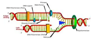
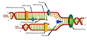
The result of DNA replication is two DNA molecules consisting of one new and one old chain of nucleotides. This is why DNA replication is described as semi-conservative, half of the chain is part of the original DNA molecule, half is brand new. DNA replication requires other enzymes in addition to DNA polymerase, including DNA primase, DNA helicase, DNA ligase, and topoisomerase.

rr
Image credit-
DNA ReplicationWikipedia

The process of replication includes-

  • Unzipping of the helical structure of the DNA molecule
  • This is done by enzyme called helicase that helps in getting the hydrogen bonds broke down by holding the base together.
  • The separating of the stand ceased a shape of Y called replication fork and acts as a template.
  • One of the strands is direction to 3’ to 5’ called the leading stands and the other is away called the lagging stands. Thus both the stands are differently replicated.
  • Then a short part called primer comes to plat helping the lagging part bind acting as the staring part.
  • DNA polymerase the binds the leading and works with it adding the complementary stands.
  • The process of DNA replication is continuous.

Polymerase

The researches take the help of the power that this enzyme holds to copy the molecules in tubes with polymerase chain reaction.

The basic role of this enzyme is to efficiently and by all accuracy help in replicating the genome so that it shall ensure the originality of the genetic data and it’s faithful to transfer via generation. It is useful during the process of DNA replication.

It is useful in gathering the molecules of both DNA and RNA by tallying the template stands and using the method of base paring interaction or also by using the half ladder replication process of RNA. It also can take part in polymerase chain reaction. Taq DNA polymerase is the most common enzyme used for PCR amplification. This enzyme is extremely heat resistant with a half-life of 40 minutes at 95°C. 

This is an enzyme that can be either is independent or dependent of template. The classification of it can be based on its structure or functions. As mentioned in the model of base paring by Watson and Crick, they helps in catalyzing the synthesis of both RNA and DNA after paring via complement base to the parent template. On April 16, 1956, about 60 years ago, Arthur Kornberg and his team of biochemists were the first to isolate and later characterize the enzyme which is now known as DNA polymerase I.

pp
Image credit- PolymeraseWikipedia

It is not just useful in getting the replication of DNA done but also helps in repair and in some times also helps in getting the cell differentiated. It helps in getting the synthesis of polydeoxyribonucleotides from the mono-deoxyribonucleoside triphosphates in DNA.

DNA polymerase is an essential component for PCR as to its key role in synthesizing new DNA strands. Consequently, understanding the characteristics of this enzyme and the subsequent development of advanced DNA polymerases is critical for adapting the power of PCR for a wide range of biological applications

Also Read:

The Intricate Structure of Cytosine: A Comprehensive Exploration

structure of cytosine

Cytosine, a fundamental nucleobase found in both DNA and RNA, has a well-defined and extensively studied structure. As a pyrimidine derivative, cytosine consists of a six-membered ring with two nitrogen atoms and four carbon atoms. Its molecular weight is approximately 111.12 g/mol, and it contains an amino group (-NH2) attached to the C4 carbon atom and a carbonyl group (-C=O) attached to the C2 carbon atom.

The Hydrogen Bonding Capabilities of Cytosine

Cytosine is a polar molecule, which allows it to form hydrogen bonds with other nucleobases. Specifically, cytosine forms two hydrogen bonds with guanine, its complementary base in DNA. The bond length between the N1 nitrogen atom in cytosine and the N3 nitrogen atom in guanine is approximately 0.29 nm, while the bond length between the O2 oxygen atom in cytosine and the N1 nitrogen atom in guanine is approximately 0.28 nm.

Cytosine Modifications: Expanding the Structural Diversity

structure of cytosine

Cytosine can undergo various chemical modifications, which can alter its structure and chemical properties. These modifications include:

Methylation

The methylation of cytosine at the C5 carbon atom results in the formation of 5-methylcytosine. This modification has been linked to diverse biological processes and disease states. The molecular weight of 5-methylcytosine is approximately 125.14 g/mol, which is slightly higher than that of cytosine due to the addition of a methyl group (-CH3).

Hydroxymethylation

Another modification is hydroxymethylation, which leads to the formation of 5-hydroxymethylcytosine. This modification is involved in the cytosine demethylation pathway and may have distinct biological roles. The molecular weight of 5-hydroxymethylcytosine is approximately 139.16 g/mol, which is higher than that of cytosine due to the addition of a hydroxymethyl group (-CH2OH).

Formylation and Carboxylation

In addition to methylation and hydroxymethylation, cytosine can also undergo formylation and carboxylation modifications. These modifications can further diversify the structural and chemical properties of cytosine, potentially impacting its biological functions.

Affinity Tagging of Cytosine

Cytosine can be modified by the addition of affinity tags, such as biotin, which can enable the enrichment and isolation of DNA fragments containing the modification or modifications of interest. The incorporation of biotin tags can be achieved using enzymatic methods, such as those employed in the modular affinity-labeling approach.

Analytical Approaches for Cytosine Modification Analysis

The various modifications of cytosine can be quantified and analyzed using a range of analytical approaches, including:

  1. Mass Spectrometry: This technique can provide precise quantification of cytosine modifications, such as methylation and hydroxymethylation, at the single-nucleotide level.

  2. Quantitative PCR (qPCR): This method can be used to measure the relative abundance of specific cytosine modifications in DNA samples.

  3. Sequencing: Advanced sequencing technologies, such as bisulfite sequencing and oxidative bisulfite sequencing, can map the distribution and patterns of cytosine modifications across the genome.

Biological Significance of Cytosine Modifications

The modifications of cytosine have been linked to diverse biological processes and disease states. For example, the methylation of cytosine has been associated with gene silencing, genomic imprinting, and the development of various cancers. The hydroxymethylation of cytosine, on the other hand, has been implicated in the cytosine demethylation pathway and may play a role in epigenetic regulation and cellular differentiation.

Conclusion

The structure of cytosine is a fundamental aspect of molecular biology, and its various modifications add to the complexity and diversity of this essential nucleobase. Understanding the intricate details of cytosine structure and its modifications is crucial for unraveling the underlying mechanisms of gene expression, epigenetic regulation, and disease pathogenesis. The analytical approaches discussed in this blog post provide valuable tools for researchers to explore the dynamic and multifaceted nature of cytosine in biological systems.

References:
– In situ Quantification of Cytosine Modification Levels in Human Cell Lines. NCBI. https://www.ncbi.nlm.nih.gov/pmc/articles/PMC10366683/
– Genome-Wide Discriminatory Information Patterns of Cytosine DNA Methylation. NCBI. https://www.ncbi.nlm.nih.gov/pmc/articles/PMC4926471/
– DNA Cytosine. ScienceDirect. https://www.sciencedirect.com/topics/chemistry/dna-cytosine
– Cytosine Derivative. ScienceDirect. https://www.sciencedirect.com/topics/pharmacology-toxicology-and-pharmaceutical-science/cytosine-derivative
– Modular affinity-labeling of the cytosine demethylation base excision repair pathway in DNA. Nature. https://www.nature.com/articles/s41598-020-76544-x

9+ Function of Cytosine: Why, How and Detailed facts

jj

Cytosine is considered one among the nucleotides bases in both of the nucleic acids mainly DNA and RNA.

Cytosine is of the four bases in DNA and RNA with the function of cytosine being controlling of the hereditary material for the living cells and also in some serving as a coenzyme that shall act in conjunction with the enzymes in body.

The bases can be a derivative of both pyrimidine and purine. Cytosine on the other hand is a derivative of pyrimidine along with thymine or uracil and is seemed to be an aromatic heterocyclic ring which have two of the substitutes linked up. There is an amine group attached at the 4th place and a keto group linked at the second position.

Cytosine is a pair of guanine and is vital in playing a metabolite for human. It is a metabolite of the Escherichia coli and a referral for mouse metabolite along with also for Saccharomyces cerevisiae. Cytosine helps in controlling many of the work in chemical reaction and getting data changed.

All of the bases do have a nucleoside of their own and the one for cytosine is called the cytidine. Considering the function of cytosine and the base pairing in the model of Watson and Crick, cytosine is seen to make there of the hydrogen bonds along with another base called the guanine that is the purine derivative. Aromaticity is a chemical property in which a conjugated ring of unsaturated bonds, lone pairs, or empty orbitals exhibit a stabilization stronger than would be expected by the stabilization of conjugation alone.

Cytosine can be found as a portion of DNA or RNA or even as a portion of any nucleotide. As a part of cytidine triphsophate it can play a part of co enzyme and can let the start for conversion of adenosine diphosphate to adenosine triphosphate. Cytosine is seemed to pair up with guanine in both the acids. In organic chemistry, a substituent is an atom or group of atoms substituted in place of a hydrogen atom on the parent chain of a hydrocarbon.

function of cytosine
Image credit-
CytosineWikipedia

Structure of Cytosine

As a base of nitrogen, cytosine is made of three of the nitrogen atoms. It is also made of one carbon ring that makes it is a base of pyrimidine.

On the other hand purine is made of two carbon rings. Cytosine is referred to as pyrimidine along with thymine in DNA and the rest two are stated to be purine in DNA. RNA also has two of the pyrimidine, they are uracil and cytosine.

Inside the DNA the base of thymine and adenine are there in similar percentage and do always link together with each other. It then leaves the rest of them being cytosine to pair up with the guanine which is a double ring structure. Cytosine pairs with guanine in both DNA and RNA. Cytosine can sometimes loose electrons by mistake and become uracil. This is known as spontaneous deamination. If DNA repair enzymes such as uracil glycosylase do not repair the damage by cleaving uracil in DNA, a point mutation may result.

Adenine (A) and Guanine (G) both have a double ring structure and are called purines. Thymine (T) and Cytosine (C) both have a single ring structure and are called pyrimidines. Because of their structural similarity, we usually refer the nine-member double rings adenine and guanine as purines, and six-member single-ring thymine, uracil, and cytosine are pyrimidines.

At the time when cytosine is one of the strands in the nucleic acid, the rest of the strands will be definite in having guanine to show up its bond and match. They are companions as they fit perfect collaborating with three bonds of hydrogen. Cytosine can also transfer easily into other bases and thus call the wild card base. Nucleotides containing cytosine can power chemical reactions and are involved in signaling inside the cell.

DNA is said to store information that helps in making and building of the cell. The transmission of the code is a task and the data for all the genetic transfer is stored in cytosine. It is replicated while the cell gets a duplicate copy and then makes the cell to divide. Heterocyclic compounds are organic compounds (those containing carbon) that contain a ring structure containing atoms in addition to carbon—such as sulfur, oxygen, or nitrogen—as part of the ring.

jj
Image credit-
Heterocyclic compoundsWikipedia

Function of cytosine

In the meteorites, there has been no traces of the base cytosine and thus its suggestion states that the first stands of the DNA or RNA had to be seen else to take the building block. 

Cytosine can be said to be a part of the nucleotide which is a molecule having a sugar and one or more of the phosphates. The time when the nucleotide gets connected they form the DNA and RNA said to the nucleic acids.

Cytosine recently found use in quantum computation. The first time any quantum mechanical properties were harnessed to process information took place on August 1st. It is termed to be a third in terms of codon in the RNA, cytosine is the synonym for uracil and are said to be interchangeable as known for its third base. When it is found to be in the second position in any codon, it is changeable.

An example for this can said to be UCU, the UCG, UCA and UCC and all of them are serine despite any one of them being the third base. There is also an enzymatic deamination that is active in the cytosine. Cytosine can be said to be 5-methycytosine by the APOBEC family of the cytosine. Cytosine-C5 DNA MTases catalyze the transfer of the methyl group from a cofactor molecule S-adenosyl-l-methionine. Purine biosynthesis differs from pyrimidine biosynthesis in that purines are formed first as a nucleotide, whereas pyrimidines are formed first as a free base

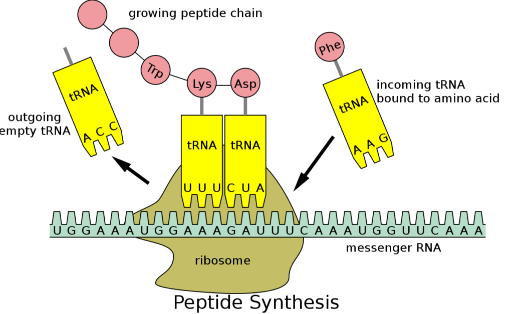
Codon in genetics is a rule set that is utilized for all the living ce;;s to convert the information encoded inside the genetic material and convert into proteins. The transfer in obtained by ribosome that links to the protein genic amino acid that is specific to the messenger RNA and uses the transfer RNA to carry them. Cytosine undergoing modifications are often located in clusters which are therefore repetitive in nature. Short-read based methods are unable to uniquely map short reads to repetitive regions, resulting in the under representation of these important regions.

It can be both useful and harmful for all the implications imposed on several processes of the cell as well as for the evolution in the organism. The outcome of the deamination onto the 5-hydromethylcytosine on the rest part seems to be hard and harmful to deal with. The coding of it is same to that of the entire organism. It is a pyrimidine base C4H5N3O that codes genetic information in the polynucleotide chain of DNA or RNA — compare adenine, guanine, thymine, uracil.

mmm
Image credit- Messenger RNAWikipedia

The function of cytosine is-

  • Each of the cytosine takes part in building of code for the cell
  • It links with the other side guanine base and can also get itself changed to uracil. Cytosine forms three hydrogen bonds with guanine in DNA and RNA. This device, however, is unstable and can transform into uracil.
  • Mostly inside the cell cytosine has a linked up extra group of methyl as a chemical
  • They can also get into modifying of its bases that shall carry the epigenetic data.
  • It can also be a carrier in having their energy carried out and also being a cofactor of CTP.
  • Cytosine can help carry the genetic data for the molecules they have.
  • Aromaticity is a chemical property in which the stability of a conjugated ring of unsaturated bonds, lone pairs, or empty orbitals is greater than can be predicted from conjugation alone.
  • Cytosine forms the nucleoside cytidine when it binds to ribose, and deoxyribose forms deoxycytidine when it binds to deoxyribose.
  • The methylation of this in cytosine is said to get the genes regulated to help them turn themselves on and off whenever needed.
  • It’s a pyrimidine nucleobase, pyrimidone, and aminopyrimidine all rolled into one. The molecule has a planar shape, and in the DNA double helix, cytosine forms three hydrogen bonds with Guanine

Also Read: