35+ Heterotrophs Examples: Detailed Explanations And Images

This article will focus on the following heterotrophs examples, along with a brief description and some images to help you learn more about each one.

All animals, humans, protozoa, fungi, and some iron-reducing bacteria are included in heterotrophs examples. 

The word “Heterotroph” is derived from two Greek words: Hetero, which denotes ‘other’, and Trophe, which denotes ‘nourishment’. So, heterotrophs are organisms that get their energy and nutrients from other sources (organisms or plants). 

These organisms belong to primary, secondary, and tertiary consumers of the food chain. As a response, they feed on the food chain’s primary, secondary, and tertiary producers. On the other hand, heterotrophs get their food from other sources of organic carbon, primarily animal and plant matter, to maintain ecosystem equilibrium.

As the second and third levels of organisms in the ecosystem, heterotrophs are classified as;

herbivores(Organisms that rely solely on plant products for their survival)

carnivores(organisms that only eat the flesh of other organisms for nutrition)

omnivores(Organisms that feed on the flesh of other animals as well as plant products)

and detritivores/decomposers(Soil microorganisms that eat dead plants and animals and feces). 

These organisms, unlike autotrophs, are unable to synthesize their own food; therefore, they must rely on autotrophs for nourishment. Soil microorganisms that eat dead plants and animals, as well as feces.

Photoheterotrophs(organisms that obtain their light energy and therefore must ingest carbon from other organisms because they are incapable of operating natural carbon dioxide from the atmosphere) 

and Chemoheterotrophs(organisms that rely on other species for their carbon and energy) are two subgroups of heterotrophs. The energy source distinguishes these two subgroups that they consume as food.

Heterotrophs Examples

All animals, humans, protozoa, fungi, and some iron-reducing bacteria are included in heterotrophs examples. 

1. Humans as Heterotrophs Examples

  • Humans are heterotrophs since they are unable to synthesize their own food; hence they rely on primary producers and other animals for their food and nutrients.
  • Depending on their food preferences, humans can be vegetarians (eat solely plant products) or non-vegetarians (consume meat from other animals).
  • So, humans are omnivores who consume and process food made from plant products (vegetables, fruits, etc.) and meat from other animals and animal products such as milk, honey, and so on.
  • Nowadays, Humans can choose to be vegans who are pure vegetarians, which means they do not consume animal products such as milk, honey, or meat in their meals. Hence, they only rely on plant products.
Humans as heterotrophs Examples
Humans as heterotrophs Examplesistock

Animals as Heterotrophs Examples

  • All animals are heterotrophs because they do not synthesize their own food and lack chlorophyll pigments, which are required for photosynthesis.
  • Heterotrophs include all herbivores, carnivores, and omnivores. They are the most important heterotrophic group in the ecosystem’s food chain. They get all of their food from primary producers. This category of example includes everything from a rat to an elephant. 

Let’s have a look at some heterotrophic animals.

2. Birds

Birds are animals having wings that rely on primary producers and some living organisms for food.

3. Aquatic Animals

Fish, octopuses, snails, and other aquatic animals are heterotrophs, meaning they eat plants and other aquatic animals for nourishment.

Aquatic animals as a Heterotrophs Example
Aquatic animals as a Heterotrophs Example – istock

4. Goat (Herbivore)

Goats are herbivores because they are heterotrophic animals that eat only plant items.

5. Lion (Carnivore)

Lions are land animals that are also carnivores, meaning they devour the meat of other animals for food.

6. Bear (Omnivore)

Bears are also heterotrophic land animals and omnivores, meaning they eat both plants and other animals for food.

7. Insects

Insects are the most abundant group of organisms in this ecosystem, with over one million species. They consume plant material, degraded organic matter, and other organisms’ blood. Insects are predators, which means they eat other little species smaller than themselves.

Insects as a Heterotroph examples
Insects as a Heterotroph examples- wikipedia

8. Ferrous Iron-Reducing Bacteria

  • Ferrous iron-reducing bacteria generate energy by metabolizing reduced iron to oxidized iron compounds under anaerobic conditions. 
  • The energy gained from this process is subsequently used for carbon source uptake and metabolism.

9. Cyanobacteria (Photoheterotrophic)

  • The Cyanobacteria are photoheterotrophic bacteria in nature exhibiting high photosynthetic capacity and minimal growth needs. 
  • These bacteria live in soggy wet settings, feeding on the organic substances produced by primary aquatic species. 

Fungi as Heterotrophs Examples

Fungi are an organo-heterotrophic group of organisms that rely on dead and decaying items for nutrition and energy.

10. Mushrooms

All mushrooms are parasitic, which means they feed on rotting and dead matter. They act as decomposers, which are important for the environment because they clean the ecosystem and keep it in balance.

Mushroom as a heterotroph
Mushroom as a heterotroph – Wikipedia

11. Yeast

Yeasts are fungi that feed on sugar. Yeast is a heterotrophic microorganism that depends completely on supplies from all other organisms for their metabolism.

12. Molds

Mold, also known as Hyphomycetes, is a living creature that acts as a decomposer. They lack chlorophyll and are unable to synthesize their own nutrition. The molds consume bread, rotting fruits, cheese, and other items.

13. Stinkhorns

Stinkhorns eat rotting or dead organic plant matter inside the soil. They operate as a decomposer and contribute to a cleaner habitat on the planet.

14. Truffles

The truffles fungi are ectomycorrhizal fungi. It feeds on the tree’s sugars, which are generated during photosynthesis. 

15. Rusts

Rust fungi are obligate parasitic fungi that are widespread. It eats corn, legumes, cereals, wheat, maize, and other crops.

Rusts (Fungus
Rusts (Fungus) – wikipedia

16. Smuts

Smuts are fungi that feed on maize plants, infecting and destroying them after feeding.

17. Mildews

Mildew is a fungus belonging to the Erysiphales order. It eats cellulose and other plant matter.

Protozoa as Heterotrophs Examples

Many protozoa are heterotrophic in nature, meaning they eat bacteria, fungi, algae, and yeast to survive.

18.  Paramecium

Paramecium is a single heterotrophic organism. Bacteria, tiny protozoa, yeast, and algae are all common foods.

19. Amoeba

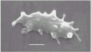
Some amoebae are predators that feed on protists and bacteria, while others are detritivores that feed on dead organic matter.
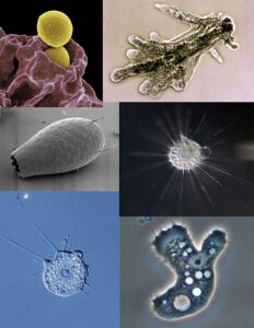
Amoeba
Amoeba- Wikipedia

20. Trypanosoma

Trypanosomes are protozoa that eat by taking nutrients from the host’s bodily fluids across their outer membrane.

21. Euglena

Euglena is single-celled protozoans, and their food is in the form of tiny, microscopic microorganisms. Interestingly, it also feeds itself. 

Heterotrophic Plant Examples

Apart from heterotrophic organisms, the environment comprises heterotrophic plants that do not produce their own food through photosynthesis. The nourishment for these heterotrophic plants derives from external sources. Plants that are parasitic or saprophytic may undergo this category. 

Heterotrophic plants are very different from autotrophic plants. Heterotrophic plants do not have chlorophyll or photosynthetic pigments required for photosynthesis for synthesizing food.

Heterotrophic plants are classified into five categories based on the nourishment they receive. And they include- Plant parasites, Symbionts, Epiphytes, Saprophytes, and Insectivorous Plants. 

So, the following are some Heterotrophic Plant Examples under these five categories.

Plant Parasites as Heterotrophic Plant Examples

22.  Viscum Album

Viscum Album is a hemiparasite tree that absorbs water and nutrients from various other trees. 
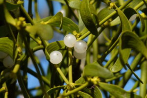
Viscum Album
Viscum Album – Wikipedia

23. Stinking Corpse Lily

Stinking Corpse Lily lacks roots and leaves and thus lacks chlorophyll, all required for photosynthesis. As a result, they must rely on parasitism to receive nutrients and water.

24.  Orobanche Ramosa

Orobanche ramosa is a parasitic plant that feeds on the nutrients of other plants. It is a heterotrophic plant since it drains nutrients from its roots and without leaves and chlorophyll, which they lack in performing photosynthesis.

Symbiotic Plants as Heterotrophic Plant Examples

25.  Psilotum

Psilotum does not have leaves or true roots but relies on encroaching rhizomes for support. On the other hand, the stems are receptacles that contain photosynthetic and transporting tissue. However, it is a symbiotic plant with myco-heterotrophic and is nourished by endophytic fungus.

26.  Marigold

Marigold is a flowering heterotrophic plant that also acts as a symbiont. The heterotrophic marigold plant and its host plant profit from their relationship in symbiosis, as marigolds bloom with tomatoes, Brussels sprouts, cauliflower, and other plants when planted together in a pot.

27.  Rosemary

Rosemary is a heterotrophic symbiont plant that may grow alongside carrots, radish, sage, and other plants, forming a symbiotic relationship in which both plants benefit. It gets its nutrients from other plants.

Epiphytic Plants as Heterotrophic Plant Examples

28.  Orchids

About 70% of orchids are epiphytic in nature, which means they grow on other plants and absorb their nutrients from that host plant for growth and nourishment.

29. Bromeliads

Bromeliads are epiphytic heterotrophs that attach themselves to the outsides of other live plants. When they attach themselves to other trees, they gain more nutrients and light exposure for photosynthesis; however, they do not directly extract nutrients from other trees.
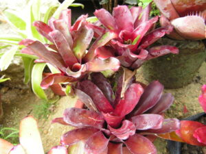
Bromeliad
Bromeliad – Wikipedia

30.  Mosses

Mosses are another type of epiphytic plant that is heterotrophic in nature. It eats plant secretions or dead tissues, such as the skin peeled cells produced in the early stages of root formation.

Saprophytic Plants as Heterotrophic Plant Examples

31.  Ghost Plant

Indian pipe, popularly known as ghost plants, is a heterotrophic saprophyte in nature. Because it lacks chlorophyll, it does not transform energy from the sun to nutrition the way green plants generally do. Saprophytic plants, such as ghost plants, feed themselves by draining the sap from another plant.

32.  Corallorhiza Orchids

Corallorhiza Orchids are completely myco-heterotrophic saprophytes since they rely on the mycorrhizal fungi that surround their roots for nutrition.
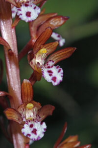
Corallorhiza Orchids
Corallorhiza Orchids – wikipedia

33.  Burmannia

Burmannia is a saprophytic heterotrophic plant that feeds on its surroundings. It feeds itself by absorbing nutrients from host saprophytes adhered to its bod

Insectivorous Plants as Heterotrophic Plant Examples

34.  Venus Flytrap

The Venus flytrap is a carnivorous autotrophic insectivorous plant. However, it is considered a heterotrophic plant due to its complex nature. It has the ability to generate energy from sunlight. The flies and insects are beneficial for their nutrition, but they are unnecessary for their existence.

35.  Drosera Capensis

Drosera Capensis is a partial heterotrophic insectivorous plant that derives nitrogen from insects that roam in its surroundings by eating them. And their leaves, including chlorophyll, conduct photosynthesis for their nourishment. As a result, it is partly autotrophic and partly heterotrophic.
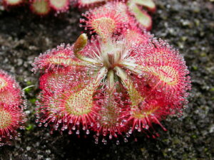
Drosera
Drosera – Wikipedia

36.  Common Butterwort

Since common butterwort is an insectivorous plant, it is heterotrophic in nature. It contains bright, appealing flowers that attract insects and attractive yellow-green leaves that exude a sticky fluid that catches insects which the plant itself then eats.

Also Read:
23 Autotrophs Examples: Detailed Explanations And Images

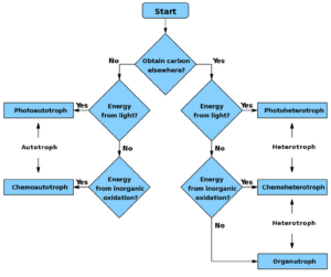
AutoHeteroTrophs flowchart 300x251 1

Here, we will be discussing about different examples of autotrophs.

Autotrophs(where Auto means Self and Trophe means Feeding) are lifeforms that prepare their own food with the help of water, sunlight, carbon dioxide, and other chemical substances. To know about it more, go through the autotrophs examples down below.

Now, let us understand the concept more by relevant examples that are based on these two food preparation processes by the autotrophic organisms.

Some examples of autotrophs that utilize photosynthesis mode:-

Some examples of autotrophs that utilize chemosynthesis mode:-

Autotrophs(where Auto means Self and Trophe means Feeding) are lifeforms that prepare their own food with the help of water, sunlight, carbon dioxide, and other chemical substances. To know about it more, go through the autotroph examples down below.

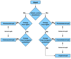
Flow Chart of autotroph and heterotroph process
Flow Chart of autotroph and heterotroph process- wikipedia

They are generally referred to as producers because they can produce their own food. There are various types of autotrophs in the ecosystem, each with its own way of preparing food. Photosynthesis and Chemosynthesis are two of the food preparation processes. 

As a result, most autotrophs (all green-leaved plants) prepare their nutrition through photosynthesis, which converts light energy from the sun into a nutrient called glucose. It is then by converting water from the soil and carbon dioxide from the environment. These are also known as phototrophs.\

On the other hand, rare autotrophs use the chemosynthesis process to synthesize food rather than relying on solar energy. Instead, they use chemical reactions to produce food, like frequently mixing hydrogen sulfide or methane with oxygen. Chemosynthetic organisms (or Chemotrophs) survive in harsh environments where toxic chemicals required for oxidation are plentiful.

Plants are the major suppliers of food. Plants synthesize nutrients from various elements acquired from the air and the soil. This series of elements also includes nitrogen. Plants use protein biosynthesis to acquire nitrogen from the soil. 

Photosynthesis (Phototrophs) Examples:

1) Green Algae

Photosynthesis produces nutrients for the organism’s growth in the cells of green algae. Photosynthesis needs the participation of both light and carbon dioxide. Green algae absorb sunlight using chlorophyll, a green component that gives them their green color.

Green Algae as autotrophs examples
Green Algae as autotrophs examples from wikipedia

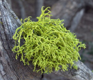
2) Lichens 

It produces its food and is not reliant on other organisms. It also functions as a heterotroph. But because of its symbiotic association with algae and fungi, its plant body is entirely covered in green chlorophyll, making it a photoautotrophic organism. In contrast, lichen is a source of energy for various heterotrophs.

Lichens
Lichens from source – wikipedia

3) Grass

The grass is green in color because it has chloroplasts present in its cell, which are responsible for photosynthesis. However, it is regarded as a primary producer and a phototroph as a field plant.

4) Pinnularia 

It is a form of planktonic algae that is phototrophic because it has chloroplasts, which allow it to photosynthesize.

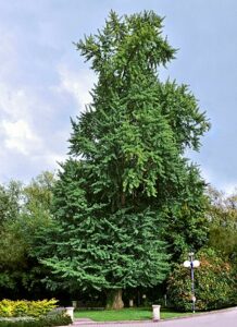
5) Gingko biloba

It is a phototrophic gymnosperm with green leaves with chloroplasts. It is also a single living species.

Gingko Biloba
Gingko Biloba from source- wikipedia

6) Mango (dicotyledonous plant)

It is a photoautotrophic plant, meaning it can prepare its food using chlorophyll and is not reliant on others for nutrition.

7) Plant of Bougainvillea

Although it is covered in pinkish blooms, it is a dicotyledonous plant with green leaves containing chlorophyll. That indicates a photoautotrophic plant.

8) Ferns

Ferns are primarily photoautotrophs or light-loving plants. They use light as a source to produce organic molecules like product glucose.

9) Liverworts (Bryophyte)

More than over 9,000 varieties of small nonvascular spore-producing plants found in the environment are classified as liverwort. As a result, they exhibit an autotrophic method of nutrition.

Liverworts
Liverworts – Wikipedia

10) Cyanobacteria (Photosynthetic Bacteria)

Cyanobacteria is a photoautotrophic prokaryote with a broad and diverse number of organisms. A specific combination of pigments defines their potential to undertake photosynthesis and respiration.

11) Phytoplankton

Phytoplankton includes everything from photosynthesizing bacteria to algae – cyanobacteria. Autotrophs are small organisms that dwell in the ocean.

12) Dinoflagellates

Dinoflagellates can be autotrophic, heterotrophic, or mixed in their mode of nutrition. About 50% of these species are photosynthetic, yet most are predatory.

Autotrophic microorganisms that are chemoautotrophs are extremophile bacteria. They thrive in severe environments where light cannot easily pass through. The fundamental source of plant nourishment is the spontaneous fixation of atmospheric carbon dioxide into simple sugars. 

Chemosynthesis (Chemotrophs) Examples:

1) Methanococcus

Methanogens or bacteria produce huge amounts of methane during the decomposition of organic matter by the process of Chemosynthesis. Methanococcus is a type of methanogen which is also an autotroph.

2) Methanospirillum

It is another form of methanogen that does not require light to generate its food and instead relies on chemical compounds to convert inorganic to organic materials.

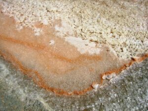
3) Dunaliella Salina

It’s a halophile, a halophilic green microalga that Chemosynthesis its food as an obligate autotroph.

Dunaliella Salina
Dunaliella Salina from wikipedia

4) Wallemia Ichthyophaga

It is one of three fungal species in the genus Wallemia; they have moderate nutritional requirements but require a lot of sodium ions for development and metabolism, which is why they are called chemotrophs.

5) Thermoplasma

These are both thermophilic and acidophilic, which means they can grow in high temperatures and low pH environments. So, they consume food through the chemosynthesis process.

Thermoplasma
Thermoplasma from wikipedia

6) Sulfolobus

It belongs to the Thermoacidophiles family. Sulfolobus is a facultative autotrophic genus that thrives at 70°C to 87°C with a pH of 2. 

7) Nitrospira

They are chemo-autotrophic organisms, making their food by converting nitrogen into ammonia or other forms. They collect nitrogen from the atmosphere and use it to generate energy through oxidation processes.

8) Nitrosomonas

Nitrifying bacteria break down ammonia, the most reduced form of nitrogen in the soil, to nitrate as the most oxidized form.

9) Beggiotoa

Sulfur-oxidizing bacteria are colorless and have high efficiency in food production. Reduced sulfur compounds are frequently generated because of anaerobic heterotrophic respiration with sulfate. However, some waterways receive significant sulfide inputs from underground.

Beggiotoa
Beggiotoa from wikipedia

10) Chromatiaceae (purple-sulfur bacteria)

It prepares its nutrition by converting sulfur and components to sulfates using light energy in an o2-free environment.

11) Acidihalobacter Properus

Yet it is another chemotroph, as it uses chemical substances such as purple-sulfur bacteria to synthesize its own nutrition.

12) Sphaerotilus

An iron-oxidizing bacteria gets its energy from oxidizing ferrous iron in the water. The underwater periphyton Sphaerotilus natans is connected with contaminated water.

Please click to learn more about Chemoautotrophs Examples.

Also Read:

Does the Nucleus Have a Double Membrane?

does nucleus have double membrane

The nucleus of a eukaryotic cell is surrounded by a double-membraned nuclear envelope (NE), which is composed of two distinct membranes: the inner nuclear membrane (INM) and the outer nuclear membrane (ONM). These membranes are separated by the perinuclear space, which is continuous with the lumen of the endoplasmic reticulum (ER). The NE is perforated … Read more

Are Lysosomes Organelles? A Comprehensive Guide

are lysosomes organelles

Lysosomes are membrane-bound organelles found in eukaryotic cells that play a crucial role in cellular homeostasis by breaking down and recycling various biomolecules. They are highly dynamic organelles involved in several cellular functions beyond their well-known degradative function, including nutrient sensing, intracellular signaling, and metabolism.

Morphological Analysis of Lysosomes

Morphological analysis of lysosomes can be done using various methods such as:

  1. Manual Outlining of the Cell and Nuclear Membrane: This method involves manually tracing the cell and nuclear membranes to define the boundaries of the cell and nucleus, which can then be used to quantify the distribution and morphology of lysosomes within the cell.

  2. Automatic Segmentation: Automated image analysis techniques can be used to segment and identify lysosomes within cells, providing quantitative data on lysosome morphology, positioning, motility, and function.

  3. Calculating the Fraction of Total Lysosomes per Subcellular Region of Interest: This method involves dividing the cell into different subcellular regions and calculating the fraction of total lysosomes present in each region, providing insights into the spatial distribution of lysosomes within the cell.

These methods can provide valuable quantitative data on various aspects of lysosome biology, including their size, shape, and distribution within the cell.

Lysosomal Positioning

are lysosomes organelles

Lysosomal positioning can be assessed by calculating the fraction of signal in the peri-Golgi area, defined by a Golgi marker such as giantin. This method involves measuring the percentage of total lysosome marker fluorescence intensity in the region surrounding the Golgi apparatus, providing information on the distribution of lysosomes in relation to this organelle.

The peri-Golgi region is an important area for lysosome biogenesis and trafficking, as newly formed lysosomes are often transported from the Golgi to their final destination within the cell. By quantifying the fraction of lysosomes in this region, researchers can gain insights into the dynamics of lysosome positioning and their relationship with the Golgi apparatus.

Lysosomal Morphometric Assays

Lysosomal morphometric assays can be performed using high-content image analysis techniques, such as the Opera Phenix High-Content Screening System (PerkinElmer). These assays can be used to calculate the percentage of cells showing a clustered lysosomal distribution versus cells showing lysosomes correctly distributed throughout the cell.

This method provides quantitative data on lysosomal distribution in normal or pathological conditions, allowing researchers to identify changes in lysosomal organization that may be associated with various disease states or cellular perturbations.

Lysosome Immunopurification

Lysosome immunopurification can be used to characterize the full molecular inventory of lysosomes using modern ‘omics’ technologies, such as mass spectrometry-based proteomics. This method involves isolating and purifying lysosomes from cells, followed by the identification and quantification of the proteins present within these organelles.

By using this approach, researchers can obtain comprehensive data on the protein composition of lysosomes, including the identification of novel lysosomal proteins and the characterization of their functions. This information can provide valuable insights into the diverse roles of lysosomes in cellular processes and their involvement in various disease pathways.

Lysosomal Enzyme Activity Measurements

Lysosomal enzyme activity can be measured using imaging-based protocols that provide insights at the cellular level. These methods can be used to quantify functions essential to lysosomal biology, including:

  1. β-glucosidase Enzymatic Cleavage: Measuring the activity of this enzyme, which is involved in the breakdown of glucosylceramide, can provide information on lysosomal function and its potential dysregulation in diseases such as Gaucher’s disease.

  2. Active Cathepsin D: Cathepsin D is a lysosomal protease that plays a crucial role in protein degradation. Quantifying its activity can offer insights into the proteolytic capacity of lysosomes.

  3. pH Regulation: Maintaining the acidic pH within lysosomes is essential for their proper function. Imaging-based protocols can be used to measure lysosomal pH in real-time, providing data on the pH regulation mechanisms within these organelles.

These methods can generate quantitative data on lysosomal enzyme activity and pH regulation, which are essential for understanding the overall function and homeostasis of lysosomes in various cellular contexts.

Conclusion

In summary, lysosomes are highly dynamic organelles involved in a wide range of cellular functions, including nutrient sensing, intracellular signaling, and metabolism, in addition to their well-known role in the degradation and recycling of biomolecules.

Researchers have developed a variety of techniques to study the morphology, positioning, distribution, and enzyme activity of lysosomes, providing valuable quantitative data on these organelles. These methods include manual outlining, automatic segmentation, lysosomal positioning analysis, high-content image analysis, immunopurification, and enzyme activity measurements.

By employing these advanced techniques, researchers can gain a deeper understanding of the complex roles of lysosomes in cellular homeostasis and their involvement in various disease processes. This knowledge can ultimately lead to the development of new therapeutic strategies targeting lysosomal dysfunction.

References

  1. Ballabio, A., & Bonifacino, J. S. (2020). Lysosomes as dynamic regulators of cell and organismal homeostasis. Nature Reviews Molecular Cell Biology, 21(2), 101-118.
  2. Settembre, C., Fraldi, A., Medina, D. L., & Ballabio, A. (2013). Signals from the lysosome: a control centre for cellular clearance and energy metabolism. Nature Reviews Molecular Cell Biology, 14(5), 283-296.
  3. Xu, H., & Ren, D. (2015). Lysosomal physiology. Annual Review of Physiology, 77, 57-80.
  4. Perera, R. M., & Zoncu, R. (2016). The lysosome as a regulatory hub. Annual Review of Cell and Developmental Biology, 32, 223-253.
  5. Luzio, J. P., Pryor, P. R., & Bright, N. A. (2007). Lysosomes: fusion and function. Nature Reviews Molecular Cell Biology, 8(8), 622-632.

Do Cell Membranes Have Lipids?

do cell membrane have lipids

Cell membranes are essential components of all living cells, and they are primarily composed of lipids. These lipids play a crucial role in the structure, function, and regulation of the cell membrane. In this comprehensive blog post, we will delve into the details of the lipid composition of cell membranes, their specific types, and the importance of these lipids in maintaining cellular homeostasis.

The Lipid Composition of Cell Membranes

Cell membranes are composed of a diverse array of lipids, with the primary lipid classes being phospholipids, sphingolipids, and sterols. These lipids are arranged in a bilayer structure, with the hydrophilic (water-loving) head groups facing the aqueous environments on both sides of the membrane, and the hydrophobic (water-fearing) fatty acid tails forming the interior of the membrane.

Phospholipids

Phospholipids, also known as glycerophospholipids, are the most abundant lipids in cell membranes, making up approximately 60% of the total lipid content in mammalian cells. These amphipathic molecules consist of a glycerol backbone, two fatty acid tails, and a phosphate-containing head group. The most common phospholipids found in cell membranes are:

  1. Phosphatidylcholine (PC): Accounts for 40-50% of the total phospholipids in the plasma membrane.
  2. Phosphatidylethanolamine (PE): Comprises 20-50% of the total phospholipids, with a higher concentration in the inner leaflet of the membrane.
  3. Phosphatidylserine (PS): Represents 5-10% of the total phospholipids, with a higher concentration in the inner leaflet.
  4. Phosphatidylinositol (PI): Accounts for 5-10% of the total phospholipids and plays a role in signal transduction.

The specific composition of phospholipids can vary depending on the cell type and the cellular function.

Sphingolipids

Sphingolipids are another class of lipids found in cell membranes, with sphingomyelin (SM) being the most abundant. Sphingolipids are characterized by a sphingosine backbone instead of a glycerol backbone. Sphingomyelin is particularly abundant in the outer leaflet of the plasma membrane, where it can account for up to 50% of the total phospholipids.

Sterols

Sterols, such as cholesterol, are non-polar lipids that play a crucial role in regulating the fluidity and permeability of cell membranes. Cholesterol is the most abundant sterol in mammalian cell membranes, making up approximately 40% of the total lipid content in the plasma membrane of red blood cells. Cholesterol is known to distribute asymmetrically in the membrane, with a higher concentration in the outer leaflet.

Lipid Asymmetry in Cell Membranes

do cell membrane have lipids

The lipid composition of the inner and outer leaflets of the cell membrane is not uniform. This asymmetric distribution of lipids is essential for maintaining the structural and functional integrity of the membrane.

In the case of red blood cell (RBC) membranes, the outer leaflet is primarily composed of sphingomyelin (SM) and phosphatidylcholine (PC), which together account for more than 85% of the phospholipids. The inner leaflet, on the other hand, has a lower percentage of SM and PC (less than 25%) and a higher percentage of phosphatidylethanolamine (PE) and phosphatidylserine (PS), with more than 45% and more than 25%, respectively.

This asymmetric distribution of lipids is maintained by the action of specialized enzymes, such as flippases and floppases, which actively transport lipids between the two leaflets. This asymmetry is crucial for various cellular processes, including signal transduction, membrane trafficking, and the regulation of membrane-associated proteins.

Importance of Lipids in Cell Membranes

The lipids in cell membranes serve several critical functions:

  1. Structural Integrity: The lipid bilayer provides the structural foundation for the cell membrane, allowing it to maintain its shape and integrity.
  2. Permeability and Transport: The lipid composition of the membrane regulates its permeability, controlling the movement of molecules in and out of the cell.
  3. Signaling and Membrane Dynamics: Lipids, such as phosphoinositides and sphingolipids, play crucial roles in signal transduction and the regulation of membrane dynamics, including vesicle formation and fusion.
  4. Membrane Fluidity: The balance of saturated and unsaturated fatty acids, as well as the presence of cholesterol, determines the fluidity of the membrane, which is essential for various cellular processes.
  5. Membrane Protein Function: Lipids can interact with and modulate the activity of membrane-bound proteins, such as ion channels and receptors.

Quantifying Lipid Composition

To determine the lipid composition of cell membranes, various analytical techniques can be employed, including:

  1. Mass Spectrometry: This powerful technique can provide a detailed analysis of the phospholipid species and their relative abundances within the membrane.
  2. Thin-Layer Chromatography (TLC): TLC can be used to separate and quantify the different classes of lipids present in the membrane.
  3. Nuclear Magnetic Resonance (NMR) Spectroscopy: NMR can be used to identify and quantify the different lipid species based on their unique chemical signatures.

By quantifying the lipid composition of cell membranes, researchers can gain valuable insights into the cellular responses to various stimuli, as well as the role of lipids in the development and progression of diseases.

Conclusion

In summary, cell membranes are composed of a diverse array of lipids, including phospholipids, sphingolipids, and sterols. These lipids are essential for the structural integrity, permeability, and functional regulation of the cell membrane. The asymmetric distribution of lipids within the membrane is crucial for maintaining cellular homeostasis and enabling various cellular processes. Understanding the lipid composition of cell membranes is a crucial aspect of cell biology and has important implications in the fields of biochemistry, physiology, and medicine.

References:

  1. Antonio Blanco, Gustavo Blanco, in Medical Biochemistry, 2017.
  2. NCBI – Lipid Asymmetry in Biological Membranes
  3. Nature – Lipid Asymmetry in the Plasma Membrane
  4. NCBI – Lipid Composition of Cell Membranes
  5. BioNumbers – Lipid Abundance in Membranes
  6. Verkleij AJ, Zwaal RF, Comfurius P, Roelofsen B. The asymmetry of the human erythrocyte membrane with respect to lipid composition. Biochim Biophys Acta. 1973 Jan 29;316(2):201-218. doi: 10.1016/0005-2736(73)90178-1. PMID: 4685149.
  7. Lorent JH, Klaasen M, van Meer G, Post JA. Lipid asymmetry of the plasma membrane of human erythrocytes as determined by mass spectrometry. Biochim Biophys Acta. 2007 Feb;1768(2):1129-1138. doi: 10.1016/j.bbalip.2006.11.006. Epub 2006 Dec 12. PMID: 17167559.

Fungi Cell Wall and Plant Cell Wall: A Comprehensive Exploration

fungi cell wall and plant cell wall

The fungal cell wall and plant cell wall are intricate structures that play crucial roles in the survival, growth, and function of their respective organisms. These cell walls are composed of a complex network of polysaccharides, proteins, and other molecules, each with unique properties and functions. Understanding the composition and architecture of these cell walls is essential for various fields, including microbiology, plant biology, and biotechnology.

Fungal Cell Wall: A Fortress of Strength and Versatility

The fungal cell wall is a dynamic and multifunctional structure that provides structural support, protects against osmotic pressure, and facilitates cell-cell recognition and adhesion. The primary components of the fungal cell wall are:

  1. Chitin: A linear polymer of N-acetylglucosamine, chitin is the primary load-bearing component of the fungal cell wall. It forms rigid and insoluble microfibrils that contribute to the overall strength and rigidity of the cell wall.

  2. β-Glucans: These branched polymers of glucose are the most abundant polysaccharides in the fungal cell wall. They are classified into three types: α-1,3-glucan, β-1,3-glucan, and β-1,6-glucan. α-1,3-glucan provides rigidity and hydrophobicity, while β-1,3-glucan and β-1,6-glucan form a well-hydrated and relatively mobile matrix that embeds the chitin fibrils and α-1,3-glucans.

  3. Mannoproteins: These glycoproteins are covalently linked to β-glucans and are found on the cell surface. They play a crucial role in cell-cell recognition and adhesion, as well as in the regulation of cell wall permeability.

The fungal cell wall’s unique composition and architecture allow it to adapt to various environmental stresses and fulfill diverse functions. Solid-state NMR spectroscopy has revealed a two-domain distribution of molecules in the cell walls of the pathogenic fungus Aspergillus fumigatus: a relatively rigid and inner portion composed of glucans and chitins, and an extremely mobile outer shell composed of mannoproteins and α-1,3-glucan.

This architectural arrangement enables the fungal cell wall to reshape its molecular structure to survive through different external stresses, such as changes in pH, temperature, or the presence of antifungal agents. Additionally, the 13C linewidth of fungal polysaccharides has been found to be comparable to that of the matrix polysaccharides in the fast-growing primary plant cell walls, but narrower than that of rigid cellulose microfibrils, indicating a higher degree of molecular mobility and flexibility.

Plant Cell Wall: A Sturdy and Dynamic Structure

fungi cell wall and plant cell wall

In contrast to the fungal cell wall, plant cell walls are primarily composed of cellulose, hemicellulose, and pectin. These components work together to provide structural support, protect the plant cells, and facilitate cell-cell communication and signaling.

  1. Cellulose: This linear polymer of glucose is the main load-bearing component of the plant cell wall. Cellulose microfibrils are arranged in a parallel fashion and are embedded in a matrix of other polysaccharides, providing the cell wall with its characteristic strength and rigidity.

  2. Hemicellulose: A heterogeneous group of polysaccharides, hemicellulose is associated with cellulose and provides additional structural support to the cell wall. Hemicellulose includes xylans, mannans, and glucans, which can interact with cellulose and pectin to form a complex network.

  3. Pectin: A complex mixture of polysaccharides, pectin is found in the middle lamella and the primary cell wall. Pectin plays a crucial role in cell-cell adhesion and signaling, as well as in the regulation of cell wall porosity and permeability.

The plant cell wall’s composition and architecture differ significantly from the fungal cell wall. While the fungal cell wall is more plastic and can reshape its molecular structure, the plant cell wall is more rigid and dehydrated upon maturation. This difference is largely due to the presence of cellulose, a flexible and hydrophilic polymer, in the plant cell wall, as opposed to the rigid and hydrophobic chitin found in the fungal cell wall.

Solid-state NMR spectroscopy has revealed that the 13C linewidth of plant cell wall polysaccharides, such as cellulose microfibrils, is generally broader than that of the fungal polysaccharides, indicating a lower degree of molecular mobility and flexibility.

Comparative Analysis: Insights from Solid-state NMR Spectroscopy

Solid-state NMR spectroscopy has emerged as a powerful tool for the study of the molecular architecture and dynamics of both fungal and plant cell walls. This technique has provided valuable insights into the structural and functional differences between these two types of cell walls.

  1. Molecular Architecture: Solid-state NMR studies on the pathogenic fungus Aspergillus fumigatus have revealed a two-domain distribution of molecules in the cell wall, with a relatively rigid and inner portion composed of glucans and chitins, and an extremely mobile outer shell composed of mannoproteins and α-1,3-glucan. In contrast, the plant cell wall exhibits a more uniform distribution of cellulose microfibrils embedded in a matrix of hemicellulose and pectin.

  2. Molecular Dynamics: The 13C linewidth of fungal polysaccharides has been found to be comparable to that of the matrix polysaccharides in the fast-growing primary plant cell walls, but narrower than that of rigid cellulose microfibrils. This suggests a higher degree of molecular mobility and flexibility in the fungal cell wall compared to the plant cell wall.

  3. Structural Adaptability: The fungal cell wall’s ability to reshape its molecular architecture in response to various environmental stresses and to fulfill diverse functions is a testament to its structural adaptability. In contrast, the plant cell wall is more rigid and dehydrated upon maturation, reflecting its primary role in providing structural support and protection to the plant cells.

These insights from solid-state NMR spectroscopy have significantly advanced our understanding of the composition, architecture, and dynamics of fungal and plant cell walls, paving the way for further research and potential applications in fields such as microbiology, plant biology, and biotechnology.

References:

  1. “Molecular architecture of fungal cell walls revealed by solid-state NMR” (https://www.ncbi.nlm.nih.gov/pmc/articles/PMC6048167/)
  2. “A molecular vision of fungal cell wall organization by functional genomics and solid-state NMR” (https://www.ncbi.nlm.nih.gov/pmc/articles/PMC8566572/)
  3. “Study of fungal cell wall evolution through its monosaccharide composition” (https://www.sciencedirect.com/science/article/pii/S2468233024000094)
  4. “Fungal Cell Wall – an overview | ScienceDirect Topics” (https://www.sciencedirect.com/topics/biochemistry-genetics-and-molecular-biology/fungal-cell-wall)
  5. “The Fungal Cell Wall: Structure, Biosynthesis, and Function” (https://journals.asm.org/doi/10.1128/microbiolspec.funk-0035-2016)
  6. “Plant cell wall composition” (https://www.sciencedirect.com/topics/agricultural-and-biological-sciences/plant-cell-wall-composition)
  7. “Cellulose microfibrils in plant cell walls” (https://www.sciencedirect.com/topics/agricultural-and-biological-sciences/cellulose-microfibrils)
  8. “Hemicellulose structure and function” (https://www.sciencedirect.com/topics/agricultural-and-biological-sciences/hemicellulose)
  9. “Pectin structure and function” (https://www.sciencedirect.com/topics/agricultural-and-biological-sciences/pectin)

Do Eukaryotic Cells Have a Cell Wall?

do eukaryotic cells have a cell wall

Eukaryotic cells, which include plant, animal, and fungal cells, are characterized by the presence of a membrane-bound nucleus and other organelles. While the presence of a cell wall is a defining feature of plant and fungal cells, it is not a universal characteristic of all eukaryotic cells. In this comprehensive guide, we will delve into the intricacies of cell walls in eukaryotic cells, exploring their composition, function, and the variations observed across different cell types.

The Presence of Cell Walls in Eukaryotic Cells

As mentioned in the initial response, the presence of a cell wall is not a universal feature of eukaryotic cells. Cell walls are primarily found in two main groups of eukaryotic organisms: plants and fungi.

Plant Cells and Cell Walls

Plant cells are the quintessential example of eukaryotic cells with a well-developed cell wall. The plant cell wall is a rigid, extracellular structure located outside the plasma membrane, providing structural support, protection, and shape maintenance to the cell.

The plant cell wall is primarily composed of the following key components:

  1. Cellulose: The primary structural component of the plant cell wall, cellulose is a polysaccharide made up of long, linear chains of β-1,4-linked glucose molecules. Cellulose microfibrils, which are long, thin, and crystalline, provide the cell wall with its characteristic rigidity and strength.

  2. Hemicellulose: A diverse group of polysaccharides, including xylan, mannan, and glucan, that interact with and cross-link cellulose microfibrils, further enhancing the structural integrity of the cell wall.

  3. Pectin: A complex polysaccharide that helps regulate the porosity and hydration of the cell wall, as well as facilitating cell-to-cell adhesion.

  4. Lignin: A complex aromatic polymer that is deposited in the secondary cell wall, providing additional strength and resistance to the cell wall, particularly in woody plant tissues.

The thickness of the plant cell wall can vary significantly, ranging from 0.1 to 10 μm for the primary cell wall and up to 20 μm for the secondary cell wall. The specific thickness depends on the plant species, cell type, and developmental stage.

Fungal Cells and Cell Walls

Fungal cells, like plant cells, also possess a cell wall as a defining feature. However, the composition of the fungal cell wall differs from that of plant cells. The primary component of the fungal cell wall is chitin, a polysaccharide made up of N-acetylglucosamine units.

In addition to chitin, the fungal cell wall may also contain other polysaccharides, such as:

  1. Glucans: β-1,3-glucans and β-1,6-glucans, which provide structural support and rigidity to the cell wall.
  2. Mannans: Polysaccharides composed of mannose units, which can be found in the outer layer of the cell wall.
  3. Glycoproteins: Proteins covalently linked to carbohydrate moieties, which can play a role in cell wall organization and function.

The thickness of the fungal cell wall can vary depending on the species and growth conditions, typically ranging from 100 to 400 nm.

Eukaryotic Cells Lacking Cell Walls

While plant and fungal cells possess a cell wall, the majority of animal cells, including human cells, do not have a cell wall. Instead, animal cells rely on the plasma membrane for protection, structural support, and shape maintenance.

The absence of a cell wall in animal cells is a key distinguishing feature between eukaryotic cell types. Animal cells are more flexible and can change shape more readily, as they are not constrained by a rigid cell wall structure.

Functional Roles of Cell Walls in Eukaryotic Cells

do eukaryotic cells have a cell wall

The presence of a cell wall in plant and fungal cells confers several important functional roles, which are crucial for the survival and well-being of these eukaryotic organisms.

Structural Support and Rigidity

The primary function of the cell wall is to provide structural support and rigidity to the cell. The cellulose microfibrils and other polysaccharides in the plant cell wall, as well as the chitin and glucans in the fungal cell wall, work together to create a strong, protective barrier around the cell.

This structural integrity is essential for maintaining the shape and size of the cell, preventing it from bursting or collapsing due to the high internal osmotic pressure. The cell wall also helps to resist mechanical stresses and environmental pressures, such as wind, rain, and pathogen attacks.

Protection and Barrier Function

The cell wall acts as a physical barrier, protecting the delicate plasma membrane and the internal organelles of the cell from various environmental threats, such as:

  1. Pathogens: The cell wall provides a physical barrier against the invasion of bacteria, viruses, and other pathogens, helping to prevent infection and maintain the cell’s integrity.
  2. Mechanical Damage: The cell wall shields the cell from physical damage, such as abrasion, puncture, or compression, which could otherwise compromise the cell’s structure and function.
  3. Osmotic Stress: The cell wall helps to regulate the flow of water and solutes into and out of the cell, protecting it from osmotic shock and maintaining the appropriate internal pressure.

Cell Growth and Division

The cell wall plays a crucial role in the growth and division of plant and fungal cells. As the cell expands during growth, the cell wall must also expand and remodel to accommodate the increasing size of the cell.

In plant cells, the primary cell wall is laid down during the early stages of cell growth, while the secondary cell wall is deposited later, providing additional strength and support as the cell matures.

During cell division, the cell wall must be carefully remodeled and reorganized to allow for the separation of the daughter cells, ensuring that each new cell is equipped with a functional cell wall.

Cell-Cell Adhesion and Communication

The cell wall also facilitates cell-cell adhesion and communication in plant and fungal tissues. The pectin and glycoproteins present in the cell wall can mediate the attachment of neighboring cells, forming a cohesive tissue structure.

This cell-cell adhesion is particularly important in plant tissues, where the cell wall helps to maintain the structural integrity of the plant and allows for the coordinated transport of water, nutrients, and signaling molecules between cells.

Variations in Cell Wall Composition and Structure

While the presence of a cell wall is a common feature of plant and fungal cells, the specific composition and structure of the cell wall can vary significantly among different eukaryotic organisms and cell types.

Variations in Plant Cell Walls

Plant cell walls exhibit a high degree of diversity in their composition and structure, which can be influenced by factors such as plant species, cell type, and developmental stage.

  1. Primary Cell Wall: The primary cell wall is typically thinner and more flexible, composed primarily of cellulose, hemicellulose, and pectin. The relative proportions of these components can vary among different plant species and cell types.

  2. Secondary Cell Wall: The secondary cell wall is deposited after the primary cell wall and is generally thicker and more rigid. It is characterized by the presence of lignin, which provides additional strength and resistance to the cell wall.

  3. Specialized Cell Walls: Certain plant cell types, such as those found in the xylem and sclerenchyma tissues, can have highly specialized cell walls with increased lignification, providing enhanced structural support and water transport capabilities.

Variations in Fungal Cell Walls

Similar to plant cell walls, the composition and structure of fungal cell walls can also exhibit significant variations among different fungal species and cell types.

  1. Chitin Content: The amount of chitin present in the fungal cell wall can vary, with some species having a higher proportion of chitin, while others may have a more diverse array of polysaccharides.

  2. Glucan Composition: The specific types of glucans (β-1,3-glucans and β-1,6-glucans) and their relative abundance can differ among fungal species, contributing to the unique properties of the cell wall.

  3. Glycoprotein Diversity: The composition and distribution of glycoproteins within the fungal cell wall can also vary, influencing the cell wall’s surface properties and interactions with the external environment.

  4. Multilayered Structure: In some fungi, the cell wall can have a multilayered structure, with distinct inner and outer layers composed of different polysaccharides and glycoproteins.

These variations in cell wall composition and structure can have significant implications for the physical, chemical, and biological properties of the cell, as well as the overall function and behavior of the eukaryotic organism.

Conclusion

In summary, the presence of a cell wall is not a universal feature of all eukaryotic cells, but rather a defining characteristic of plant and fungal cells. The cell wall plays a crucial role in providing structural support, protection, and facilitating various cellular processes, such as growth, division, and communication.

While the basic components of the cell wall, such as cellulose in plants and chitin in fungi, are shared across these eukaryotic organisms, the specific composition and structure of the cell wall can vary significantly among different species and cell types. These variations contribute to the diverse range of functions and adaptations observed in the eukaryotic domain.

Understanding the intricacies of cell walls in eukaryotic cells is essential for a comprehensive understanding of cellular biology, as well as for applications in fields such as plant and fungal biotechnology, agriculture, and medicine.

References

  1. Taiz, L., Zeiger, E., Møller, I. M., & Murphy, A. (2015). Plant Physiology and Development (6th ed.). Sinauer Associates.
  2. Klis, F. M., Boorsma, A., & De Groot, P. W. (2006). Cell wall construction in Saccharomyces cerevisiae. Yeast, 23(3), 185-202.
  3. Bowman, S. M., & Free, S. J. (2006). The structure and synthesis of the fungal cell wall. BioEssays, 28(8), 799-808.
  4. Cosgrove, D. J. (2005). Growth of the plant cell wall. Nature Reviews Molecular Cell Biology, 6(11), 850-861.
  5. Gow, N. A., Latge, J. P., & Munro, C. A. (2017). The fungal cell wall: structure, biosynthesis, and function. Microbiology Spectrum, 5(3).

Are Chromosomes Prokaryotic? A Comprehensive Guide

are chromosomes prokaryotic

Prokaryotic cells, such as bacteria and archaea, possess a unique type of chromosome that differs significantly from the chromosomes found in eukaryotic cells. Understanding the structure, organization, and properties of prokaryotic chromosomes is crucial for understanding the fundamental biology of these organisms. In this comprehensive guide, we will delve into the intricacies of prokaryotic chromosomes, exploring their characteristics, variations, and the latest research findings.

The Circular Nature of Prokaryotic Chromosomes

Prokaryotic chromosomes are typically circular in shape, unlike the linear chromosomes found in eukaryotic cells. This circular structure is a defining feature of prokaryotic genetic material and plays a crucial role in its organization and replication. The circular nature of prokaryotic chromosomes allows for efficient packaging and segregation during cell division, ensuring the faithful transmission of genetic information to daughter cells.

One notable exception to this rule is the bacterium Vibrio cholerae, which possesses two circular chromosomes. This unique characteristic highlights the diversity within the prokaryotic domain and the adaptability of these organisms to various environmental conditions and evolutionary pressures.

The Nucleoid: The Home of Prokaryotic Chromosomes

are chromosomes prokaryotic

Prokaryotic chromosomes are not enclosed within a membrane-bound nucleus, as is the case in eukaryotic cells. Instead, they are located in a region of the cytoplasm called the nucleoid. The nucleoid is a distinct area within the prokaryotic cell where the genetic material is concentrated and organized.

The nucleoid is not a membrane-bound organelle but rather a dynamic and highly structured region that undergoes various conformational changes to accommodate the large and complex prokaryotic chromosome. The organization and compaction of the chromosome within the nucleoid are facilitated by specialized proteins called nucleoid-associated proteins (NAPs), which play a crucial role in the supercoiling and looping of the DNA molecule.

Supercoiling and Compaction: Fitting the Chromosome into the Tiny Prokaryotic Cell

Prokaryotic chromosomes are much larger than the cells that contain them, often several times the size of the cell itself. To fit this massive genetic material within the limited space of the prokaryotic cell, the chromosome undergoes a process called supercoiling.

Supercoiling is a form of DNA compaction where the DNA molecule is twisted and coiled upon itself, creating a highly compact and organized structure. This process is facilitated by the aforementioned nucleoid-associated proteins (NAPs), which bind to the DNA and introduce negative supercoils, effectively reducing the overall volume occupied by the chromosome.

The degree of supercoiling can vary among different prokaryotic species, with some exhibiting a higher degree of compaction than others. This variation in supercoiling patterns can be influenced by factors such as environmental conditions, growth phase, and the specific NAPs present in the cell.

Haploid Nature of Prokaryotic Chromosomes

Prokaryotic cells are classified as haploid (1n), meaning they typically possess a single chromosome. This is in contrast to eukaryotic cells, which are diploid (2n) and have a homologous pair of chromosomes.

Even in cases where a prokaryotic cell, such as Vibrio cholerae, has two circular chromosomes, these chromosomes are not considered a homologous pair. Instead, they are distinct and independent genetic elements that contribute to the overall genetic diversity and adaptability of the organism.

Variability in Chromosome Size and Gene Content

The size and gene content of prokaryotic chromosomes can vary significantly among different species and even within the same genus. For example, the chromosome of Escherichia coli, a well-studied bacterium, is approximately 4.6 million base pairs in length and contains around 4,290 genes. In contrast, the chromosome of Mycoplasma genitalium, a small parasitic bacterium, is only 580,000 base pairs long and has a mere 482 genes.

This variability in chromosome size and gene content reflects the diverse evolutionary adaptations and specialized functions of different prokaryotic organisms. Larger chromosomes often contain a greater number of genes, allowing for a more complex and versatile metabolic and physiological repertoire. Smaller chromosomes, on the other hand, may be found in organisms with more specialized or streamlined genomes, such as parasitic bacteria.

GC Content Variation in Prokaryotic Chromosomes

The composition of prokaryotic chromosomes, in terms of the relative abundance of guanine (G) and cytosine (C) nucleotides, can also vary significantly among different species. This characteristic, known as GC content, can range from as low as 25% to as high as 75% in different prokaryotic organisms.

The GC content of a chromosome can have important implications for its stability, melting temperature, and the overall organization of the genetic material. Organisms with high GC content tend to have more stable and compact chromosomes, as the stronger G-C base pairing requires more energy to separate the DNA strands.

Interestingly, the GC content of a prokaryotic chromosome can also serve as a valuable marker for identifying and classifying different species, as it is often a relatively stable and conserved feature within a given taxonomic group.

Implications for Experimental Design and Data Analysis

The unique properties of prokaryotic chromosomes, such as their circular shape, variable size, gene content, and GC composition, have important implications for experimental design and data analysis in the field of prokaryotic genomics and molecular biology.

For example, a study evaluating software tools for prokaryotic chromosomal interaction domain identification found that the GC content and the density of restriction sites along the chromosome should be considered when planning experiments and choosing appropriate software for data processing. The study also highlighted the importance of accounting for the coverage and resolution of the contact map, as some domain calling algorithms may be more suitable for prokaryotic datasets with varying sequencing depths and resolutions.

By understanding the nuances of prokaryotic chromosomes, researchers can make more informed decisions about experimental approaches, data analysis methods, and the interpretation of their findings, ultimately leading to a deeper understanding of these fundamental genetic structures and the organisms that harbor them.

Conclusion

Prokaryotic chromosomes are remarkable genetic structures that exhibit unique characteristics, such as their circular shape, location within the nucleoid, and the mechanisms of supercoiling and compaction. The variability in chromosome size, gene content, and GC composition among different prokaryotic species highlights the adaptability and diversity of these organisms.

Understanding the properties of prokaryotic chromosomes is crucial for advancing our knowledge of microbial biology, genomics, and the development of biotechnological applications. By incorporating the latest research findings and considering the specific features of prokaryotic chromosomes, researchers can design more effective experiments, analyze data more accurately, and gain deeper insights into the fundamental workings of these essential genetic elements.

References

  1. Magnitov, M. D., Kuznetsova, V. S., Ulianov, S. V., Razin, S. V., & Tyakht, A. V. (2021). Benchmark of software tools for prokaryotic chromosomal interaction domain identification. Bioinformatics, 37(10), 1508-1516.
  2. Rocha, E. P. (2008). The precarious prokaryotic chromosome. PMC, 4011006.
  3. Chattoraj, D. K., & Roy, H. K. (2008). Genome packaging in prokaryotes: the circular chromosome. Nature Education, 1(1), 10.
  4. Visible Body. (n.d.). Prokaryotic Chromosomes. Retrieved from https://www.visiblebody.com/learn/biology/dna-chromosomes/prokaryotic-chromosomes
  5. Sauvonnet, N., et al. (1998). Diversity of prokaryotic chromosomal proteins and the origin of the nucleosome. Cellular and Molecular Life Sciences, 54(9), 1350-1364.

25 Plant-Like Protists Examples: With Their Characteristics

protista examples

Algae, seaweeds, diatoms, and dinoflagellates are the main group of single-celled and multicellular plant-like protists examples. Since algae contain chlorophyll, these plant-like protists are able to produce their own food through the process of photosynthesis. These plant-like protists are known to feature structures known as pyrenoids in their chloroplast.

  1. Rhodophyceae
  2. Bangiophyceae
  3. Dinoflagellates
  4. Sea lettuce
  5. Kelp
  6. Cryptomonads or Fire algae
  7. Chrysophyceae or golden alga
  8. Xanthophyceae
  9. Euglena deses
  10. Diatoms
  11. Irish moss
  12. Gracilaria
  13. Florideophyceae
  14. Delesseria sanguinea
  15. Marimo
  16. Ulva intestinalis
  17. Caulerpa Racemosa
  18. Laminaria
  19. Sargassum
  20. Sphacelariales
  21. Myriogramme
  22. Botrydiaceae
  23. Vaucheria
  24. Euglena gracilis
  25. Euglena geniculata

1.    Rhodophyceae

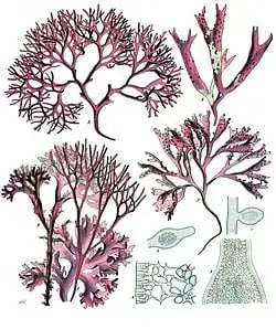
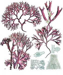
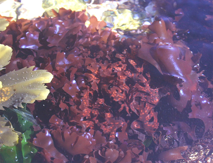
Cyanidioschyzon merolae belongs to the Rhodophyceae family of red algae protists. It is a marine alga that can be found in maritime habitats. Since it includes chlorophyll a and d, lutein, beta-carotene, myxoxanthin, violaxanthin, and fucoxanthin, it is generally red in color. These eukaryotic cells have a filamentous structure and are sometimes membranous.

Red algae or Rhodophyceae as plant like protists examples
Red algae or Rhodophyceae – Wikipedia

2.    Bangiophyceae

The formal definition of Bangiophyceae is a paraphyletic class of red algae. Due to the obvious lack of identifiable morphological traits and the species’ assumed morphological flexibility, taxonomic confirmation has proven to be challenging.

3. Dinoflagellates

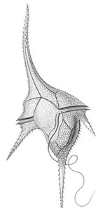
Dinoflagellates are a group of unicellular protists classified as either plants or animals because they have not yet been identified. These can be found both in freshwater and marine water habitats.

Dinoflagellate as plant like protists examples
Dinoflagellate as plant like protist example- Wikipedia

4. Sea lettuce

Sea lettuce, often known as green nori, is an edible alga that belongs to the genus Ulva and is found mostly in the ocean. It’s a macro alga protist that varies in hue from light to dark green and is held together by circular rigid support. Their thallus is shaped like a leaf and is flattened.

5. Kelp

Kelp is a brown alga that can be found in seawater and is also a seaweed protist. It is classified as a protist since it has multiple types of cells and is multicellular. Despite its plant-like aesthetic, it is far dissimilar from being a plant. It is still not considered a plant but rather a brown alga. However, the enormous kelp is a complicated species.

6. Cryptomonads or Fire algae

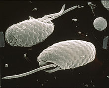
Cryptomonads, also known as Fire algae, is a type of single-celled algae found in several freshwater, marine, and brackish environments. Cryptomonads are a distinct phylum of protists because most species are photosynthetic and motile. They do have flagella, which supports the movement.

Cryptomonad as plant like protists examples
Cryptomonad Wikipedia

7. Chrysophyceae or golden alga

The Chrysophyceae family, often known as unicellular golden algae, is primarily found in freshwater environments. They have specialized flagella that allow them to move across the surface of freshwater. Diverse structural components can be found on the surface of developed cysts, which can help identify species. This species is classified as a protist.

8. Xanthophyceae

Xanthophyceae is a family of yellow-green algae classified as a protist since they have a nucleus and other membrane-bound components. Each structure has a particular structure because it performs a certain function. In particular, algae-like Xanthophyceae, photosynthetic life forms of the order Protista, are found in water bodies.

9. Euglena deses

Euglena deses is a member of the Euglena genus. It is a dominating freshwater eukaryote species that can be found in various habitats, but primarily in freshwater. These are single-celled organisms with a flagellum that assists in their movement in the water. 

It is a protist that contains photosynthetic chloroplast in its cell body. Although Euglena has both plant and animal features, most of its characteristics imply that it is a plant.

10. Diatoms

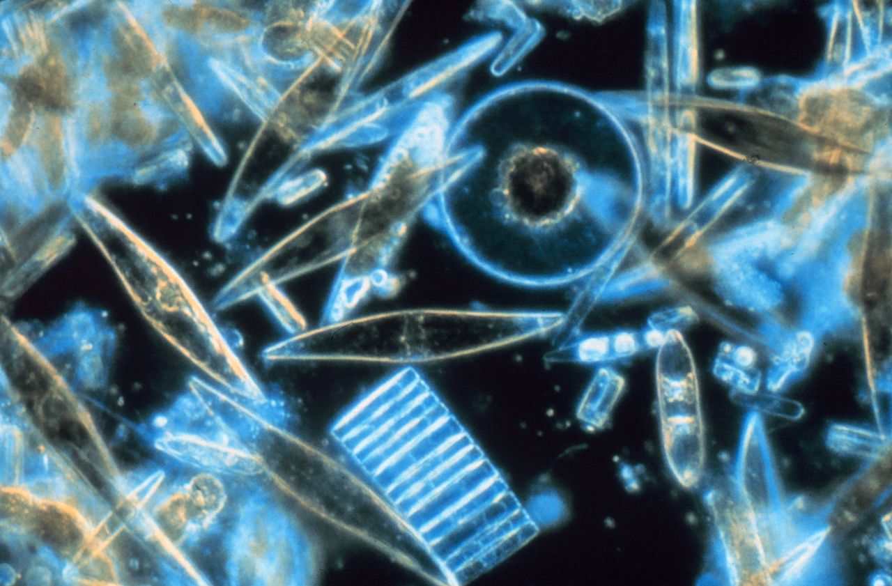
Diatoms are single-celled algae, more precisely microalgae, with photosynthetic abilities. These can be found in both freshwater and marine water habitats, as well as in most damp areas.

Diatoms can also contribute a significant part to the nutrient cycles of mineral and energy resources. They are plant-like protists that survive alone or in groups, such as in chains, swirls, or zig zags.

Diatoms as plant like protists examples
Diatoms – Wikipedia

11. Irish moss

Irish moss, often known as Chondrus crispus, is a species of red algae that grows on rocks from the medium aquatic environment to the shallow coastal zone and in the ocean water. It can thrive in the absence of sunlight. It is a protist plant that is also an autotroph.

Irish Moss as plant like protists examples
Irish Moss – Wikipedia

12. Gracilaria

Gracilaria is red algae belonging to the genus Rhodophyta. These types of algae are edible. Although they are mainly found in marine environments, they are also very important from an economic point of view. It can contribute economically to the production of protists and agar.

13. Florideophyceae

Florideophyceae is a red alga with a distinctive triphasic generational alternation in their life cycle. These are protists and eukaryotes. These can be found in marine and freshwater habitats from the Arctic to the Tropics. It also provides a shelter for a variety of aquatic species.

14. Delesseria sanguinea

Delesseria sanguine is a type of red algae or seaweed that belongs to the Rhodophyta division. These protists can be found in maritime environments. They possess a bright red everlasting alga that rises from a discoid holdfast with flattened leaf-like red spikes.

15. Marimo

Marimo, commonly known as a moss ball, is a freshwater ball-shaped alga. It’s an ornamental plant that’s commonly kept in aquariums. Protists are organisms that can produce their food through photosynthesis. They can survive in low-light environments.

Marimo as plant like protists examples
Marimo – Wikipedia

16. Ulva intestinalis

Ulva intestinalis is a form of green algae found in the oceans. Other names include sea lettuce, grass kelp, and so forth. These are motile because they are biflagellate. 

It contributes to the raising and cultivation of crops all over the globe. It is a protist that belongs to the kingdom of Protista.

17. Caulerpa Racemosa

Caulerpa racemose, often known as sea grapes, is a green alga found in marine environments. These are both edible and protist. For their survival, they rely on autotrophic nourishment.

18. Laminaria

Brown algae or seaweed, Laminaria, is a species of brown algae. It lives in marine habitats and is economically significant. It’s edible, and people use it in a variety of recipes. It is a protist and belongs to the kingdom of Protista.

19. Sargassum

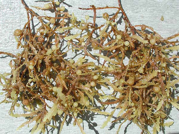
Sargassum is a brown microalga that grows in saltwater environments. It contains a heavily branching thallus with sunken berry-like floats that allow it to float on the water’s edge.

Sargassum as plant like protists examples
Sargassum – Wikipedia

20. Sphacelariales

Sphacelariales is a brown alga whose thallus is filamentous and branching in most instances. It can be seen all year on the bottom beach and below the low-water mark. It is a protist since it belongs to the kingdom of Protista.

21. Myriogramme

Myriogramme is a genus of red algae belonging to the Rhodophyta division. It’s a protist with a vivid red appearance.

22. Botrydiaceae

Botrydiaceae is a family of yellow-green algae belonging to the kingdom of Protista. These can be found in a damp environment.

23. Vaucheria

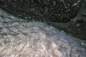
Vaucheria is a yellow-green alga that is commonly referred to as water felt. There are only two genera in the Vaucheriaceae family, one of which is this kind. Its zoospores are extensive and multinucleate, with numerous pairs of uneven flagella. Since they belong to the kingdom of Protista, they are protists.

24. Euglena gracilis

Euglena gracilis is a single-celled alga that belongs to the genus Euglena and the euglenids. Its potential to manufacture bioproducts could be very valuable commercially. It is a photosynthetic protist that can move and seek food in various ways.

25. Euglena geniculata

Euglena geniculata is a single-celled alga from the Euglena genus. It is a single-celled photosynthetic unicellular protist that can migrate and eat.

Conclusion:

Algae are the most common protists that resemble plants. Single-celled diatoms and multicellular seaweed are also among them. Algae, like plants, have photosynthetic pigments called chlorophyll and use photosynthesis to produce food. Green algae, Brown algae, Red algae, Fire algae, Golden-brown algae, Yellow-green algae, Diatoms, Dinoflagellates, and Euglenoids are some types.

Also Read:

Does Mitosis Occur in Plant Cells?

does mitosis occur in plant cells

Mitosis is a fundamental cellular process that occurs in both plant and animal cells, playing a crucial role in growth, development, and tissue repair. In the context of plant cells, mitosis is primarily limited to the meristematic tissue, which is found at the tips of roots and shoots. This specialized tissue is responsible for the growth and expansion of the plant, and mitosis is the mechanism by which new cells are generated.

Understanding Mitosis in Plant Cells

Mitosis is a highly regulated process that involves the precise duplication and segregation of genetic material, followed by the division of the cell’s cytoplasm to form two genetically identical daughter cells. In plant cells, the process of mitosis is similar to that observed in animal cells, with a few key differences.

Phases of Mitosis in Plant Cells

The mitotic process in plant cells can be divided into the following phases:

  1. Interphase: During this phase, the cell prepares for division by replicating its genetic material (DNA) and accumulating the necessary resources for cell division.
  2. Prophase: The chromosomes condense, and the nuclear envelope breaks down, allowing the mitotic spindle to form.
  3. Metaphase: The chromosomes align at the center of the cell, with their centromeres attached to the mitotic spindle.
  4. Anaphase: The sister chromatids separate and move towards the opposite poles of the cell.
  5. Telophase: The nuclear envelope reforms, and the cell begins to divide.
  6. Cytokinesis: The cytoplasm of the cell is divided, forming two genetically identical daughter cells.

Cell Plate Formation in Plant Cells

One of the key differences between mitosis in plant and animal cells is the formation of the cell plate during cytokinesis. In animal cells, a cleavage furrow forms and deepens, eventually dividing the cell into two. In plant cells, however, a cell plate is formed in the middle of the cell, which eventually develops into a cell wall, separating the two daughter cells.

The formation of the cell plate is a complex process that involves the following steps:

  1. Vesicle Fusion: Golgi-derived vesicles containing cell wall precursors fuse at the center of the cell, forming the cell plate.
  2. Cell Plate Expansion: The cell plate expands outwards, towards the cell walls, eventually fusing with the existing cell walls to form the new cell wall.
  3. Cell Wall Maturation: The cell wall matures, with the deposition of cellulose, hemicellulose, and other structural components.

This unique process of cell plate formation is a hallmark of mitosis in plant cells and is essential for the maintenance of the plant’s structural integrity and the separation of daughter cells.

Regulation of Mitosis in Plant Cells

does mitosis occur in plant cells

Mitosis is a highly regulated process, with a number of checkpoints that ensure proper division and chromosome separation. In plant cells, these checkpoints are particularly important, as errors in mitosis can lead to developmental abnormalities and genetic disorders.

Mitotic Checkpoints in Plant Cells

Some of the key mitotic checkpoints in plant cells include:

  1. Spindle Assembly Checkpoint: This checkpoint ensures that the mitotic spindle is properly assembled and that the chromosomes are correctly attached to the spindle.
  2. Metaphase-to-Anaphase Checkpoint: This checkpoint ensures that all chromosomes are properly aligned at the metaphase plate before the sister chromatids are allowed to separate.
  3. Cytokinesis Checkpoint: This checkpoint ensures that the cell plate is properly formed and that the cytoplasm is divided equally between the two daughter cells.

These checkpoints are regulated by a complex network of signaling pathways and regulatory proteins, which work together to ensure the fidelity of the mitotic process.

Quantifying Mitosis in Plant Cells

There are a number of quantifiable data points that can be used to measure mitosis in plant cells. These include:

  1. Mitotic Index: The mitotic index is the percentage of cells in a given tissue that are undergoing mitosis at a particular time. This can provide insight into the rate of cell division and growth.
  2. Mitotic Duration: The duration of each phase of mitosis can be measured, providing information about the timing and regulation of the process.
  3. Mitotic Frequency: The frequency of mitotic events can be measured in different tissues and developmental stages, further elucidating the patterns of cell division and growth.

These quantifiable data points can be used to study the dynamics of mitosis in plant cells, as well as to identify any abnormalities or disruptions in the process.

Conclusion

Mitosis is a crucial process in plant cells, occurring primarily in the meristematic tissue and playing a key role in growth and development. The process is highly regulated, with a number of checkpoints and quantifiable data points that can be used to measure its frequency and duration. Understanding the intricacies of mitosis in plant cells is essential for advancing our knowledge of plant biology and for developing strategies for improving plant growth and productivity.

References:

  1. Biology EOI Flashcards | Quizlet. (n.d.). Retrieved from https://quizlet.com/22339456/biology-eoi-flash-cards/
  2. Mitosis in plants: how far we have come at the molecular level? (2002). Retrieved from https://www.sciencedirect.com/science/article/abs/pii/S1369526602002972
  3. Where does mitosis actively take place in plants? – Socratic. (2017). Retrieved from https://www.socratic.org/questions/where-does-mitosis-actively-take-place-in-plants
  4. Müller, S. (2019). Cytokinesis in plant cells. Current Biology, 29(9), R358-R359.
  5. Eleftheriou, E. P. (2013). The plant cell wall. In Plant Anatomy (pp. 63-92). Springer, Berlin, Heidelberg.