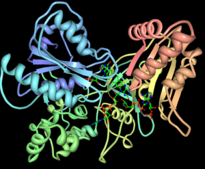
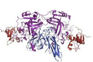
Do Bacteria Do Glycolysis? A Comprehensive Guide

do bacteria do glycolysis

Bacteria, like many other living organisms, rely on the fundamental metabolic pathway of glycolysis to generate energy and essential metabolic intermediates. Glycolysis, a central catabolic process, involves the conversion of glucose into pyruvate, yielding ATP and NADH in the process. This pathway is a crucial component of cellular respiration and plays a vital role in the energy metabolism of bacteria.

Glycolysis in Bacteria: An Overview

Glycolysis is a ubiquitous metabolic pathway found in the majority of bacteria, serving as the primary route for glucose catabolism. During glycolysis, glucose is broken down into two molecules of pyruvate, generating a net of 2 ATP and 2 NADH molecules. This process occurs in the cytoplasm of bacterial cells and is the first step in the larger metabolic network that includes the tricarboxylic acid (TCA) cycle and oxidative phosphorylation.

Enzymes Involved in Bacterial Glycolysis

Glycolysis in bacteria is catalyzed by a series of enzymes that facilitate the stepwise conversion of glucose to pyruvate. The key enzymes involved in this pathway include:

  1. Hexokinase: Catalyzes the phosphorylation of glucose to glucose-6-phosphate.
  2. Phosphoglucose isomerase: Converts glucose-6-phosphate to fructose-6-phosphate.
  3. Phosphofructokinase: Phosphorylates fructose-6-phosphate to fructose-1,6-bisphosphate.
  4. Aldolase: Cleaves fructose-1,6-bisphosphate into two triose phosphates, glyceraldehyde-3-phosphate and dihydroxyacetone phosphate.
  5. Glyceraldehyde-3-phosphate dehydrogenase: Oxidizes glyceraldehyde-3-phosphate to 1,3-bisphosphoglycerate, generating NADH.
  6. Phosphoglycerate kinase: Transfers a phosphate group from 1,3-bisphosphoglycerate to ADP, producing ATP and 3-phosphoglycerate.
  7. Phosphoglycerate mutase: Converts 3-phosphoglycerate to 2-phosphoglycerate.
  8. Enolase: Dehydrates 2-phosphoglycerate to phosphoenolpyruvate.
  9. Pyruvate kinase: Catalyzes the conversion of phosphoenolpyruvate to pyruvate, generating ATP.

Regulation of Glycolysis in Bacteria

Bacterial glycolysis is subject to various regulatory mechanisms to ensure efficient energy production and maintain metabolic homeostasis. Some key regulatory mechanisms include:

  1. Allosteric regulation: Certain glycolytic enzymes, such as phosphofructokinase and pyruvate kinase, are regulated by allosteric effectors, which can either activate or inhibit their activity based on the cellular energy status.
  2. Transcriptional regulation: The expression of glycolytic genes can be modulated by transcriptional regulators, allowing bacteria to adapt their glycolytic capacity to environmental conditions and nutrient availability.
  3. Post-translational modifications: Glycolytic enzymes can be subject to various post-translational modifications, such as phosphorylation, acetylation, or ADP-ribosylation, which can alter their activity and regulate the flux through the glycolytic pathway.
  4. Metabolite sensing: Bacteria can sense the availability of key glycolytic intermediates, such as fructose-1,6-bisphosphate or pyruvate, and use this information to fine-tune the activity of glycolytic enzymes and the overall flux through the pathway.

Glycolysis in Bacterial Metabolism

do bacteria do glycolysis

Glycolysis plays a central role in the overall metabolism of bacteria, serving as a hub for various metabolic pathways and providing essential precursors for biosynthesis.

Coupling of Glycolysis with Other Metabolic Pathways

Glycolysis is closely integrated with other metabolic pathways in bacteria, including:

  1. Tricarboxylic acid (TCA) cycle: The pyruvate generated from glycolysis can be further oxidized in the TCA cycle, generating additional ATP and reducing equivalents (NADH and FADH2) for the electron transport chain.
  2. Pentose phosphate pathway: Some of the glycolytic intermediates, such as glucose-6-phosphate, can be diverted to the pentose phosphate pathway, which generates NADPH and precursors for nucleotide and amino acid biosynthesis.
  3. Gluconeogenesis: Under certain conditions, such as growth on non-carbohydrate substrates, bacteria can reverse the glycolytic pathway to synthesize glucose from non-carbohydrate precursors, a process known as gluconeogenesis.

Metabolic Flexibility of Bacteria

Bacteria exhibit remarkable metabolic flexibility, allowing them to adapt their glycolytic capacity and energy metabolism to various environmental conditions and nutrient sources. This flexibility is crucial for their survival and proliferation in diverse ecological niches.

For example, some bacteria can switch between glycolysis and gluconeogenesis, depending on the available carbon sources. This metabolic flexibility is often observed in bacteria like Escherichia coli, which can grow on glucose as well as acetate or other non-carbohydrate substrates.

Quantitative Analysis of Glycolysis in Bacteria

Advances in experimental techniques and computational modeling have enabled researchers to obtain quantitative data on the enzyme and metabolite abundances in bacterial glycolysis and gluconeogenesis.

Coarse-Grained Modeling of Glycolysis and Gluconeogenesis

A coarse-grained model of glycolysis and gluconeogenesis in E. coli has been developed and calibrated using published metabolite and proteomics data. This model has provided valuable insights into the reorganization of metabolism under different growth conditions, such as glucose and acetate as sole carbon sources.

The model was able to reach distinct steady states for glycolytic and gluconeogenic conditions, demonstrating the ability to capture the metabolic switches between these two pathways. Despite the simplifications of the metabolic and regulatory networks, the model was found to well-describe the reorganization of metabolism, highlighting the importance of understanding the underlying biochemical constraints and flux sensing mechanisms.

Experimental Techniques for Monitoring Glycolysis

Various experimental techniques have been employed to quantify glucose uptake, lactate excretion, and the flux through glycolytic and related metabolic pathways in bacterial cultures. These techniques include:

  1. Glucose uptake and lactate excretion measurements: The rates of glucose uptake and lactate excretion can be determined by analyzing the concentrations of these metabolites in the culture medium over time.
  2. Stable isotope tracers: Glucose tracers, such as [U-13C 6]-glucose, [1,2-13C 2]-glucose, [2-13C 1]-glucose, and [5-3H]-glucose, can be used to measure the flux through glycolysis and related pathways with varying degrees of detection precision.
  3. Metabolomics and proteomics: Advanced analytical techniques, such as mass spectrometry, can be employed to quantify the abundances of glycolytic enzymes and metabolites, providing a comprehensive view of the metabolic state of bacterial cells.

These quantitative approaches, combined with computational modeling, have enabled researchers to gain a deeper understanding of the regulation and dynamics of glycolysis in bacteria, as well as its integration with other metabolic pathways.

Conclusion

In summary, bacteria do indeed perform glycolysis, a fundamental metabolic pathway that is crucial for their energy production and biosynthetic processes. Glycolysis in bacteria is a highly regulated process, involving a series of enzymes and subject to various control mechanisms to ensure efficient energy metabolism and adaptation to environmental conditions.

The integration of glycolysis with other metabolic pathways, such as the TCA cycle and gluconeogenesis, highlights the metabolic flexibility of bacteria, allowing them to thrive in diverse ecological niches. Advances in experimental techniques and computational modeling have provided valuable insights into the quantitative aspects of glycolysis in bacteria, paving the way for a deeper understanding of their metabolic dynamics and potential applications in biotechnology and medicine.

References:

  1. Severin Josef Schink, Dimitris Christodoulou, Avik Mukherjee, Edward Athaide, Viktoria Brunner, Tobias Fuhrer, Gary Andrew Bradshaw, Uwe Sauer, and Markus Basan. Glycolysis-gluconeogenesis switches in Escherichia coli are driven by biochemical constraints of flux sensing. Molecular Systems Biology, 2022.
  2. Tara TeSlaa and Michael A. Teitell. Techniques to Monitor Glycolysis. PMC, 2022.
  3. Glycolysis – an overview | ScienceDirect Topics. ScienceDirect, 2022.
  4. Chitra Thakur and Fei Chen. Linking metabolism to epigenetic reprogramming in stem cells. Seminars in Cancer Biology, 2019.

Do Bacteria Have Cell Walls?

do bacteria have cell walls

Bacteria are microscopic, single-celled organisms that are ubiquitous in our environment, playing crucial roles in various ecosystems and human health. A fundamental aspect of bacterial biology is the presence or absence of a cell wall, which has significant implications for their structure, function, and interactions with the surrounding environment. In this comprehensive blog post, we will delve into the intricacies of bacterial cell walls, exploring their composition, classification, and the techniques used to study their mechanical properties.

The Composition and Structure of Bacterial Cell Walls

Bacterial cell walls are typically composed of a complex and unique substance called peptidoglycan, also known as murein. Peptidoglycan is a polymer consisting of long glycan strands cross-linked by short peptide chains, forming a rigid and sturdy structure that surrounds the bacterial cell membrane. This peptidoglycan layer provides several essential functions for the bacterial cell, including:

  1. Maintaining Cell Shape: The peptidoglycan layer helps maintain the characteristic shape of bacterial cells, whether they are spherical (cocci), rod-shaped (bacilli), or spiral (spirilla).
  2. Protecting against Osmotic Lysis: The peptidoglycan layer acts as a barrier, preventing the bacterial cell from undergoing osmotic lysis (bursting) due to the difference in osmotic pressure between the cell’s interior and the external environment.
  3. Providing Structural Integrity: The cross-linking of the peptidoglycan strands creates a strong and resilient cell wall that protects the bacterial cell from mechanical stress and damage.
  4. Controlling Permeability: The peptidoglycan layer selectively allows the passage of certain molecules, such as nutrients and waste products, while restricting the entry of harmful substances.

The composition and structure of the peptidoglycan layer can vary significantly among different bacterial species, contributing to their diverse characteristics and adaptations.

Gram-Positive and Gram-Negative Bacteria

do bacteria have cell walls

Bacteria can be classified into two major groups based on their cell wall structure: gram-positive and gram-negative. This classification is determined by the Gram staining procedure, a differential staining technique that relies on the differences in the composition and thickness of the cell wall.

Gram-Positive Bacteria

Gram-positive bacteria have a thick peptidoglycan layer, typically 20-80 nanometers (nm) in thickness, which accounts for a significant portion of their cell wall. In addition to the peptidoglycan layer, gram-positive bacteria also have teichoic acids, which are polymers of ribitol or glycerol phosphate that are covalently linked to the peptidoglycan. Examples of gram-positive bacteria include Staphylococcus, Streptococcus, and Bacillus.

Gram-Negative Bacteria

Gram-negative bacteria have a relatively thin peptidoglycan layer, typically 2-7 nm in thickness, which is surrounded by an additional outer membrane. This outer membrane contains lipopolysaccharides (LPS), which are complex molecules that contribute to the overall structural integrity and permeability of the cell wall. Examples of gram-negative bacteria include Escherichia coli, Salmonella, and Pseudomonas.

The differences in cell wall structure between gram-positive and gram-negative bacteria have important implications for their susceptibility to antibiotics, interactions with the host immune system, and overall pathogenicity.

Techniques for Studying Bacterial Cell Mechanics

The mechanical properties of bacterial cell walls have been the subject of extensive research, as they play a crucial role in the survival, adaptation, and interactions of these microorganisms. Several advanced techniques have been employed to study the structure, composition, and mechanical properties of bacterial cell walls, including:

  1. Ultraperformance Liquid Chromatography-Mass Spectrometry (UPLC-MS): This technique is used to analyze the chemical composition and cross-linking of the peptidoglycan layer, providing insights into the structural features of the cell wall.

  2. Atomic Force Microscopy (AFM): AFM allows for the direct measurement of the mechanical properties of individual bacterial cells, such as their stiffness, elasticity, and adhesion forces.

  3. Electron Cryotomography: This imaging technique enables the high-resolution visualization of the three-dimensional structure of the bacterial cell wall, including the arrangement and organization of the peptidoglycan layer.

  4. Genetic Screens: By studying the genetic factors and molecular pathways involved in the synthesis, modification, and regulation of the bacterial cell wall, researchers can gain a deeper understanding of the underlying mechanisms that govern its mechanical properties.

These advanced techniques have revealed fascinating insights into the diversity and complexity of bacterial cell walls, highlighting their crucial role in the survival and adaptation of these microorganisms.

The Influence of Experimental Conditions on Bacterial Cell Mechanics

The mechanical properties of bacterial cells can be significantly influenced by the experimental conditions under which they are studied. Factors such as dehydration, treatment with chelating agents, and the ionic strength of the media can all have a profound impact on the measured stiffness and elasticity of bacterial cells.

  1. Dehydration: When bacterial cells are dehydrated, the peptidoglycan layer can undergo structural changes, leading to a drastic increase in cell stiffness. This is due to the loss of water molecules, which can alter the cross-linking and organization of the peptidoglycan network.

  2. Chelating Agents: Certain chelating agents, such as ethylenediaminetetraacetic acid (EDTA), can disrupt the ionic interactions within the bacterial cell wall, causing a decrease in cell stiffness. This is because the chelating agents can remove divalent cations, such as calcium and magnesium, which are important for maintaining the structural integrity of the peptidoglycan layer.

  3. Ionic Strength: The ionic strength of the media used in experiments can also significantly influence the measured mechanical properties of bacterial cells. Changes in the concentration of ions, such as sodium, potassium, and chloride, can alter the electrostatic interactions within the cell wall, leading to variations in cell stiffness and elasticity.

Understanding the impact of these experimental conditions is crucial when interpreting the results of studies on bacterial cell mechanics, as it helps researchers distinguish between intrinsic cellular properties and those that are influenced by the experimental setup.

Conclusion

In conclusion, the presence of cell walls is a defining characteristic of bacteria, playing a crucial role in their structure, function, and interactions with the environment. The cell walls of bacteria are typically composed of a unique substance called peptidoglycan, which can be classified into two main groups: gram-positive and gram-negative. The mechanical properties of bacterial cell walls have been extensively studied using advanced techniques such as UPLC-MS, AFM, electron cryotomography, and genetic screens, providing valuable insights into the biology, biophysics, and genetics underlying this fundamental aspect of bacterial biology. Additionally, the influence of experimental conditions, such as dehydration, chelating agents, and ionic strength, on the measured mechanical properties of bacterial cells highlights the importance of carefully considering these factors when interpreting research findings.

As the field of bacterial cell mechanics continues to evolve, the knowledge gained from these studies will undoubtedly contribute to our understanding of bacterial adaptation, pathogenicity, and the development of novel antimicrobial strategies. By delving deeper into the intricacies of bacterial cell walls, researchers can unlock new avenues for advancing our knowledge of these ubiquitous and fascinating microorganisms.

References:

  1. Bacterial Cell Mechanics – PMC – NCBI (ncbi.nlm.nih.gov)
  2. Determination of bacterial surface charge density via saturation of adsorbed ions (sciencedirect.com)
  3. Bacteria: Cell Walls – General Microbiology – Oregon State University (open.oregonstate.education)
  4. The Bacterial Cell Wall – Microbiology – NCBI Bookshelf (nih.gov)
  5. Peptidoglycan structure and architecture – ScienceDirect (sciencedirect.com)
  6. Gram-Positive vs. Gram-Negative Bacteria: What’s the Difference? (healthline.com)
  7. Atomic Force Microscopy of Bacterial Cells – PMC (ncbi.nlm.nih.gov)
  8. Electron Cryotomography of Bacterial Cells and Viruses – PMC (ncbi.nlm.nih.gov)
  9. Genetic Screens for Cell Wall Mutants in Gram-Positive Bacteria – PMC (ncbi.nlm.nih.gov)
  10. The Influence of Ionic Strength on the Mechanical Properties of Bacterial Cells – PMC (ncbi.nlm.nih.gov)

15 + Oxidoreductase Enzyme Example: Detailed Facts

Glutathione reductase 300x264 1

Oxidoreductase enzymes catalyze the transfer of electrons from the reductant to the oxidant. These enzymes utilize NADP+ or NAD+ as cofactors.

Transmembrane oxidoreductase enzymes are found in electron transport chains of bacteria, chloroplast, and mitochondria. Some of these enzymes are also anchored to the peripheral membrane with the help of a transmembrane helix. A considerable number of oxidoreductase enzymes are widely studied in biochemistry.

Oxidoreductase enzyme examples:

Let us discuss the above-mentioned oxidoreductase enzymes in detail.

Aromatase

Aromatase found in vertebrates is a key enzyme for the biosynthesis of estrogen from androstenedione and estradiol from testosterone. In humans, aromatase is present throughout the body and localized to the endoplasmic reticulum of the cell.

Choline oxidase

The choline oxidase enzyme catalyzes the reaction between two molecules of oxygen gas and choline to form two molecules of hydrogen peroxide and betaine aldehyde. Betaine aldehyde is an organic compound used by plants to adapt to temperature differences.

Laccase

Laccase is a multicopper oxidase enzyme. It is found in fungus and used for dye adsorption from polluted environments. Laccases also play a significant role in the paper, food, and textile industries.

Dihydrofolate reductase

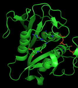
The dihydrofolate reductase enzyme is present in all organisms and responsible for catalyzing the reduction of dihydrofolate to tetrahydrofolate.

oxidoreductase enzyme example
3D structure of dihydrofolate reductase from Wikimedia

This enzyme is used for synthesizing inhibitors used for cancer therapeutics.

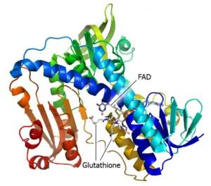
Glutathione reductase

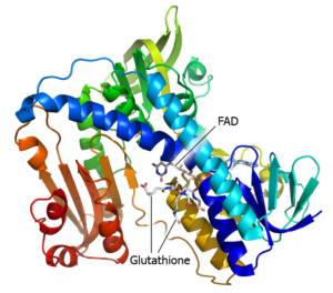
The glutathione reductase enzyme generates two molecules of reduced glutathione from oxidized glutathione.

Glutathione reductase
3 D structure of glutathione reductase from Wikimedia

Glutathione reductase plays a key role in preventing human cells from oxidative stress.

Glyceraldehyde-3-Phosphate Dehydrogenase

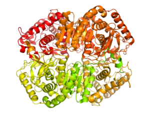
Glyceraldehyde-3-phosphate dehydrogenase is an important enzyme in glycolysis. It breaks down glucose for energy production.

2 Glyceraldehyde 3 phosphate dehydrogenase 3GPD wpmp
3 D structure of Glyceraldehyde-3-Phosphate Dehydrogenase from Wikimedia

It also plays a prominent role in plants and algae for the fixation of carbon dioxide into carbohydrates.

HMG-CoA Reductase

HMG-CoA reductase is an essential enzyme for cholesterol biosynthesis. Its activity is inhibited by statins. HMG-CoA reductase can be identified in the endoplasmic reticulum membrane.

Lactate Dehydrogenase

Lactate dehydrogenase is an essential enzyme for anaerobic respiration. It converts pyruvate to lactic acid in the absence of oxygen.

1170px Lactate Dehydrogenase B
3 D structure of lactate dehydrogenase from Wikimedia

Also, it anaerobically converts NADH to NAD+.

Monoamine oxidase b

Monoamine oxidase b enzyme present in the outer membrane of mitochondria catalyzes the oxidative deamination of serotonin. To prevent neurological disorders, monoamine oxidase b is inhibited by deprenyl.

NADPH Cytochrome P450 Oxidoreductase

NADPH Cytochrome P450 Oxidoreductase enzyme was first identified by Horecker in 1950.

1095px Cytochrome P450 Oxidoreductase aus 2BN4 pdb
3 D structure of NADPH Cytochrome P450 Oxidoreductase from Wikimedia

It plays an important role in embryogenesis and also increases the sensitivity of cancer cells to anticancer drugs.

Nitric Oxide Synthase

In cell communication and cell signaling, nitric oxide synthase plays a significant role. It catalyzes the formation of nitric oxide from L-arginine.

2-Oxoglutarate Dehydrogenase

2-oxoglutarate dehydrogenase is a multienzyme complex consisting of three monomers, viz; E1, E2, and E3. This enzyme plays a prominent role in lysine degradation, citric acid cycle, and tryptophan metabolism.

Phenylalanine hydroxylase

In the liver, phenylalanine is converted to tyrosine with the help of the phenylalanine hydroxylase enzyme. Dysregulation of this enzyme results in phenylketonuria due to the conversion of phenylalanine to phenylpyruvate.

Sulfite Oxidase

Sulfite oxidase enzyme is found in the liver, heart, and kidney and localized to cell mitochondria.

Sulfite oxidase
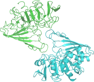
3 D structure of sulfite oxidase from Wikipedia

Sulfite oxidase is a homodimer that has two similar monomers. A point mutation in this enzyme causes neurological disorders.

Thioredoxin Reductase

Thioredoxin reductase enzyme is a member of the family of pyridine nucleotide disulfide oxidoreductases. It’s a homodimer with each subunit composed of alpha helices and beta sheets. It is identified in both prokaryotes and eukaryotes.

Urate Oxidase

Urate oxidase enzyme plays an important role in purine degradation. It catalyzes the oxidation of uric acid, thereby preventing uric acid buildup. It is not found in humans and higher apes.

Xanthine oxidoreductase

Xanthine oxidoreductase is a dimeric enzyme found in bacteria and eukaryotes. It is available in two interconvertible forms: xanthine oxidase and xanthine dehydrogenase.

What are oxidoreductase enzymes?

Oxidoreductase enzymes are one of the six major classes of enzymes, responsible for catalyzing oxidation and reduction reactions. The molecule donating electron also known as oxidant or electron donor gets oxidized and the molecule accepting electron also known as reductant or electron acceptor gets reduced in a redox reaction. The term ‘redox’ is interchangeably used for the oxidation-reduction reaction.

Oxidoreductase enzyme structure

Oxidoreductase enzyme structure mainly includes dimeric forms. Like all other enzymes, the primary structure of the oxidoreductase enzyme consists of amino acid sequences.

Oxidoreductase enzyme function

Oxidoreductase enzymes function as important biocatalysts in the pharmaceutical and agricultural sectors. These enzymes are involved in the synthesis of several therapeutic drugs such as: 3,4-dihydroxylphenyl alanine (DOPA) for treating Parkinson’s disease and boceprevir to treat chronic hepatitis C infection. Oxidoreductase enzymes present in plats play a crucial role in quantitative and qualitative productivity of crops. Also, glucose oxidase found in fungal species are used as preservatives in dairy products.

Also Read:

15 + Microbial Enzyme Example: Detailed Facts

3 D Structure of Alpha amylase 300x144 1

Microbial enzymes are potential biocatalysts that are extensively used in various industries such as fermentation, food, agricultural, chemical and pharmaceutical. These enzymes derived from different microorganisms are used to produce 0ver 500 commercial products.

A substantial number of microbial enzymes have found their wide use across different industries throughout the world. Bacteria, fungi, and yeasts are the selected microorganisms that are globally studied for the biosynthesis of a wide range of microbial enzymes.

Microbial enzyme examples:

In the following section, we will learn about the above-mentioned enzymes in detail.

α-amylase

α-amylase is specifically used for starch conversion. It is capable of hydrolyzing internal α-1,4-glycosidic linkages present in low molecular weight oligo- and polysaccharides. α-amylases are widely used in the brewing and baking industry.

microbial enzyme example
Structure of α-Amylase from Wikimedia

It is also used as a digestive aid in the pharmaceutical industry.

Glucoamylase

Glucoamylases also known as saccharifying enzymes are used in the food industry for the production of high-glucose syrups. They also find their use in the baking industry for the production of high-quality baked products. This enzyme plays a key role in the production of soya sauce and the reduction of alcohol content in the beer.

Protease

Proteases hydrolyzing peptide bonds found in proteins are well-known biocatalysts in the dairy industry. They reduce allergenic traits of milk products, enhance cheese flavor and accelerate cheese ripening.

Lactase (β-galactosidase)

The use of lactase in milk-based products aids in reducing lactose intolerance in people. Hydrolysis of lactose with lactase significantly improves the creaminess of ice creams. Lactase also plays a crucial role in lowering lactose-related biological oxygen demand during cheese production.

Lipase

Lipases are commercially used in the dairy industry for enhancing cheese flavor. It is also used to modify the aroma of wine and to improve cocoa butter quality. Moreover, this enzyme is known to be widely used in the biofuel, detergent, and leather industries.

Phospholipase

Phospholipases are used for the production of oils by breaking down phospholipids into fatty acids. These enzymes are also used in the degumming of vegetable oils.

Esterase

Esterases play a significant role in the alcoholic beverage and food industries. Feruloyl esterase contributes to the fruity aroma in foods and beverages by the biosynthesis of ferulic acid.

Cellulase

Cellulases are popular biocatalysts used in the juice industry for increasing yield. These enzymes enhance the aroma and taste of citrus fruits by reducing their bitterness. Cellulases are used in wine production to improve quality and color.

Xylanase

Xylanases known to cleave xylans are extensively used in bread making as they increase bread volume and improve bread quality. Its use also improves the taste and texture of biscuits. These enzymes improve extraction in juice production thereby increasing juice yield.

Pectinase

Pectinases capable of hydrolyzing glycosidic bonds in pectic polymers are used in juice production. This enzyme removes the turbidity of naturally derived fruit juices and improves drink flavor and color of the drink.

Glucose oxidase

Glucose oxidase belongs to the oxidoreductase enzyme family. It is widely used in the baking, pharmaceutical, and food industry. The use of this enzyme prevents the rotting of food by improving the shelf life of food products. It is also used to remove oxygen in order to increase storage life.

Laccase

Laccases are used in beverage and food industries to modify color appearance. It finds its use to improve the storage life of beer by removing oxygen in the final step of beer production. Interestingly, laccases are used in the cork stopper manufacturing industry.

Catalase

Catalase finds its use in the fabric industry for the removal of excess hydrogen peroxide from fabric. It is used for food preservation and the elimination of peroxide from milk. Also, catalase prevents oxidation in food wrappers.

Peroxidase

Peroxidase is used for producing flavor in the food industry. It plays a key role in managing environmental pollution by treating industrial phenolic effluents. It is also used as a biosensor.

α-acetolactate dehydrogenase

α-acetolactate dehydrogenase speeds up beer maturation by removing α-acetolactate and α-Aceto-α-hydroxybutyrate. Surprisingly, beer maturation takes 2 to 12 weeks without this enzyme, whereas the use of α-acetolactate dehydrogenase results in maturation within 24 hours.

Asparaginase

Asparaginase is majorly used in the pharmaceutical industry as an anticancer agent. It catalyzes the breakdown of asparagine an essential amino acid for the growth of cancerous cells.  

Debittering enzymes – naringinase

Naringinase breaks down naringin responsible for bitterness in citrus fruits. The use of naringinase helps in debittering fruit juices.

What are microbial enzymes?

Microbial enzymes discovered from microorganisms are used as organic catalysts for the large-scale manufacture of commercially used products. These enzymes possessing special characteristics such as the capability to function under abnormal conditions are designed using protein engineering, metagenomics, and biochemical reaction engineering.

Microbial enzyme structure

Like all other enzymes, the primary structure of microbial enzymes is made up of amino acids that are linked together by peptide bonds in a linear chain.

Also Read:

19 + Saprophytic Bacteria Examples:Detailed Facts Around It

Pseudomonas fluorescens Gram Stain on Microscope Slide 300x300 1

In this article, we will explore interesting facts about different saprophytic bacteria.

Saprophytic bacteria play a significant role in sustaining the nutrient cycle. The term saprophyte is derived from the Greek words ‘sapros’ and ‘phyton’ indicating the one who decays plant matter. These bacteria find a prominent use in several biotechnological applications. Let us learn about a few saprophytic bacteria examples.

Saprophytic bacteria examples:

In the following section, we have discussed in detail the above-mentioned saprophytic bacteria.

Cytophaga hutchinsonii

C. hutchinsoniiis a gram-negative, aerobic, soil bacterium first classified in 1929 by Sergei Winogradsky. It is known to degrade crystalline cellulose to glucose with the help of multiple degrading enzymes. This bacterium shows gliding motility without involving flagella.

Escherichia coli

E. coli feed on different meat and food products in nature, and those when consumed by humans cause serious food-borne infections.

Zymomonas mobilis

Z. mobilis is capable of fermenting glucose, sucrose, and fructose to produce carbon dioxide and ethanol. It can grow at 25-30 0C.

Acetobacter aceti

Acetobacter aceti is an economically valuable obligate anaerobic bacterium. It plays a significant role in the production of vinegar by converting the ethanol present in wine or cider into acetic acid. It can grow at 25-30 0C and 5.4 to 6.3 pH.

Clostridium aceto-butylicum

C. aceto-butylicum played a key role during World War I in 1914. It was extensively used for the production of acetone, a solvent required to produce smokeless gunpowder ‘cordite’. This bacterium has also found its use in pharmaceutical research for the delivery of therapeutic drugs in the body.

Geobacillus stearothermophilus

G. stearuothermophilus aerobically oxidizes carbon monoxide.

Clostridium thermosaccharolyticium

C. thermosaccharolyticium produces ethanol and acetic acid by fermenting pentose and hexose carbohydrates.

Staphylococcus saprophyticus

S. saprophyticus is known to be associated with urinary tract infection and are incapable of reducing nitrate.

Achromobacter xylosoxidans

Achromobacter xylosoxidans is a non-fermenting aerobic bacterium.

Alcaligenes faecalis

Alcaligenes faecalis is a nitrifying bacterium. It can generate nitrate and nitrite by oxidizing ammonia.

Bacillus subtilis

B. subtilis increases the ammonia-nitrogen content of the water body by decomposing nitrogen-rich organic litter.

Mycobacterium marinum

M. marinum is a slow-growing pathogenic bacterium.

Micrococcus antarcticus

M. antarcticus is a cold-adapted, gram-positive bacterium and is reported to cause pneumonia.

Pseudomonas aeruginosa

P. aeruginosa is capable of decomposing hydrocarbons. It is found in both man-made and natural environments.

Pseudomonas syringae

P. syringae grows in the phylloshpere as a saptrotroph. It exhibits its pathogenicity by invading a plant, forming biofilm, and overcoming host resistance.

Pseudomonas fluorescens

P. fluorescens breaks down the dead organic matter present in water bodies and soil.

saprophytic bacteria examples
Pseudomonas fluorescens from Wikimedia

Streptomyces species

Species from the genus Streptomyces are well-known decomposers. They produce geosmin while feeding on plant matter. Geosmin provides an earthy smell.

Azotobacter species

The species of the genus Azotobacter are free-living bacteria capable of fixing nitrogen.

Rhizobium

Rhizobia bacteria help the plants to utilize a ready form of nitrogen by fixing atmospheric nitrogen gas.

Thiobacillus ferrooxidans

T. ferrooxidans is capable of oxidizing soluble ferrous iron at 2.5 pH.

Magnetospirillum magneticum

M. magneticum produces magnetic materials such as biogenic magnetite which acts as a biomarker of environmental change. This bacterium aids in establishing environmental history.

What are saprophytic organisms?

Saprophytic organisms feed on non-living organic matter. They play a vital role in increasing the mineral content of the soil by decomposing dead plants and animals and converting the food waste into the compost bins. The mineral-rich soil around the saprophytic organisms allows the growth of healthy plants. The saprophytic organisms cannot prepare their own food because of their inability to perform photosynthesis. Several fungi and some flowering plants and bacteria belong to this group.

Frequently Asked Questions

Why saprophytes are important?

Saprophytes maintain the nutrients cycle efficiently by feeding on dead organic matter and making the nutrients available to plants in a ready form.

What are the examples of saprophytic organisms?

Examples of saprophytic organisms include: bacteria, fungi, mushrooms, moulds, earthworms, etc.

Is yeast a saprophyte?

Yeast is a saprophyte that feeds on dead and decaying organisms.

What are the examples of saprophyte plants?

Examples of saprophytic plants include Indian pipe, mushrooms, Mycorrhizal fungi, etc.

How saprophytes obtain their food?

Saprophytes decompose the dead organic matter by extracellular digestion. They secrete digestive substances in their surroundings and break down the organic matter.

Why saprophytes cannot prepare their own food?

Saprophytes cannot perform photosynthesis due to the lack of chlorophyll, hence they need to feed on dead substances.

Do saprophytes clean the environment?

Yes, saprophytes clean the environment by decomposing dead matter.

Also Read:

19 + Facultative Bacteria Examples: Detailed Explanations and Images

E. coli Bacteria 16578744517 2 300x217 1

In this article, we will explore different facultative bacteria and their images.

An array of facultative bacteria has been extensively used in various industrial processes and also plays a prominent role in sustaining ecology. Apart from that their versatility allows them to survive in injured tissues having inadequate blood supply.  

Facultative bacteria examples:

Facultative aerobic bacteria examples:

Escherichia coli

E. coli is a gram-negative, facultative, rod-shaped (1.1–1.5 μm wide by 2–6 μm long), aerobic bacterium. It has a reputation for causing food contamination in warm-blooded organisms as it commonly resides in the small intestine of endotherms contributing substantially to the gut microbiome. Due to the availability of genome sequences of numerous E. coli strains, it is largely used for molecular cloning and is interestingly familiar as a ‘molecular biologist toolbox’ amongst researchers.

facultative bacteria examples
Scanning electron micrograph of Escherichia coli from Wikimedia

Its rapid growth and easy scale-up process make it a good choice for an expression host in the biotechnology industry, where it is used for the large-scale production of recombinant proteins for therapeutics.

Klebsiella pneumoniae

K. pneumoniae is an encapsulated, gram-negative, rod-shaped (0.5 μm wide by 2 μm long), facultative aerobic bacterium.

860px Klebsiella pneumoniae Bacterium 13383468143
Klebsiella pneumoniae Bacterium from Wikimedia

This non-motile bacterium is strongly associated with pneumonia due to its inhabitancy in the human mucosal surfaces. In an oxygen-deprived environment, this naturally occurring bacterium has the ability to fix nitrogen.

Proteus mirabilis

P. mirabilis is a rod-shaped (2 μm long), facultative aerobic, gram-negative bacterium. It is well known for its swarming ability across solid or semi-solid surfaces with the help of flagella.

proteus mirabilis 8a8ded
Colonies of Proteus mirabilis from Picryl

Also, the bacterium is known to produce high levels of urease leading to the alkalinity of urine which is a common cause of kidney failure. Interestingly, P. mirabilis is not reported to have detrimental effects on poultry animals.

Listeria monocytogenes

L. monocytogenes is popularly known as listeriosis causing pathogenic, facultative anaerobic bacterium. It successfully passes through three major barriers in the host viz; the intestines, the blood-brain barrier, and the feto-placenta barrier thereby causing infections in high-risk individuals. Ongoing studies have used this bacterium as a vector in gene delivery therapies.

Staphylococcus epidermidis

S. epidermidis is a spherical-shaped (0.5 – 1.5 µm in diameter), facultative anaerobic, gram-positive bacterium. It is a part of human skin flora and is thought to provide benefits to the human host by out-competing more virulent pathogens.

Staphylococcus epidermidis Bacteria 5613984108
Staphylococcus epidermidis Bacteria from Wikimedia

However, its opportunistic nature in patients receiving medical devices causes orthopedic device-related infections which lead to the tag ‘accidental pathogen’.

  Streptococcus agalactiae

S. agalactiae is a gram-positive, ovoid (0.5-1.0 X 1.0-2.0 µm), facultative anaerobic bacterium. It is a virulent organism that causes serious health issues such as; pneumonia and meningitis in new-born.

Colonia de streptococcus agalactiae
Streptococcus agalactiae from Wikimedia

Streptococcus pneumonia

S. pneumoniae is a gram-positive, spherical (0.5 to 1.25 µm in diameter), facultative anaerobic bacterium.

1200px Streptococcus pneumoniae A causative bacteria of meningitis
Streptococcus pneumonia from Wikimedia

Apart from causing pneumococcal infection in the upper respiratory tract, it has been harnessed for a range of applications in biotechnology.

Streptococcus mutans

S. mutans is a round-shaped (0.5 to 0.75 µm), facultative anaerobic, gram-positive bacterium. It is commonly found in the human oral cavity and has evolved to increase its virulence by enhancing its ability to survive in a low pH environment.

Yersinia pestis

Y. pestis is a gram-negative, rod-ovoid (0.5-0.8 µm width 1-3 µm length), facultative anaerobic bacterium.

7316086176 d95bc87f19 c
Yersinia pestis from Flickr

It is widely known to cause plague. Also, in response to the production of antibodies and phagocytosis, it suppresses the host immune system.

Staphylococcus saprophyticus

S. saprophyticus is a spherical-shaped (1 µm in diameter), facultative anaerobic, gram-positive, bacterium. It is commonly found in the female genital tract and is a causative agent of urinary tract infections in 17-27 years old.

Streptococcus salivarius

S. salivarius is a grampositive, spherical (2 µm in diameter), facultative anaerobic bacterium. It has a therapeutic effect against oral infections as it produces antimicrobial peptides that prevent the growth of more virulent strains of Streptococcus in the oral environment.

Streptococcus sanguinis

S. sanguinis is a coccus-shaped, gram-positive, facultative anaerobic bacterium. It is commonly found in dental plaque and has a tendency to enter the bloodstream leading to colonization of the heart valves.

Cutibacterium acnes

C. acnes is a rod-shaped (0.4-0.7 µm width; 3-5 µm length), slow-growing, gram-positive, facultative anaerobic bacterium. Its habitat includes healthy adult skin and endophytes of plants. In the North American and European populations, it is reported to show resistance against a range of antibiotics such as; erythromycin, azithromycin, and doxycycline.

Shewanella oneidensis

S. oneidensis isolated from Lake Oneida is a facultative bacterium capable of reducing metal ions. It is well known for the synthesis of several nanoparticles such as silver. Also, it is a potent candidate for use in wastewater treatment.

Streptococcus bovis

S. bovis is a facultative anaerobic, gram-positive bacterium. Its habitat includes the alimentary tract and gastrointestinal tract of ruminants and humans, respectively.

Streptococcus mitis

S. mitis is a spherical-shaped, gram-positive, facultative anaerobic bacterium. Its habitat includes the oral cavity and gastrointestinal tract along with the female genital tract. Interestingly, S. mitis survived for over two years on the Surveyor 3 of the American uncrewed Surveyor program indicating its ability to sustain radiation exposure without a nutrient source.

Streptococcus thermophilus

S. thermophilus is a gram-positive, round-shaped (0.7-0.9 µm) fermentative facultative anaerobic bacterium. It is also known as a lactic acid bacterium due to its use in the production of yogurt. Yogurt and cheese containing live cultures of S. thermophilus are easily digested by lactose-intolerant people.

Staphylococcus hominis

S. hominis is a gram-positive, spherical (1-2 mm), facultative anaerobic bacterium. It contributes to body odor by producing thioalcohol compounds.

Staphylococcus haemolyticus

S. haemolyticus is a gram-positive, coccus shaped (0.8-1.3 µm), facultative anaerobic bacterium. It is an opportunistic pathogen and forms a part of the human skin flora.

Staphylococcus lugdunensis

S. lugdunensis is a gram-positive, spherical (2-4 mm in diameter), facultative anaerobic bacterium. It is a causative agent of arthritis.

Streptococcus anginosus

S. anginosus is a gram-positive, facultative anaerobic bacterium. It is a part of the human bacteria flora. Colonies of this bacterium often smell like butterscotch or caramel.

Streptococcus dysgalactiae

S. dysgalactiae is a gram-positive, facultative anaerobic bacterium. It is popularly found in the alimentary tract and genital tract of both humans and animals.

Staphylococcus capitis

S. capitis is a gram-positive, spherical (0.5-1.5 µm in diameter), facultative anaerobic bacterium. Although it forms a major part of the human biome, it can be pathogenic in immunocompromised people.

Streptococcus constellatus

S. constellatus is a spherical-shaped (0.5-1 µm in diameter), facultative anaerobic, gram-positive bacterium. It is normally found in the oral cavities and upper respiratory tracts.

Staphylococcus warneri

S. warneri is a non-motile, spherical (2-4 mm) facultative bacterium.

Frequently Asked Questions

What are facultative bacteria?

Facultative bacteria are popularly known as both facultative aerobic or anaerobic bacteria. These are the most flexible type of bacteria with an ability to adapt to extreme environmental conditions. These organisms have the potential to survive in both oxygen-rich and oxygen-deficient environment.

In the presence of oxygen, they make ATP by aerobic respiration, while they use fermentation or anaerobic respiration in an oxygen-deficient environment.

Also Read:

19+Spiral Bacteria Examples: Detailed Facts Around It

leptospira interrogans 2034321 960 720 300x232 1

Let us learn about a few spiral bacteria.

Spiral bacteria are gram-negative, spiral-shaped, motile prokaryotes belonging to the genus Spirillum. Since the creation of this genus in 1832 by Gottfried Ehrenberg, a German zoologist, numerous species having similar morphological appearances began to be discovered over time. These bacteria are commonly found in aquatic habitats and are capable to swim with the help of flagella.

Spiral bacteria examples:

In the following section, we will discuss the spiral bacteria examples in detail.

Spirillum volutans

S. volutans, one of the largest species of bacteria is found in both fresh water and stagnant water.

Helicobacter pylori

H. pylori is commonly found in the stomach and is known to cause several infections in humans. It is a helix-shaped (3µm long, 0.5 µm diameter) bacterium. H. pylori requires oxygen in lower concentrations than in the atmosphere.

Treponema pallidum

T. pallidum is popularly known to cause syphilis in humans and can be observed under a light microscope by only using darkfield illumination.

Leptospira interrogans

L. interrogans is a gram-negative, obligate aerobic spirochete. Two periplasmic flagella on its surface provide its motility and are commonly found in tropical regions. The species name ‘interrogans’ can be explained by the fact that these cells have hooked ends that resemble a question mark.

spiral bacteria examples
Leptospira interrogans from Pixabay

L. interrogans can survive at a temperature range of 28 0C to 30 0C and a pH of 7.4. These prokaryotes are host-associated in nature and tend to grow in the blood of hosts from where they spread throughout the body.

Borrelia recurrentis

B. recurrentis is widely known to cause relapsing fever in Ethiopia. It is transmitted from person to person via body louse.

Campylobacter jejuni

C. jejuni is a helical-shaped, gram-negative, nonfermenting, motile bacterium. It is a pathogenic bacterium and responsible for causing life-threatening food poisoning in Europe and in the US. It colonizes the digestive tract of many bird species and is also isolated from kangaroo feces. In laboratories, it is cultured at 42 0C which is the normal avian body temperature.

Treponema carateum

T. carateum is a causative agent of pinta, a skin infection in the children of tropical American countries.

Leptospira biflexa

L. biflexa is a non-pathogenic member of the genus Leptospira.

Borrelia burgdorferi

B. burgdorferi is a widely studied spirochaete and one of the causative agents of Lyme disease in humans. It is identified to have long cells with flexible cell walls. This organism can survive in low oxygen concentration and the presence of eleven periplasmic flagella set at each polar end allow them to move across high viscous media.

Treponema vincentii

T. vincentii is a motile spirochaete found in chronic periodontitis.

Leptospira kirschneri

L. kirschneri is a gram-negative, obligate, aerobic species and causes leptospirosis in pigs.

Borrelia afzelii

B. afzelii is named after Swedish dermatologist Arvid Afzelius and can infect various species of vertebrates and invertebrates.  

Treponema denticola

T. denticola is a gram-negative, oblicate aerobic, motile spirochaete bacterium. It survives in a diverse oral microbial community.

Borrelia duttoni

B. duttoni is named after Joseph Everett Dutton, who died of relapsing fever in 1905. This bacterium is a causative agent of relapsing fever.

Treponema paraluiscuniculi

T. paraluiscuniculi is a spiral-shaped, gram-negative bacterium and causes Rabbit Syphilis in the laboratory, pet, and wild rabbits.

Spirillum winogradskyi

S. winogradskyi is a gram-negative spiral bacterium found in the wastewater treatment plant.  

Borrelia turicatae

B. turicatae is predominantly found in the Southwestern United States, where its host organisms are mostly sick dogs.

Borrelia anserina

B. anserina is a helical spiral bacterium and can be grown in anaerobic conditions. It’s a pathogenic agent for poultry birds.

Treponema socranskii

T. socranskii is a helical, motile, anaerobic bacterium. It is commonly found in the oral cavity in the space between the teeth and gums of periodontitis patients.

Borrelia lusitaniae

B. lusitaniae is a pathogenic, gram-negative, spirochaete bacterium. Originating from Portugal, it has spread to European and North African parts. The host organism for this species is the common wall lizard whose migration is very limited leading to its availability to certain sites.

Treponema azotonutricium

T. azotonutricium is the first termite gut spiral bacterium to be isolated.

Types of spiral bacteria

On the basis of cell thickness, cell flexibility, and motility, spiral bacteria can be classified into three types:

Vibrio

Vibrios are commonly known as comma-shaped, facultative anaerobic bacteria belonging to the family Vibrionaceae. These are gram-negative, highly motile organisms due to the presence of polar flagella. Vibrios have a requirement of salt and can grow over a wide range of temperatures (20 0C to >40 0C). They are commonly found in aquatic and marine habitats.

Several Vibrio species are associated with a number of infections in humans. These infections are divided into two major groups: cholera and non-cholera infections. V. cholerae, the causative agent of cholera enters the human through contaminated food or water. V. parahaemolyticus, and V. vulnificus, are the two non-cholera Vibrio species responsible for causing vibriosis. The above-mentioned three Vibrio species are well documented human pathogens. Another Vibrio species, V. anguillarum is found in diseased fishes.

Spirillum

Spirilla are spiral-shaped bacteria belonging to the family Spirillaceae. Morphologically, spirilla are identified as gram-negative, motile, helical, rigid cell with a bunch of flagella at polar ends. These prokaryotes are aquatic except for S. minus, which is found in the blood of healthy rodents and can be transmitted to man causing rat-bite fever. They are usually found in organic matter-rich stagnant fresh water.

Aquaspirillum and Ocenospirillum are two other categories of spirilla bacteria. Aquaspirillum is commonly found in stagnant freshwater habitats such as ditches, canal water, and pond water. These bacteria are very small in size and cannot grow in saline environment due to low salt tolerance. Most of the species in this group use respiratory metabolism, but a few of them have shown the ability to grow anaerobically. Ocenospirillum can grow only in marine environments by forming coccoid bodies that are characterized by thin walls and resistant to lysis when placed in distilled water.  

Spirochaete

Spirochaetes are gram-negative, motile, spiral bacteria having endocellular flagella. They are commonly found in liquid environment such as, mud water, and blood. Several species of this group are known to cause severe human infections: syphilis, Lyme disease, and relapsing fever. Most of these organisms are free-living and anaerobic.

They are also found as metabolic symbionts of insects and parasites on animals. These prokaryotes can effectively translocate through viscous media and tissue barriers owing to endoflagella also known as axial filaments that provide the organism’s rotational flexibility.

Frequently Asked Questions

What are spiral bacteria?

Spiral bacteria are spiral-shaped, one of the three major morphologically categorized prokaryotes along with spherical cocci and rod-shaped bacilli. These organisms can be obligate and facultative in nature in context to their oxygen dependency.

What advantage does the spiral shape provide to bacteria?

Spiral bacteria like Campylobacter jejuni and Helicobacter pylori display decreased virulence when the genes contributing to the spiral shape were knocked out.

What is the purpose of the flagellum in spiral bacteria?

Flagellum aids in movement and chemotaxis in spiral bacteria.

How spiral bacteria are transmitted?

Spiral bacteria are generally transmitted through contaminated water or uncooked seafood or vegetables.

Also Read: