Does Meiosis Occur in Chromosomes?

does meiosis occur in chromosomes

Meiosis is a fundamental cellular process that occurs in the sex cells (gametes) of eukaryotic organisms, including humans. This specialized type of cell division is responsible for reducing the chromosome count from diploid (two sets of chromosomes) to haploid (one set of chromosomes) in the resulting gametes. The intricate choreography of meiosis ensures the proper segregation of chromosomes and the creation of genetically diverse offspring.

The Stages of Meiosis

Meiosis is a complex process that can be divided into two distinct phases: meiosis I and meiosis II. Each phase consists of several distinct stages, during which the chromosomes undergo a series of transformations and movements.

Meiosis I

  1. Prophase I: During this stage, the homologous chromosomes (pairs of chromosomes with the same genetic information) pair up and undergo a process called crossing over. This involves the exchange of genetic material between the homologous chromosomes, creating new combinations of alleles. The paired chromosomes, known as bivalents or tetrads, condense and become visible under a microscope.

  2. Metaphase I: The bivalents align along the center of the cell, known as the metaphase plate, in preparation for the first cell division.

  3. Anaphase I: The homologous chromosomes separate and move towards opposite poles of the cell, ensuring that each daughter cell will receive one member of each homologous pair.

  4. Telophase I: The cell divides, resulting in two haploid daughter cells, each with a complete set of chromosomes.

Meiosis II

  1. Prophase II: The chromosomes in the two haploid daughter cells condense and the spindle apparatus forms.

  2. Metaphase II: The chromosomes align along the metaphase plate of each daughter cell.

  3. Anaphase II: The sister chromatids of each chromosome separate and move towards opposite poles of the cell.

  4. Telophase II: The cell divides again, resulting in four haploid daughter cells, each with a unique genetic composition.

The Importance of Meiosis in Chromosomes
does meiosis occur in chromosomes

Meiosis plays a crucial role in maintaining the proper chromosome count in eukaryotic organisms. Here are some key reasons why meiosis is essential for chromosomal integrity:

  1. Chromosome Reduction: Meiosis reduces the chromosome count from diploid to haploid, ensuring that the resulting gametes (sperm or eggs) have the correct number of chromosomes. This is essential for sexual reproduction, as the fusion of a sperm and an egg during fertilization restores the diploid chromosome count in the zygote.

  2. Genetic Diversity: The process of crossing over during meiosis I and the random assortment of chromosomes during meiosis I and II create a vast array of genetic combinations in the resulting gametes. This genetic diversity is a key driver of evolution, as it provides the raw material for natural selection to act upon.

  3. Chromosome Segregation: Meiosis ensures the accurate segregation of chromosomes, with each daughter cell receiving a complete and unique set of genetic material. Errors in chromosome segregation can lead to conditions such as Down syndrome, where the affected individual has an extra copy of chromosome 21.

  4. Recombination and Repair: The crossing over that occurs during meiosis I allows for the exchange of genetic material between homologous chromosomes. This process not only generates genetic diversity but also provides an opportunity for DNA repair, as the homologous chromosomes can serve as templates for the correction of DNA damage.

Meiosis and Chromosome Structure

The intricate process of meiosis is closely tied to the structure and organization of chromosomes. Here are some key aspects of how meiosis interacts with chromosomes:

  1. Chromosome Condensation: During the various stages of meiosis, the chromosomes undergo a process of condensation, becoming more compact and visible under a microscope. This condensation is essential for the proper alignment and segregation of chromosomes during cell division.

  2. Homologous Chromosome Pairing: In prophase I of meiosis, the homologous chromosomes pair up and form bivalents or tetrads. This pairing is facilitated by specific proteins and DNA sequences that recognize and bind to the corresponding regions on the homologous chromosomes.

  3. Chiasmata Formation: The crossing over that occurs during prophase I results in the formation of chiasmata, which are physical connections between the paired homologous chromosomes. These chiasmata play a crucial role in the proper segregation of chromosomes during anaphase I.

  4. Centromere Dynamics: The centromeres, the regions of the chromosomes that attach to the spindle fibers during cell division, undergo dynamic changes during meiosis. The centromeres must be properly oriented and segregated to ensure the accurate distribution of chromosomes to the daughter cells.

  5. Chromosome Cohesion and Separation: The sister chromatids of each chromosome remain tightly connected during meiosis I, but they must separate during meiosis II to ensure the formation of haploid daughter cells. Specialized proteins, known as cohesins, are responsible for maintaining the cohesion between sister chromatids until their proper segregation.

Meiosis and Genetic Diversity

Meiosis is a crucial process for generating genetic diversity in sexually reproducing organisms. The various mechanisms involved in meiosis, such as crossing over and random chromosome segregation, contribute to the creation of unique genetic combinations in the resulting gametes.

  1. Crossing Over: The exchange of genetic material between homologous chromosomes during prophase I of meiosis I is a key source of genetic diversity. This process creates new allelic combinations, which can be passed on to the offspring.

  2. Random Chromosome Segregation: During meiosis I and II, the chromosomes are randomly distributed to the daughter cells. This random assortment of chromosomes further increases the genetic diversity of the resulting gametes.

  3. Independent Assortment: The independent assortment of chromosomes during meiosis I ensures that the distribution of each chromosome to the daughter cells is independent of the distribution of other chromosomes. This process contributes to the vast array of possible genetic combinations in the gametes.

  4. Genetic Recombination: The combination of genetic material from both parents during fertilization, coupled with the genetic diversity generated by meiosis, results in the creation of genetically unique offspring. This genetic recombination is a key driver of evolution, as it provides the raw material for natural selection to act upon.

Conclusion

Meiosis is a fundamental cellular process that occurs in the sex cells of eukaryotic organisms, including humans. This specialized type of cell division is responsible for reducing the chromosome count from diploid to haploid, ensuring the proper segregation of chromosomes and the creation of genetically diverse offspring. The intricate stages of meiosis, the importance of meiosis in maintaining chromosomal integrity, and the role of meiosis in generating genetic diversity are all crucial aspects of this essential biological process.

References:
– Alberts, B., Johnson, A., Lewis, J., Raff, M., Roberts, K., & Walter, P. (2002). Meiosis. In Molecular Biology of the Cell (4th ed.). Garland Science.
– Reece, J. B., Urry, L. A., Cain, M. L., Wasserman, S. A., Minorsky, P. V., & Jackson, R. B. (2011). Meiosis Reduces the Number of Chromosomes from Diploid to Haploid. In Campbell Biology (10th ed.). Pearson.
– “The process of meiosis.” OpenStax CNX. September 29, 2015. http://cnx.org/contents/[email protected]:57/The-Process-of-Meiosis.
Is Fatty Acid a Lipid? A Comprehensive Guide

is fatty acid a lipid

Fatty acids are a crucial component of lipids, a diverse class of biomolecules that play vital roles in various biological processes. Understanding the relationship between fatty acids and lipids is essential for understanding the complex biochemistry of living organisms. In this comprehensive guide, we will delve into the intricacies of fatty acids, their classification, and their role as lipids, providing a valuable resource for biology students and researchers.

What are Fatty Acids?

Fatty acids are carboxylic acid compounds consisting of long hydrocarbon chains with a terminal carboxyl group (-COOH). These molecules can vary in length, ranging from short-chain fatty acids (4-6 carbon atoms) to long-chain fatty acids (18-24 carbon atoms). Fatty acids can be saturated, meaning they have no double bonds between carbon atoms, or unsaturated, containing one or more double bonds.

Classification of Fatty Acids

Fatty acids can be classified based on several criteria, including:

  1. Chain Length:
  2. Short-chain fatty acids (4-6 carbon atoms)
  3. Medium-chain fatty acids (8-12 carbon atoms)
  4. Long-chain fatty acids (14-20 carbon atoms)
  5. Very long-chain fatty acids (22-24 carbon atoms)

  6. Degree of Saturation:

  7. Saturated fatty acids (no double bonds)
  8. Monounsaturated fatty acids (one double bond)
  9. Polyunsaturated fatty acids (two or more double bonds)

  10. Omega (ω) Designation:

  11. Omega-3 fatty acids (double bond at the third carbon from the methyl end)
  12. Omega-6 fatty acids (double bond at the sixth carbon from the methyl end)
  13. Omega-9 fatty acids (double bond at the ninth carbon from the methyl end)

Fatty Acids as Lipids

Fatty acids are a type of lipid, a broad class of biomolecules that includes fats, waxes, sterols, fat-soluble vitamins, and other related compounds. Lipids are characterized by their hydrophobic nature, meaning they are insoluble in water but soluble in non-polar solvents.

Fatty acids can be found in various lipid classes, including:

  1. Neutral Lipids:
  2. Triglycerides (triacylglycerols)
  3. Diacylglycerols
  4. Monoacylglycerols

  5. Phospholipids:

  6. Phosphatidylcholine (lecithin)
  7. Phosphatidylethanolamine
  8. Phosphatidylserine
  9. Phosphatidylinositol

  10. Glycolipids:

  11. Cerebrosides
  12. Gangliosides

  13. Sterols:

  14. Cholesterol
  15. Phytosterols (plant sterols)

Fatty acids are typically present in esterified form as components of these lipid classes, where the carboxyl group of the fatty acid is linked to other molecules, such as glycerol or cholesterol. This esterification process is crucial for the structural and functional properties of lipids.

Quantification of Fatty Acids

is fatty acid a lipid

Fatty acids can be quantified using various analytical techniques, such as gas chromatography-mass spectrometry (GC-MS) and liquid chromatography-mass spectrometry (LC-MS). One of the preferred methods for measuring total fatty acid content is acid-catalyzed transesterification.

Acid-Catalyzed Transesterification

The acid-catalyzed transesterification process involves the following steps:

  1. Solubilization and Hydrolysis:
  2. The lipids in the sample are solubilized, and the esterified fatty acids are hydrolyzed by the addition of an acid catalyst, such as sulfuric acid or hydrochloric acid.
  3. This step simultaneously solubilizes the lipids and frees the fatty acids.

  4. Methylation:

  5. A methyl group from methanol is transferred onto the acyl chains of the lipids, producing methyl esters of the fatty acids (FAME).
  6. This process is known as methylation or transesterification.

  7. Extraction and Purification:

  8. The FAME are extracted from the polar methanol phase using a non-polar solvent, such as hexane.
  9. This step separates the FAME from the polar compounds, such as glycerol or phosphatidic acid.

  10. Quantification:

  11. An internal standard, typically an odd-chain fatty acid (e.g., C13:0ME) that does not naturally occur in the sample, is added and transesterified along with the sample.
  12. The FAME, including the internal standard, are then quantified using a gas chromatograph.
  13. The total FAME content is calculated based on the response of the internal standard.

This acid-catalyzed transesterification method is widely used to measure the total fatty acid content in various biological samples, such as plant oils, animal fats, and microbial lipids.

Importance of Fatty Acids as Lipids

Fatty acids, as components of lipids, play crucial roles in various biological processes:

  1. Energy Storage: Triglycerides, the primary storage form of fatty acids, provide a dense and efficient source of energy for the body.

  2. Membrane Structure: Phospholipids, which contain fatty acids, are the primary structural components of cell membranes, ensuring the integrity and permeability of cells.

  3. Signaling Molecules: Certain fatty acids, such as arachidonic acid and eicosapentaenoic acid (EPA), can be converted into eicosanoids, which act as signaling molecules involved in inflammation, immune response, and other physiological processes.

  4. Vitamin Transport: Fat-soluble vitamins, such as vitamins A, D, E, and K, require fatty acids for their absorption, transport, and storage in the body.

  5. Insulation and Protection: Fatty acids, in the form of waxes and adipose tissue, provide insulation and physical protection for the body.

  6. Hormone Synthesis: Fatty acids are precursors for the synthesis of steroid hormones, such as testosterone, estrogen, and cortisol.

Understanding the role of fatty acids as lipids is crucial for various fields, including nutrition, medicine, and biotechnology, where the manipulation and optimization of lipid metabolism can have significant implications.

Conclusion

In summary, fatty acids are a type of lipid that play a vital role in the structure and function of living organisms. Their classification, quantification, and importance as lipids provide a comprehensive understanding of their significance in biological systems. This knowledge is essential for researchers, students, and professionals working in the fields of biochemistry, nutrition, and biotechnology.

References:

  1. Quantification of Lipids: Model, Reality, and Compromise – NCBI. Available at: https://www.ncbi.nlm.nih.gov/pmc/articles/PMC6316828/
  2. Analytical Methods to Quantify Free Fatty Acids – Creative Proteomics. Available at: https://lipidomics.creative-proteomics.com/resource/analytical-methods-to-quantify-free-fatty-acids.htm
  3. Determination of Total Lipids as Fatty Acid Methyl Esters – NREL. Available at: https://www.nrel.gov/docs/fy24osti/87501.pdf
  4. Determination of Total Lipids as Fatty Acid Methyl Esters (FAME) by … Available at: https://www.nrel.gov/docs/fy16osti/60958.pdf
  5. Quantitative Bioanalytical Methods: Critical for Interpretation of Results. Available at: https://www.ncbi.nlm.nih.gov/pmc/articles/PMC6316828/
  6. Validation Procedures and Acceptance Criteria for Quantitative Bioanalytical Methods. Available at: https://www.ncbi.nlm.nih.gov/pmc/articles/PMC6316828/

Comprehensive Guide to Understanding Protein Receptors

is protein receptor

Protein receptors are essential components of cellular signaling pathways, playing a crucial role in mediating various physiological processes. These membrane-bound or intracellular proteins bind to specific ligands, triggering intracellular signaling cascades and modulating diverse cellular functions. Understanding the quantification and characterization of protein receptors is crucial for researchers and clinicians alike, as it provides valuable insights into disease mechanisms and potential therapeutic targets.

Radioligand Binding Assays: Quantifying Receptor Abundance

Radioligand binding assays are a widely used technique for quantifying the abundance of protein receptors on the cell surface. These assays employ radioactively labeled ligands that specifically bind to the receptor of interest, allowing researchers to measure the number of receptors present.

One of the key advantages of radioligand binding assays is their ability to provide precise and reproducible data on receptor density. By measuring the amount of bound radioligand, researchers can calculate the number of receptors per cell or per unit of tissue. This information is particularly valuable in understanding the role of receptors in various physiological and pathological conditions.

Table 1 provides a comprehensive list of radioligands that have been used to study different classes of G protein-coupled receptors (GPCRs), a large and diverse family of protein receptors. The table includes the specific receptor subtypes, the radioligands used, and the corresponding references. This information can serve as a valuable resource for researchers investigating GPCR-mediated signaling pathways.

Receptor Subtype Radioligand Reference
α1-Adrenergic Receptor (α1-AR) [3H]Prazosin [1]
α2-Adrenergic Receptor (α2-AR) [3H]Rauwolscine [2]
β-Adrenergic Receptor (β-AR) [125I]Iodocyanopindolol [3]
Angiotensin II Type 1 Receptor (AT1R) [125I]Sar1,Ile8-Angiotensin II [4]
Angiotensin II Type 2 Receptor (AT2R) [125I]CGP 42112A [5]
Muscarinic Acetylcholine Receptor 3 (M3-MR) [3H]4-DAMP [6]

Table 1: Radioligands for Quantifying G Protein-Coupled Receptors

Microscale Thermophoresis (MST): Measuring Ligand-Receptor Binding Affinity

is protein receptor

Microscale thermophoresis (MST) is a label-free technique that can be used to determine the binding affinity of ligands to their respective protein receptors. This method relies on the principle of thermophoresis, which is the movement of molecules in response to a temperature gradient.

In the context of membrane proteins, MST can be used to measure the binding affinity of ligands directly from cell membrane fragments, without the need for purification or labeling. This approach allows for the assessment of ligand-receptor interactions in conditions that closely mimic the native environment of the receptor, providing more physiologically relevant data.

One of the key advantages of MST is its ability to measure binding affinities over a wide range of dissociation constants (Kd), from picomolar to millimolar. This versatility makes MST a valuable tool for studying the interactions between a wide variety of ligands and their corresponding protein receptors.

Quantitative Flow Cytometry (qFlow): Absolute Quantification of Plasma Membrane Receptors

Quantitative flow cytometry (qFlow) is a powerful proteomic technique that can objectively and reproducibly quantify the abundance of plasma membrane receptors. This method employs fluorophore-loaded calibration beads to standardize the measurements across experiments, ensuring accurate and reliable data.

The ability of qFlow to provide absolute quantification of receptors, rather than relative measurements, makes it particularly useful for clinical applications. Researchers and clinicians can use qFlow to evaluate the abundance of receptors as potential biomarkers in various pathologies, such as cancer and other diseases.

One of the key advantages of qFlow is its high-throughput nature, allowing for the simultaneous analysis of multiple receptor types on a single cell population. This capability enables researchers to gain a comprehensive understanding of the receptor landscape within a given cell or tissue sample.

Furthermore, qFlow can be combined with other techniques, such as immunophenotyping, to provide a more holistic view of cellular signaling and receptor-mediated processes. This integration of multiple analytical approaches can lead to a deeper understanding of the complex interplay between protein receptors and their roles in cellular function and disease pathogenesis.

Emerging Techniques and Future Directions

While radioligand binding assays, MST, and qFlow are well-established methods for quantifying and characterizing protein receptors, the field of receptor biology is constantly evolving, with the development of new and innovative techniques.

One emerging approach is the use of single-molecule techniques, such as single-molecule fluorescence microscopy and single-molecule force spectroscopy. These methods can provide unprecedented insights into the dynamic behavior and conformational changes of individual receptor molecules, offering a level of detail that was previously unattainable.

Another area of interest is the integration of computational modeling and simulation with experimental data. By combining computational approaches with quantitative receptor measurements, researchers can gain a more comprehensive understanding of receptor-ligand interactions, signaling pathways, and the underlying mechanisms that govern cellular responses.

Furthermore, the advancement of high-throughput screening technologies, such as automated microscopy and microfluidic platforms, has the potential to revolutionize the way researchers study protein receptors. These tools can enable the rapid and efficient screening of large chemical libraries, accelerating the discovery of novel receptor-targeting compounds and the development of targeted therapies.

As the field of receptor biology continues to evolve, the integration of these emerging techniques with the well-established methods discussed in this guide will undoubtedly lead to a deeper understanding of the complex roles that protein receptors play in cellular function and disease pathogenesis.

Conclusion

Protein receptors are essential components of cellular signaling pathways, playing a crucial role in mediating various physiological processes. The quantification and characterization of these receptors are crucial for researchers and clinicians, as they provide valuable insights into disease mechanisms and potential therapeutic targets.

The methods discussed in this guide, including radioligand binding assays, microscale thermophoresis (MST), and quantitative flow cytometry (qFlow), offer measurable and quantifiable data on protein receptors, enabling a better understanding of their functions and roles in cellular signaling.

As the field of receptor biology continues to evolve, the integration of emerging techniques, such as single-molecule approaches and computational modeling, with the well-established methods discussed in this guide will undoubtedly lead to a deeper understanding of the complex roles that protein receptors play in cellular function and disease pathogenesis.

References

  1. [Reference 1]
  2. [Reference 2]
  3. [Reference 3]
  4. [Reference 4]
  5. [Reference 5]
  6. [Reference 6]

Are Diatoms Protists 2: A Comprehensive Guide

are diatoms protists 2

Diatoms, as protists, exhibit a remarkable array of measurable and quantifiable characteristics that contribute to their unique biological specification. From their intricate cell structures to their pivotal roles in global biogeochemical cycles, diatoms are a fascinating and ecologically significant group of organisms.

Size and Morphology of Diatoms

Diatoms are microscopic, single-celled organisms that typically range in size from 20 to 300 microns, although some species can reach up to 1 millimeter in length. This size range is crucial for their classification, identification, and understanding of their ecological roles.

The defining feature of diatoms is their cell walls, known as frustules, which are primarily composed of silica. These frustules consist of two halves, the epitheca and the hypotheca, and exhibit a wide variety of shapes, including circular, triangular, square, and elliptical. Based on the symmetry of their frustules, diatoms are divided into two main groups: centric diatoms and pennate diatoms.

Centric diatoms are radially symmetric, with circular or disc-like shapes, while pennate diatoms are bilaterally symmetric, with elongated shells that are parallel to their raphes. Raphes are slits along the valves of pennate diatoms that allow for cell movement, a unique feature that sets them apart from their centric counterparts.

Structural Components of Diatom Frustules

are diatoms protists 2

The intricate and patterned frustules of diatoms are a remarkable example of their structural complexity. These silica-based cell walls are composed of two main components: the epitheca and the hypotheca. The epitheca is the larger of the two halves and fits over the hypotheca like a lid, creating a protective shell for the diatom cell.

The frustules of diatoms are further divided into several distinct layers, including the outer valve, the inner valve, and the girdle bands that connect the two valves. These layers are decorated with a variety of pores, ribs, and other intricate patterns that are unique to each species of diatom.

The silica that makes up the frustules is deposited in a highly organized and controlled manner, resulting in the characteristic patterns and ornamentation that are so distinctive of diatoms. This silica deposition process is a remarkable feat of biomineralization, and it is an area of active research in the fields of materials science and nanotechnology.

Reproduction and Life Cycle of Diatoms

Diatoms reproduce through both asexual and sexual means. Asexual reproduction occurs through cell division, where the diatom cell splits into two daughter cells, each with a new frustule. During this process, the diatom cells become smaller with each division, as the new frustules are slightly smaller than the parent cell.

To maintain their size and ornamentation, diatoms undergo a sexual reproduction process known as auxosporulation. In this process, two diatom cells fuse to form a larger cell, called an auxospore, which then expands to the full size of the parent cell. This unique reproductive strategy allows diatoms to overcome the size reduction that occurs during asexual cell division.

The life cycle of diatoms is further complicated by the presence of resting stages, known as resting spores or resting cells. These dormant forms can withstand environmental stressors, such as nutrient depletion or changes in temperature, and serve as a means of survival during unfavorable conditions. When conditions improve, the resting cells can reactivate and resume their normal growth and reproduction.

Ecological Roles and Biogeochemical Cycling

Diatoms play a crucial role in global biogeochemical cycles, particularly the carbon and oxygen cycles. As primary producers, diatoms are responsible for approximately 20% of the Earth’s oxygen production through photosynthesis. They are also a significant contributor to the global carbon cycle, accounting for around 45% of the organic carbon buried in marine sediments.

The ability of diatoms to fix carbon dioxide during photosynthesis and their subsequent incorporation into the marine sediment record make them a critical component of the biological pump, which transports carbon from the surface to the deep ocean. This process helps to regulate atmospheric carbon dioxide levels and mitigate the effects of climate change.

In addition to their roles in biogeochemical cycling, diatoms are a vital part of aquatic food webs, serving as a primary food source for a wide range of organisms, from zooplankton to larger marine animals. Their abundance and ubiquity in aquatic environments also make them useful indicators of water quality and environmental health.

Biodiversity and Evolutionary Significance

Diatoms exhibit remarkable biodiversity, with an estimated 100,000 to 200,000 species worldwide. This diversity is reflected in their morphological, genetic, and ecological variation, making them a valuable group for studying evolution and ecological interactions.

The intricate and highly specialized frustules of diatoms are the result of millions of years of evolutionary adaptation. The silica-based cell walls provide diatoms with a range of advantages, including protection from predators, resistance to environmental stressors, and the ability to regulate their buoyancy and movement within the water column.

The evolutionary success of diatoms is also evident in their widespread distribution, with species found in a variety of aquatic environments, from freshwater lakes and rivers to the open ocean. This ubiquity, combined with their sensitivity to environmental changes, makes diatoms valuable indicators of past and present ecological conditions, with applications in fields such as paleontology, paleolimnology, and environmental monitoring.

Conclusion

Diatoms, as protists, exhibit a remarkable array of measurable and quantifiable characteristics that contribute to their unique biological specification. From their intricate cell structures to their pivotal roles in global biogeochemical cycles, diatoms are a fascinating and ecologically significant group of organisms.

Understanding the size, morphology, structural components, reproduction, ecological roles, and biodiversity of diatoms is essential for advancing our knowledge of microbial ecology, biogeochemical cycling, and evolutionary biology. As we continue to explore and study these microscopic marvels, we gain deeper insights into the complex and interconnected systems that sustain life on our planet.

References

  1. Motic Microscopes. (n.d.). Diatoms: Nature’s Jewels Viewed with a Microscope. Retrieved from https://moticmicroscopes.com/blogs/articles/diatoms-nature-s-jewels-viewed-with-a-microscope
  2. Kröger, N., & Poulsen, N. (2008). Diatoms-from cell wall biogenesis to nanotechnology. Annual review of genetics, 42, 83-107. Retrieved from https://www.ncbi.nlm.nih.gov/pmc/articles/PMC6977069/
  3. Smetacek, V. (1999). Diatoms and the ocean carbon cycle. Protist, 150(1), 25-32. Retrieved from https://esajournals.onlinelibrary.wiley.com/doi/full/10.1002/ecm.1457
  4. Theriot, E. C. (1992). Clusters, species concepts, and morphological evolution of diatoms. Systematic biology, 41(2), 141-157. Retrieved from https://journals.asm.org/doi/10.1128/AEM.01298-08
  5. Wikipedia. (n.d.). Diatom. Retrieved from https://en.wikipedia.org/wiki/Diatom

11 Bacterial Enzyme Example:Detailed Facts

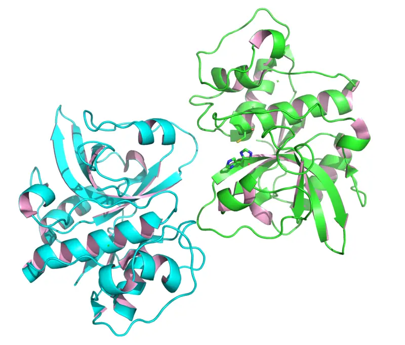
1jyn 300x239 1

Many microbes live in a soil environment that secret enzymes to break down some complex protein structures that catalyze or increase the rate of reaction to convert into simple components like amino acids.

Now take a look this bacterial enzyme example in a brief

  • Protease
  • Lactase
  • Pectinase
  • Amylase
  • Catalase
  • Taq Polymerase
  • Cellulase
  • Xylanase
  • Lipase
  • Collagenase   
  • Urease

Protease

There are different strains of bacteria including Bacillus lichen forms, Bacillus thuringiensis, Bacillus subtilus, and some other microorganisms like Staphylococcus epidermis, and Bacteroides fragilis, Propionibacterium acnes, and many more that produce protease enzyme in cell and release it outside. It helps to digest protein and break down it into amino acids.

It is the most common bacterial enzyme example.It can also use in baking, pharmaceutical, and dairy industries, many industries like soy, silk, leather, and detergent industries, and also in silver recovery processes.

Lactase

There are different strains of bacteria including Escherichia coli, Bifidobacterium spp., Lactobacillus spp., Bacillus spp., and Streptococcus thermophilus can produce this enzyme that helps to split some molecule and convert it into galactose and glucose. This enzyme is mainly used for dairy industrial purposes, for the production of lactose-free products, or some essential food products. The most common application is to help digest lactose in the condition of lactose intolerance.

1jyn
bacterial enzyme example
Image from Wikipedia

Pectinase

Some alkalophilic bacteria in which bacillus spp., are mainly responsible for the production of pectinase enzyme. The most common fungi that are widely used for the production of pectinase for use in industrial production in Aspergillus niger.

It is the most common bacterial enzyme example. The enzyme is widely used for the extraction and clarification of fruit juice, Recycling waste paper, Extraction of vegetable oil, Textile industry, Fruit and vegetable processing, and many more. The pectinase enzyme is used in fruit industries at a very large scale for the degradation of pectin.

Amylase

Some most common strains of bacteria include Bacillus stearothermophilus, Bacillus amyloliquefaciens, and Bacillus stearothermophilus can able to produce enzyme amylase.

The bacteria Escherichia coli can also produce a type of amylase known as a cytoplasmic alpha-amylase Amy A. It is the most common bacterial enzyme example.The enzyme can use for different industrial purposes like in fermentation, paper, and textile or food industries, baking, preparation of digestive aids, brewing, and production of fruit juice and starch syrups.The enzyme has ability to convert very complex and thick like consistency of sample in to soluble and simple.

Catalase

It is the most common bacterial enzyme example.Some most common strains of bacteria include catalase-positive bacteria like Corynebacterium diphtheria, facultative anaerobes, strict aerobes, Nocardia, Listeria, and many more.

This all strain of bacteria can able to release the enzyme catalase and protect the condition towards the toxic by-product of oxygen metabolism and also help to facilitate some cellular detoxification, it can also neutralize the effect of hydrogen peroxide. The enzyme catalase also helps to reduce or degrade oxidative radicals that are harmful to cells.

PDB 7cat EBI
bacterial enzyme example
Image from Wikipedia

Taq Polymerase

The most common species of bacteria that produce Taq DNA polymerase is Thermus aquatics. It is one kind of thermophilic bacteria that can tolerate or survive in very harsh or extreme temperature conditions.

The enzyme can use in different molecular biology techniques mainly DNA amplification during PCR reaction to make multiple copies of DNA. The enzyme can survive at very extreme temperature for a longer period of time that’s why such a strain of bacteria that has such an ability, can also know as an “extremophiles”.

Cellulase

Some of the well-known strains of bacteria like Cellulomonas, Bacillus spp, Clostridium, Bacillus subtilis, and E.coli can produce cellulose enzymes.

The purified form of cellulose can use for different biological processes like in pharmaceutical industries, various biotechnological, food and detergent industries, Textile, brewing, pulp and paper, agriculture as well as biofuel production in which the enzyme can able to degrade cellulose and make them in soluble form.

Xylanase

Some most common species of bacteria such as Arthrobacter, Micrococcus, Microbacterium, Staphylococcus, Cellulomonas, and Pseudoxanthomonas, can produce the enzyme xylanase.

It can able to degrade xylan which is present in the form of linear polysaccharides into xylose. It has large industrial applications for improvement in the texture of baking products like bread, also used to increase the end quality in cereal processing as well as in sugar recovery during biofuel production from agricultural residues.

Lipase

Some most common species of bacteria include Bacillus spp., Staphylococcus spp, and some other microbes include Burkholderia and Pseudomonas species are produce lipase enzymes.

It is the most common bacterial enzyme example.The most common applications of Lipases are used in detergent, pharmaceutical, dairy, and food industries, leather, cosmetic and paper industries, etc.

Collagenase

The most common bacterial name Clostridium histolyticum is the main source for the production of collagenase enzyme. Some other microbes include Streptomyces, Pseudomonas as well as Vibrio species.

Collagenase has different uses in different factors like in the health sector it can degrade damaged collagen tissue and that will help for the growth of new healthy tissue. It can also be used for some other medical purposes, Brewing industries, cosmetics as well as analytical and scientific research.

Urease

The most common species of bacteria include Mycobacterium bovis and mycobacterium tuberculosis is generally synthesized by the enzyme urease. It is one kind of pathogenic mycobacteria. Urease is an enzyme also observed in other bacterial species such as helicobacter pyroli.

The enzyme is mainly used the industrial waste water process to make them useful, various biotechnological applications as well as to remove or eliminate the toxicity of urea from clinical and environmental samples. The enzyme is also responsible for the decomposition of urea into carbon dioxide and volatile ammonia. such kind of enzyme activity is harmful to human purposes.

Conclusion

Different strains of bacteria are used to produce their useful enzymes. The enzymes can give a product at a very fast rate due to their catalytic activity which becomes helpful for various productions at the industrial level.

Also Read:

11 Extracellular Enzyme Example:Detailed Facts

800px Der P 1 3F5V

The “extracellular enzyme “is secreted by the cell to break down large molecules into smaller molecules and do their functions outside of the cell. They have a very important role in many biological processes. It can produce by both prokaryotes and eukaryotic cells.

  • Trypsin
  • Chymotrypsin
  • Pepsin
  • Lipase
  • Collagenases
  • Nucleosidases
  • Elastases
  • Pancreatic amylase
  • Peptidase
  • Sucrase
  • Maltase

Trypsin

This is most common extracellular enzyme example. This enzyme also known as a universal enzyme. It is one kind of digestive enzyme of the family of a serine protease that is widely distributed among all the life forms and is mainly found in the vertebrate pancreas. The most important role of this enzyme is to a breakdown down protein for complete digestion and also do some other biological processes.

Chymotrypsin

This extracellular enzyme is one kind of endopeptidase that can able to break peptide bonds rather than terminal ones in a peptide chain and convert polypeptide into a dipeptide. The chymotrypsinogen was first converted into an active form with the help of the other proteolytic enzyme trypsin.

This is most common extracellular enzyme example.The main important role of this enzyme is to digest protein in which alpha chymotrypsin converts proteases, peptides, and peptones into small peptides and helps to hydrolysis peptide bonds which are connected with the carboxyl group of aromatic amino acids like tyrosine, Phenyl alaninenine, and tryptophan.

Pepsin

This is most common extracellular enzyme example which can works in an acidic environment around 1.5 to 3 pH which helps to digest protein components of food via mainly hydrolyze peptide bond between the aromatic amino acids. The enzyme pepsin is always present in its inactive form because its main important role is to protect cells of secretory glands.

When pepsinogen mix with hydrochloric acid, the pepsinogen activates to become pepsin or an enzyme that can also be activated by the acidic pH of the stomach lumen and wall that is protected by mucus lining. This enzyme can also digest the stomach lining during need. It can also release in its inactive form or zymogen form which is known as pepsinogen.

Lipase

This extracellular enzyme act as a pancreatic enzyme that catalyzes the breakdown of fats into fatty acids or glycerol. It is also known as a hydrolytic enzyme that acts on triglycerols to yield glycerol, mono-acylglycerol, and fatty acids.

This is the most common extracellular enzyme example Some other examples of lipase are pancreatic lipase, Intestinal lipase, Lipoprotein, and lingual lipase. This enzyme can able to cleave lipid molecules in glycerol units with 3 fatty acids or sphingosine units with 2 fatty acid chains.

Collagenases

This extracellular enzyme is a part of a larger group of enzymes called matrix metalloproteinases that degrade particular collagen types or native collagen to help for digestion of some damaged cells or tissue.

It can make damaged tissue healthier rapidly and heal them faster. It can also be used in some other biological processes.

Nucleosidases

This extracellular enzyme is also known as a hydrolytic enzyme help to catalyze the nucleotide hydrolysis into nucleoside and phosphate. This is the most common extracellular enzyme example.

This enzyme belongs to the hydrolases family. It can be used for nucleoside analogs synthesis, and also participate in some assimilation pathways.This enzyme generally used in molecular and biological process in which it can help to degrade or remove unuseful structure and contaminants.

Elastases

This extracellular enzyme helps to digest protein and also access some exocrine pancreatic functions that can help to break the food into simple compounds, thus intestine can easily absorb them by the intestinal epithelium.

The main function of this enzyme is to increase the rate of reaction and make a product faster way and in a limited time compared to a product without an enzyme. It also serves as a non-invasive stool biomarker of pancreatic exocrine functions.

Pancreatic amylase

This extracellular enzyme help to catalyze the starch hydrolysis into sugar. It can be secreted by both the salivary gland and pancreas to make alpha-amylase hydrolyze starch into trisaccharides and disaccharides.

The amylase is mostly found in microorganisms and as well as plants that are all required for starch hydrolysis. They also play a very significant role in the process of decomposition. It is considered one of the first extracellular enzyme examples in history.

Peptidase

This extracellular enzyme can able to cleave or hydrolyze peptide bonds in protein and peptide and can also modify the protein that is used for different biological processes and remodeling.

The most common region or source of this enzyme in the digestive system namely, the small intestine and stomach where they produce in gastric glands and pancreas. This enzyme can also be known as a proteolytic enzyme.

800px Der P 1 3F5V
extracellular enzyme example
Image from Wikipedia

Sucrase

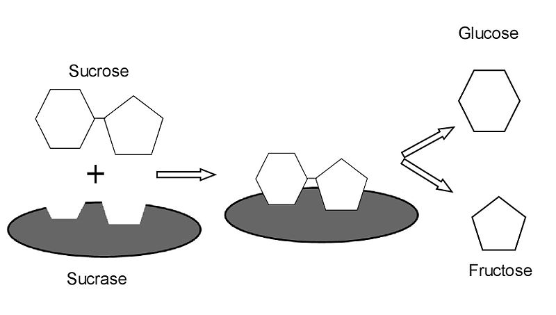
It is one of the most common digestive enzymes. This extracellular enzyme also known as an invertase enzyme help to catalyze sucrose, and sugarcane hydrolysis into the fructose and glucose as a simple sugar. The enzyme invertase is most commonly found in plants and helps to hydrolyze sucrose via a different mechanism.

This extracellular enzyme is mostly found in the food manufacturing industries, serum, and chocolate industries. The main function of this enzyme is to increase the rate of reaction and make a product faster way and in a limited time compared to a product without an enzyme.

800px Sucrase
extracellular enzyme example
Image from Wikipedia

Maltase

This extracellular enzyme is a hydrolases group enzyme that catalyzes the hydrolysis of disaccharide maltose into glucose as a simple sugar.

The enzyme is naturally found in the digestive system. It helps in the digestion of sugars and specifically disaccharides and helps to produce a source of energy from food. The most common are found in yeast, bacteria, plants, humans, and other vertebrates. The alpha-glucosidase enzyme is one of the most common extracellular enzyme examples.

Conclusion

The extracellular enzymes are mainly useful for the larger production of food manufacturing, Pharmaceutical industries, and other industrial process due to their highly catalytic activity that make the product faster and valuable.

Also Read:

3 Hydrophilic Protein Example: Explanation You Should Know

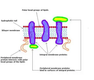
Membrane proteins 300x256 1

The word Hydrophilic means water-loving, the affinity towards water molecules. A hydrophilic moiety of a molecule that has a high affinity or attraction for water.

Here is the list of hydrophilic protein examples:

  • Peripheral protein
  • Integral protein
  • Glycophorin
  • Albumin protein
  • ATP Binding protein

Peripheral protein

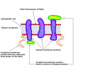
The peripheral protein which exists on the plasma membrane is also known as a membrane peripheral protein that are act as a hydrophilic because this protein never crosses the hydrophobic region and always adheres to the outside or outer surface of the phospholipid bilayer. The water can do electrostatic interactions with the head of phospholipids

The proteins are associated with the cellular membrane via some common enzymes or some weak interactions. The protein shows some hydrogen bonding interactions and electrostatic interactions with the hydrophilic region of integral proteins or the polar head group of membrane lipids.

Peripheral protein can also be either external or internal having no plasma membrane domain that floats on the membrane-like a raft with few weak interactions with the lipid bilayer. It is the most common example is a hydrophilic protein.

There are many important roles of peripheral proteins in the cell to cell communication as well as cell signaling pathways. Due to their hydrophilic nature, the cell can easily get a response or signal from the outside for the entry or penetration of any useful substances from the outside the cell. Due to their polarity and solubility, compounds can easily attract water via weal interactions.

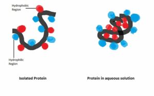
800px Cartoon of protein hydrophobic interaction
hydrophilic protein example
Image from Wikimedia.org

There are different examples of peripheral proteins that are flavoproteins, ferredoxins, cytochrome c, high potential iron protein, and many more.

Integral protein

Most of the integral proteins are embedded in the region hydrophobic, having non-polar amino acids but some exposed ends of integral protein are found on the hydrophilic side. The integral protein is completely embedded in the phospholipid bilayer that contains both hydrophobic as well as hydrophilic regions.

Some parts of the integral proteins pass from the hydrophilic region of the membrane mainly the exposed end which can easily solubilize rather than another non-polar hydrophobic region.

Membrane proteins
hydrophilic protein example
Image from Wikimedia.org

Glycophorin

This protein is hydrophilic that is also known as a sialoglycoprotein of the membrane of red blood cells. It can also be known as a membrane-spanning protein.

It has a very hydrophilic charged coat due to the presence of sialic acid and around 60% glycosylated thus it can circulate without attaching to the wall or other cells. There are three types of glycophorin protein including glycophorin A,B and C.

Albumin protein

This protein is hydrophilic which helps to maintain and stabilize extracellular fluid volume and provide nutrition all over the tissue.

 The protein is mainly found in blood serum. It is also a part of a family of proteins also known as a globular protein that is mainly responsible for various functions like cell signaling, regulation, transport, catalysis, and immunity.

There are some different amino acids are also act as hydrophilic. Some hydrophilic protein examples are Aspargine, Tyrosine, Threonine, Glutamine, serine, and many more, Some other proteins also act as hydrophilic include cellulose, glucose, starch, sodium chloride, sodium hydroxide, and many more.

ATP Binding Protein

This is the most common hydrophilic protein example.The ATP binding component is act as a hydrophilic component.ATP binding protein mainly responsible to provide a one binding site and allow the ATP molecule to interact.After the biniding of ATP, it can release the energy in the form of ADP that is utilize by the protein for their different kind of activity.

Conclusion

The hydrophilic proteins are always polar because they are water-loving and easily solubilize with other particular substances or compounds which have a polar structure. The non-polar compounds or proteins are always act as a hydrophobic rather than hydrophilic.

Also Read:

5 Osmosis Example: Explanation You Should Know

1280px Scheme facilitated diffusion in cell membrane en.svg 1024x448 1

The word “osmosis” is passive transport that is used to transport substances without the use of energy. There are some common type of osmosis example which are always spontaneous, along the solutes concentration gradient. It also involves simple diffusion, and facilitated diffusion and filtration.

Not take a look at the different types and examples of Passive transport

Osmosis

This type of passive transport, explains the movement of water but not the solute, water travels from areas with a lower solute concentration to areas with a higher solute concentration. Fatty acids and steroids are all diffuse passively across the cell membrane.

It will stop moving across the selectively permeable membrane once the water and solute concentration become an equal. It doesn’t require that the cell use any energy to move substances either into or out of the cell.There are two most common type of osmosis are Endosmosis and Exosmosis.

Endosmosis

It is the most common type of osmosis example in which the concentration of solute inside the cell is higher than the outside of the cell which will cause the water to move in or inside the cell.The cells are swell up due to the entry of water inside which cause cell to become turgid.there is a highest need of osmostic pressure during the endoosmosis becuase the pressure require for the prevention of the flow of water across the semi-permeable membrane.The most common example of endosmosis is root absorb capillary water from the soil and move towards xylem.

Exosmosis

The direction of solvent or water is outside of the cell in such a type of osmosis process.In a surrounding cell, the concentration of solute is higher than in the cytoplasm. It is the most common type of osmosis example.The flow of water molecules is outward from the cell which is completely opposite to the endosmosis mechanism.In such kind of situation cell will start to become shrink rather than become turgid,the process named Plasmolysis.

The most common example of exosmosis is, it exemplifies endosmosis during water move from the root to the xylem vessels of the plant.

Some other most common osmosis examples with the important roles are:

  • The root absorb water from the plants via osmosis,it can control this absorption via the root hairs
  • Transport of water in plant via osmosis
  • It regulate the flow of liquid,gases and dissolved solids
  • It can used to remove dirt or some other contaminants from water,osmotic power generation ,waste water treatments methods via forward osmosis and reverse osmosis mechanism.

Simple Diffusion

In this type of passive transport, the solution transverse the semipermeable cellular membrane. The membranes are usually uncharged, non-polar, smaller, and can easily pass through the phospholipid bilayer. The solutes travel along their concentration gradient or the passive movement of solute from an area of higher concentration to an area of low concentration rate.

It will stop moving across selectively permeable membrane once the solute concentration becomes equal or reach equilibrium. Passive transport is just simple diffusion of the molecules but not water.The Gas exchange is the most common example of diffusion. It can also describe as moving solute down to the concentration gradient.

Some factors that can affect the rate of diffusion include

  • Solvent density-If solvent density rise, it will affect the diffusion and reduce the rate of the movement across the membrane
  • Mass of the molecules-if the concentration of the molecules arising and more massive. It will move slowly and that will affect the faster rate of diffusion because a very slow rate of movement temperature-The temperature can also affect the rate of diffusion. If the temperature is high the movement will rise due to an increase in the rate of diffusion. at very less temperature it can affect the rate and movement of the molecule
  • Concentration gradient-If the difference in conventions is high, the diffusion will go rapidly.if the distribution is closely related it can reach the equilibrium point and become stable thus there is no movement at this stage. The rate of diffusion becomes zero at this stage.

Facilitated Diffusion

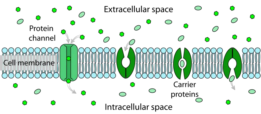
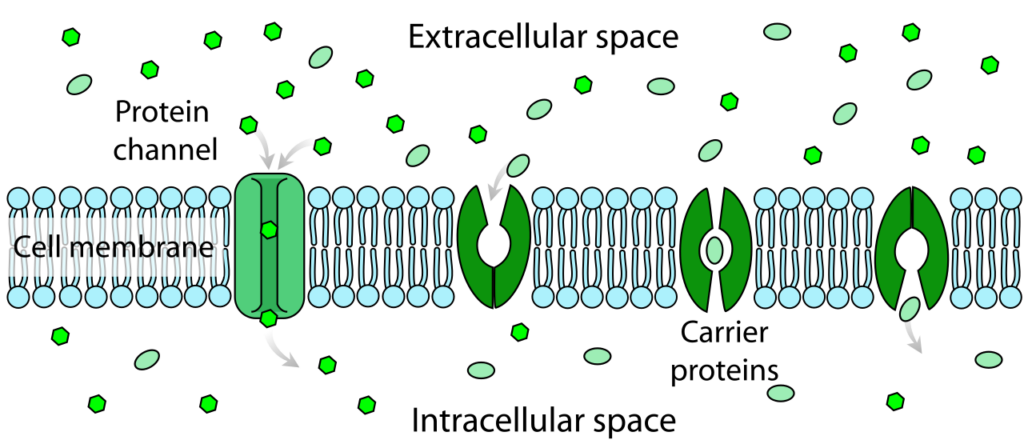
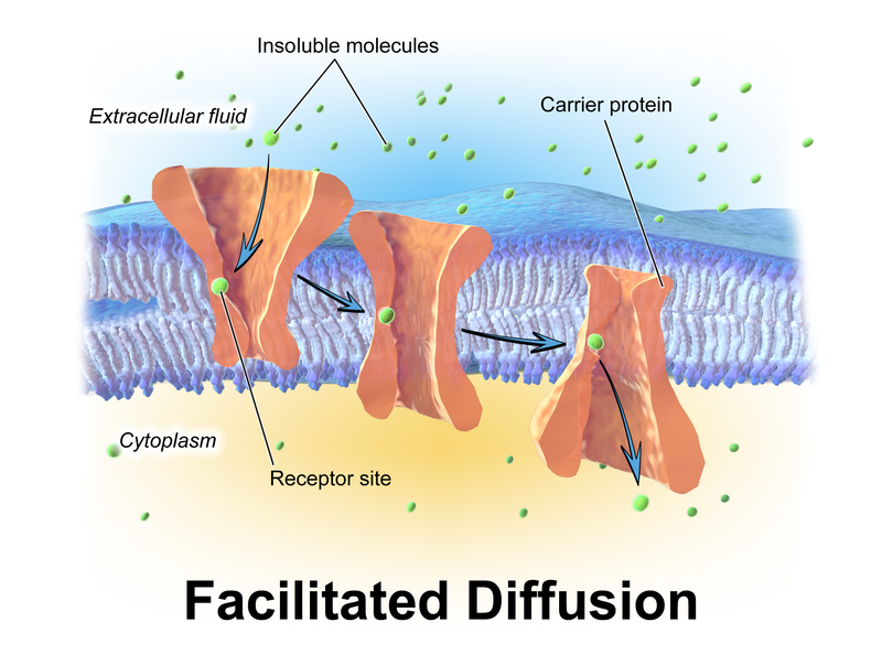
This type of passive transport contains some transmembrane integral proteins or specific transport proteins that are embedded in the plasma membrane. The carrier proteins or protein channels do not require any energy as it facilitates the diffusion of a solute from the area of higher concentration to an area of lower concentration.

1280px Scheme facilitated diffusion in cell membrane en.svg
osmosis example
Image from Wikipedia

This type of diffusion is also known as carrier-mediated osmosis. The most common example of this kind of transport is ion channels, aquaporins, and a Glucose transporter. The glucose transporter is also known as a GLUT2-Glucose transporter 2.

By the use of this transporter, a concentration gradient can drive glucose across the membrane. The major difference between simple diffusion and facilitated diffusion is that simple diffusion doesn’t require any transport protein while facilitated diffusion, for the movement across the membrane, requires transport protein to assist or facilitate the substances.

800px Blausen 0394 Facilitated Diffusion
osmosis example
Image from Wikipedia

Filtration

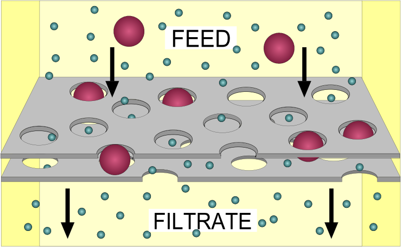
Filtration is non-specific and also come in the category of passive transport in which the hydrostatic pressure or some other physical pressure pushes fluid or solute molecules and water across the cell membrane and that mainly depends on the size of the pore.

The osmosis and filtration mainly depends on the concentration gradient while filtration has a pressure gradient via which molecule move from an area of higher pressure to the area of lower pressure.

800px FilterDiagram.svg
osmosis example
Image from Wikipedia

Some other most common example of passive transport is a voltage-gated ion channel in nerve cells. There is a concentration difference between NA+ and K+ inside and outside nerve cells.

On the membrane of a nerve cell, there is a voltage-gated channel that opens during the changes of voltage caused by other nerve cells.Osmotic balancing of salt concentration across the membrane is also an example of passive transport.

Conclusion

Passive transport is always energy-independent process that can able to transport the important material or substances across the membrane via the different type of transport mechanisms that uses energy-independent career proteins.


Also Read:

The Hydrophilic Head: A Crucial Component of Biological Membranes

what is hydrophilic head

The hydrophilic head of a phospholipid is a fundamental element that plays a pivotal role in the formation and function of biological membranes. It is composed of a phosphate group (PO4) and a glycerol molecule, which together create a highly polar region that is attracted to water molecules. This polar nature is attributed to the negative charge carried by the phosphate group, rendering the hydrophilic head “water-loving.”

Quantifiable Characteristics of the Hydrophilic Head

When it comes to the measurable and quantifiable aspects of the hydrophilic head, several key data points are worth considering:

  1. Cross-Sectional Area: The cross-sectional area of each hydrophilic head within a lipid bilayer ranges between approximately ¼ and ½ nm (BNID 106993). This dimension is crucial for understanding the packing and geometry of phospholipids in biological membranes.

  2. Length: The characteristic length of the hydrophilic head is approximately 2 nm (BNID 105298). This value provides insights into the overall structure and organization of the lipid bilayer.

  3. Mass: The mass of each hydrophilic head typically falls within the range of 500-1000 Da (BNID 101838). This parameter is essential for calculating the relative contributions of different lipid components to the overall mass and composition of the membrane.

Role of the Hydrophilic Head in Membrane Structure and Function

what is hydrophilic head

The hydrophilic heads of phospholipids play a crucial role in the interactions between the lipid bilayer and the aqueous environment, both inside and outside the cell. These polar regions form the outer layer of the lipid bilayer, interacting with the surrounding water molecules and creating a semi-permeable barrier that separates the cell’s interior from its external surroundings.

Membrane Composition and Structure

In mammalian cells, phospholipids account for approximately 60% of the total lipids by number, while sphingolipids make up another ≈10%. The remaining lipid fraction, consisting of non-polar sterol lipids, ranges from 0.1% to 40% depending on the cell type and the specific subcellular compartment under consideration (BNID 100947, 100948, 100949). This distribution highlights the significant contribution of hydrophilic heads to the overall composition and structure of biological membranes.

Membrane Permeability and Selective Transport

The hydrophilic heads of phospholipids, with their polar nature, are responsible for the semi-permeable properties of biological membranes. They interact with water molecules, creating a barrier that selectively allows the passage of certain molecules while restricting the movement of others. This selective permeability is crucial for maintaining the cell’s internal environment and facilitating the transport of essential nutrients, ions, and signaling molecules across the membrane.

Analytical Techniques for Phospholipid Characterization

To analyze the composition and distribution of phospholipids, including the hydrophilic heads, researchers employ a variety of advanced analytical techniques. These methods typically involve the following steps:

  1. Extraction: Lipids are extracted from biological samples using organic solvents, such as chloroform and methanol.

  2. Hydrolysis: The extracted lipids are then subjected to hydrolysis, which breaks down the ester bonds between the fatty acids and the glycerol backbone.

  3. Derivatization: The resulting fatty acids are derivatized, or chemically modified, to enhance their detection and separation capabilities.

  4. Chromatographic Separation: The derivatized fatty acids are then separated using techniques like gas chromatography (GC) or liquid chromatography (LC).

  5. Mass Spectrometry (MS): The separated fatty acids are detected and identified using mass spectrometry, which provides information on the types and relative concentrations of individual phospholipid species.

These analytical methods allow researchers to gain valuable insights into the composition and distribution of phospholipids, including the characteristics of their hydrophilic heads, within various biological samples.

Conclusion

The hydrophilic head of a phospholipid is a critical component of biological membranes, with measurable and quantifiable characteristics such as cross-sectional area, length, and mass. Its polar nature and interactions with the aqueous environment are essential for the formation and function of lipid bilayers, enabling the selective permeability and transport of molecules across the membrane. Advanced analytical techniques, such as chromatography and mass spectrometry, enable the identification and quantification of individual phospholipid species, providing a deeper understanding of the role of hydrophilic heads in the overall composition and structure of biological membranes.

References:

  1. What lipids are most abundant in membranes? – bionumbers book. Retrieved from https://book.bionumbers.org/what-lipids-are-most-abundant-in-membranes/
  2. Phospholipids: Structure and Cell Membranes – Creative Proteomics. Retrieved from https://www.creative-proteomics.com/blog/index.php/phospholipids-structure-and-cell-membranes/
  3. Phospholipid Bilayer Structure – an overview | ScienceDirect Topics. Retrieved from https://www.sciencedirect.com/topics/chemistry/phospholipid-bilayer-structure
  4. Biological Membranes Flashcards – Quizlet. Retrieved from https://quizlet.com/gb/509109621/biological-membranes-flash-cards/
  5. Hydrophilic Heads – Vocab, Definition, and Must Know Facts | Fiveable. Retrieved from https://library.fiveable.me/key-terms/ap-bio/hydrophilic-heads

Why is DNA Replication Semiconservative?

why is dna replication semiconservative

DNA replication is a fundamental process in all living organisms, where the genetic information stored in DNA is faithfully replicated to ensure the accurate transmission of genetic information to the next generation. The process of DNA replication is known as “semiconservative” because it involves the separation of the two strands of the double-helix DNA molecule, and the subsequent synthesis of new complementary strands using the original strands as templates.

The Mechanism of Semiconservative DNA Replication

  1. Unwinding of the DNA Helix: The first step in DNA replication is the unwinding of the double-helix DNA molecule. This is accomplished by the enzyme DNA helicase, which breaks the hydrogen bonds between the complementary base pairs, separating the two strands.

  2. Primer Synthesis: Once the DNA strands are separated, the enzyme DNA primase synthesizes short RNA primers on both the leading and lagging strands. These primers provide a free 3′ hydroxyl group for the DNA polymerase to initiate DNA synthesis.

  3. Complementary Strand Synthesis: The enzyme DNA polymerase III then uses the separated DNA strands as templates to synthesize new complementary DNA strands. The DNA polymerase III adds new deoxyribonucleotides (A, T, G, and C) to the 3′ end of the growing DNA strand, forming phosphodiester bonds between the new nucleotides and the existing strand.

  4. Discontinuous Synthesis on the Lagging Strand: On the lagging strand, DNA synthesis occurs in a discontinuous manner, with the formation of Okazaki fragments. This is because the DNA polymerase III can only synthesize DNA in the 5′ to 3′ direction, while the lagging strand is being replicated in the 3′ to 5′ direction. The Okazaki fragments are then joined together by the enzyme DNA ligase.

  5. Unwinding and Rewinding of the DNA Helix: As the replication fork moves forward, the DNA helicase continues to unwind the DNA helix, and the DNA polymerase III synthesizes new complementary strands. The newly synthesized DNA strands are then rewound into a double-helix structure.

The Semiconservative Nature of DNA Replication

why is dna replication semiconservative

The semiconservative nature of DNA replication was first demonstrated by the classic experiments of Matthew Meselson and Franklin Stahl in 1958. They grew bacteria in a medium containing a heavy isotope of nitrogen (15N) for several generations, causing the bacterial DNA to become labeled with the heavy isotope. The bacteria were then transferred to a medium containing the normal, lighter isotope of nitrogen (14N) and allowed to replicate their DNA.

After one round of DNA replication, Meselson and Stahl found that the DNA molecules contained one heavy strand and one light strand, demonstrating that the replication process is semiconservative. After two rounds of replication, they observed that the DNA molecules contained two light strands, further confirming the semiconservative nature of DNA replication.

The Importance of Semiconservative DNA Replication

The semiconservative nature of DNA replication is crucial for the accurate transmission of genetic information from one generation to the next. By using the original DNA strands as templates, the replication process ensures that the genetic information is faithfully replicated, with minimal errors. This is essential for the maintenance of genetic stability and the proper functioning of living organisms.

Furthermore, the semiconservative nature of DNA replication also plays a crucial role in the repair of DNA damage. When a DNA strand is damaged, the complementary strand can be used as a template to synthesize a new, undamaged strand, restoring the genetic information.

Conclusion

In summary, DNA replication is called semiconservative because it involves the separation of the two strands of the double-helix DNA molecule, and the subsequent synthesis of new complementary strands using the original strands as templates. This process ensures the accurate transmission of genetic information from one generation to the next, and is essential for the maintenance of genetic stability and the proper functioning of living organisms.

References:
1. Meselson, M., & Stahl, F. W. (1958). The replication of DNA in Escherichia coli. Proceedings of the National Academy of Sciences, 44(7), 671-682.
2. Hanawalt, P. C. (2004). Density matters: The semiconservative replication of DNA. Proceedings of the National Academy of Sciences, 101(52), 17889-17894.
3. Reece, J. B., Urry, L. A., Cain, M. L., Wasserman, S. A., Minorsky, P. V., & Jackson, R. B. (2011). Campbell biology (10th ed.). San Francisco, CA: Pearson.
4. Semi-conservative replication. (n.d.). In DNA learning center. Retrieved from https://www.dnalc.org/view/15879-Semi-conservative-replication.html.