Is a Chromosome an Allele?

is a chromosome an allele

Summary

Chromosomes and alleles are distinct biological entities that represent different levels of organization within the genetic makeup of an organism. While chromosomes are thread-like structures that contain genetic material, including genes and alleles, alleles are specific variants of a gene that occupy the same position or locus on a chromosome. This article delves into the key differences between chromosomes and alleles, providing a comprehensive understanding of their roles and relationships in the realm of genetics.

Understanding Chromosomes
is a chromosome an allele

Chromosomes are the thread-like structures found within the nucleus of eukaryotic cells, such as those found in plants, animals, and fungi. These structures are composed of a single, long molecule of deoxyribonucleic acid (DNA) tightly packaged with various proteins, including histones, to form a compact and organized unit.

Chromosome Structure and Composition

  • Chromosomes are typically composed of a single, continuous DNA molecule that can range from millions to billions of base pairs in length, depending on the organism.
  • The DNA molecule is wrapped around histone proteins, forming nucleosomes, which further coil and fold to create the characteristic chromosome structure.
  • Chromosomes contain not only the genes that encode proteins but also regulatory sequences, repetitive DNA, and other non-coding regions.
  • The number of chromosomes varies among different species, with humans having 23 pairs of chromosomes, for a total of 46 chromosomes.
  • Each chromosome has a distinct structure, with a centromere, two arms (a short arm and a long arm), and telomeres at the ends, which play a crucial role in chromosome stability and replication.

Chromosome Inheritance and Ploidy

  • Chromosomes are inherited in pairs, with one chromosome from each pair coming from the maternal parent and the other from the paternal parent.
  • The number of chromosome sets, or the ploidy level, can vary among different organisms. Humans are diploid, meaning they have two sets of chromosomes (2n).
  • Other organisms, such as some plants and some protists, can be polyploid, having more than two sets of chromosomes (e.g., 4n, 6n, 8n).
  • The ploidy level is an important factor in genetic diversity, as it can influence the expression and inheritance of traits.

Understanding Alleles

Alleles are the different versions of a gene that occupy the same position, or locus, on a chromosome. These variations can result in different phenotypic (physical) characteristics or traits.

Allele Variations and Genotypes

  • Each gene has two alleles, one inherited from each parent.
  • Alleles can be the same (homozygous) or different (heterozygous) for a particular gene.
  • Homozygous individuals have two copies of the same allele (e.g., AA or aa), while heterozygous individuals have two different alleles (e.g., Aa).
  • The combination of alleles for a particular gene is known as the genotype, which determines the phenotypic expression of that trait.

Allele Dominance and Expression

  • Alleles can be classified as dominant or recessive based on their relative expression in the phenotype.
  • Dominant alleles are expressed in the phenotype, even if the individual is heterozygous (Aa).
  • Recessive alleles are only expressed in the phenotype when the individual is homozygous for the recessive allele (aa).
  • The concept of dominance and recessiveness is crucial in understanding the inheritance of traits and the expression of genetic disorders.

Allele Frequency and Genetic Diversity

  • Allele frequency refers to the relative abundance of a particular allele within a population.
  • Genetic diversity within a population is influenced by the presence and frequency of different alleles for a given gene.
  • Factors such as mutation, gene flow, genetic drift, and natural selection can affect the frequency and distribution of alleles in a population over time.
  • Maintaining genetic diversity is important for the adaptability and resilience of populations in the face of environmental changes or challenges.

Chromosomes and Alleles: The Relationship

While chromosomes and alleles are distinct biological entities, they are closely related in the context of genetics and heredity.

Genes and Chromosomes

  • Genes are the fundamental units of heredity, and they are located on chromosomes.
  • Each chromosome contains numerous genes, with the specific number of genes varying depending on the organism and the size of the chromosome.
  • Genes are the DNA sequences that encode the instructions for the synthesis of proteins or other functional molecules within the cell.

Alleles and Chromosomes

  • Alleles are the different versions of a gene that occupy the same position, or locus, on a chromosome.
  • Each gene has two alleles, one inherited from each parent, and these alleles can be the same (homozygous) or different (heterozygous).
  • The combination of alleles for a particular gene determines the genotype, which in turn influences the phenotypic expression of that trait.

Chromosomes, Genes, and Alleles in Genetic Inheritance

  • During meiosis, the process of cell division that produces gametes (sperm or eggs), the chromosomes are replicated and then segregated into the daughter cells.
  • Each gamete receives one chromosome from each pair, ensuring that the offspring inherits one set of chromosomes from each parent.
  • The alleles present on the chromosomes inherited from the parents determine the genetic makeup of the offspring, influencing their physical and physiological characteristics.

Conclusion

In summary, chromosomes and alleles are distinct biological entities that play crucial roles in the genetic makeup and inheritance of organisms. Chromosomes are the thread-like structures that contain the genetic material, including genes and alleles, while alleles are the different versions of a gene that occupy the same position on a chromosome. Understanding the relationship between chromosomes, genes, and alleles is essential for comprehending the fundamental principles of genetics and the mechanisms of heredity.
References:

  1. Nature Scitable – Allele
  2. ScienceDirect – Allele
  3. Biology Online – Allele
  4. Genetics Generation – Chromosomes
  5. National Human Genome Research Institute – Chromosome
  6. Khan Academy – Alleles and Genotypes

Are Proteins Inherited? A Comprehensive Guide

are proteins inherited

Summary

Proteins are the fundamental building blocks of life, and their inheritance is a complex and fascinating topic in the field of genetics. While proteins are directly produced from the genetic information encoded in DNA, the relationship between genotype (genetic makeup) and phenotype (observable traits) is not always straightforward. This article delves into the intricacies of protein inheritance, exploring the role of cis-acting and trans-acting genetic variants, the impact of transcriptional and translational regulation, and the insights gained from recent studies on the genetic basis of protein level variation.

Understanding Protein Inheritance

are proteins inherited

The Genetic Basis of Protein Production

Proteins are the products of genes, which are themselves inherited. The genetic information encoded in DNA is first transcribed into messenger RNA (mRNA), and then translated into the corresponding protein molecules. This process of gene expression is the foundation of protein inheritance, as the genetic information passed down from parents to offspring ultimately determines the proteins that will be produced in the cells of the offspring.

The Complexity of Genotype-Phenotype Relationships

While the inheritance of proteins is directly linked to the inheritance of genes, the relationship between genotype and phenotype is not always straightforward. The expression of genes is influenced by a variety of factors, including:

  1. Transcriptional Regulation: The rate and timing of gene transcription can be regulated by transcription factors, epigenetic modifications, and other regulatory elements.
  2. Translational Regulation: The efficiency of protein synthesis can be influenced by factors such as mRNA stability, codon usage, and the availability of translation machinery.
  3. Post-Translational Modifications: Proteins can undergo various chemical modifications after translation, which can alter their structure, function, and stability.
  4. Protein-Protein Interactions: Proteins often interact with other proteins, forming complex networks that can influence their activity and localization within the cell.

These factors can lead to differences in protein expression and function, even in individuals with the same genotype. As a result, the same genotype can give rise to different phenotypes under different environmental or developmental conditions.

Cis-acting and Trans-acting Genetic Variants

In the context of protein inheritance, it is important to consider both cis-acting and trans-acting genetic variants. Cis-acting variants are located near the gene they affect and can influence the expression of that gene by affecting its transcription or translation. Trans-acting variants, on the other hand, are located elsewhere in the genome and can influence the expression of genes at distant loci.

A study published in the journal Science in 2014 analyzed the levels of 4,084 GFP-tagged yeast proteins in the progeny of a cross between a laboratory and a wild strain. The study found that:

  1. Cis-acting variants had a large effect on protein level, six times more frequently than the rest of the genome combined.
  2. Trans-acting variants contributed little to protein level variation between individual cells but explained more than 50% of the variance in the population’s average protein abundance for half of the GFP fusions tested.

This suggests that both cis-acting and trans-acting genetic variants play important roles in determining protein levels, with cis-acting variants having a more pronounced effect on individual protein expression and trans-acting variants contributing more to population-level variation.

Shared and Independent Genetic Control of Transcript and Protein Abundance

The study also found that more than half of the expression quantitative trait loci (eQTLs) contribute to changes in protein levels of regulated genes, but several protein quantitative trait loci (pQTLs) do not affect their cognate transcript levels. This indicates that there is both shared and independent genetic control of transcript and protein abundance.

Factors that can contribute to this independent genetic control include:

  1. Post-Transcriptional Regulation: Processes such as mRNA stability, translation efficiency, and protein degradation can be influenced by genetic variants that do not affect transcript levels.
  2. Post-Translational Modifications: Genetic variants can influence the enzymes and pathways responsible for post-translational modifications, leading to changes in protein function and abundance without affecting transcript levels.
  3. Protein Localization and Trafficking: Genetic variants can impact the cellular localization and trafficking of proteins, which can affect their availability and function without altering transcript levels.

Understanding the complex interplay between genetic variants, transcript abundance, and protein expression is crucial for unraveling the genetic basis of heritable traits and phenotypes.

Implications and Applications

The study of protein inheritance has important implications for various fields, including:

  1. Evolutionary Biology: Analyzing the genetic basis of protein level variation can provide insights into the evolutionary forces that shape the genotype-phenotype relationship and the emergence of new traits.
  2. Personalized Medicine: Identifying the genetic determinants of protein expression and function can help develop targeted therapies and personalized treatment approaches for genetic disorders and complex diseases.
  3. Biotechnology and Synthetic Biology: Understanding the principles of protein inheritance can inform the design and engineering of novel proteins and biological systems with desired properties.

By delving deeper into the intricacies of protein inheritance, researchers can uncover the complex mechanisms that govern the expression and function of these essential biomolecules, ultimately advancing our understanding of life and its remarkable diversity.

Conclusion

Proteins are the fundamental building blocks of life, and their inheritance is a complex and fascinating topic in the field of genetics. While proteins are directly produced from the genetic information encoded in DNA, the relationship between genotype and phenotype is not always straightforward. Factors such as transcriptional and translational regulation, post-translational modifications, and protein-protein interactions can all influence the expression and function of proteins.

In the context of protein inheritance, it is important to consider both cis-acting and trans-acting genetic variants, as well as the shared and independent genetic control of transcript and protein abundance. By studying the genetic basis of protein level variation, researchers can gain insights into the evolutionary forces that shape the genotype-phenotype relationship and develop new approaches for personalized medicine and biotechnology applications.

As our understanding of protein inheritance continues to evolve, we can expect to see exciting advancements in our ability to harness the power of these essential biomolecules and unlock the secrets of life itself.

References

  1. Estimating the information content of genetic sequence data: https://academic.oup.com/jrsssc/article/72/5/1310/7230752
  2. Quantification of Protein Levels in Single Living Cells: https://www.sciencedirect.com/science/article/pii/S2211124715013637
  3. Heritability and genetic basis of protein level variation in an outbred population: https://www.ncbi.nlm.nih.gov/pmc/articles/PMC4120089/

Is Osmosis Hypotonic? A Comprehensive Guide

is osmosis hypotonic

Osmosis is a fundamental biological process that involves the movement of water molecules through a selectively permeable membrane from a region of higher water potential (lower solute concentration) to a region of lower water potential (higher solute concentration). When the concentration of solutes on either side of the membrane differs, the solution with a lower solute concentration is considered hypotonic, while the solution with a higher solute concentration is hypertonic. In a hypotonic solution, the water potential is higher than that of the cell, causing water to move into the cell, potentially leading to its bursting if the concentration difference is significant.

Understanding Osmosis and Tonicity

Osmosis is a passive transport process that occurs due to the difference in the concentration of solutes on either side of a selectively permeable membrane. The movement of water molecules is driven by the difference in water potential, which is influenced by the concentration of solutes.

Tonicity and its Types

Tonicity refers to the relative concentration of solutes on either side of the membrane. There are three main types of tonicity:

  1. Hypotonic: The solution has a lower solute concentration than the cell, resulting in a higher water potential. This causes water to move into the cell, potentially leading to cell swelling and bursting.

  2. Hypertonic: The solution has a higher solute concentration than the cell, resulting in a lower water potential. This causes water to move out of the cell, leading to cell shrinkage and plasmolysis.

  3. Isotonic: The solution has the same solute concentration as the cell, resulting in no net movement of water across the membrane.

Factors Affecting Osmosis

Several factors can influence the rate and direction of osmosis, including:

  1. Membrane Permeability: The selective permeability of the membrane determines which molecules can pass through and at what rate.

  2. Solute Concentration: The difference in solute concentration between the two solutions drives the movement of water.

  3. Temperature: Increased temperature can enhance the kinetic energy of water molecules, leading to a faster rate of osmosis.

  4. Pressure: Applying external pressure can affect the direction and rate of osmosis, as described by the principles of osmotic pressure.

Quantifying Osmosis: Experimental Approaches

is osmosis hypotonic

To measure osmosis quantitatively, researchers often conduct experiments involving the change in mass of a substance due to the movement of water.

Potato Experiment

In this experiment, the mass of a potato is measured before and after immersion in a salt solution of varying concentrations. The resulting change in mass can be used to determine the osmotic pressure and water potential of the solution.

  1. Procedure:
  2. Weigh a potato sample and record its initial mass.
  3. Immerse the potato in a salt solution of known concentration.
  4. Measure the mass of the potato after a specified time interval.
  5. Calculate the change in mass and relate it to the osmotic pressure and water potential of the solution.

  6. Calculations:

  7. Percent change in mass = (Final mass – Initial mass) / Initial mass × 100
  8. Osmotic pressure = -RTc, where R is the gas constant, T is the absolute temperature, and c is the molar concentration of the solute.
  9. Water potential = Solute potential + Pressure potential

Dialysis Tubing Experiment

In this experiment, a dialysis tubing bag is filled with a glucose solution and immersed in a solution of varying glucose concentrations. The change in mass of the bag is used to calculate the concentration of glucose and water inside the bag, providing a quantitative measure of osmosis.

  1. Procedure:
  2. Weigh the empty dialysis tubing bag and record its initial mass.
  3. Fill the bag with a glucose solution of known concentration.
  4. Immerse the filled bag in a solution with a different glucose concentration.
  5. Measure the mass of the bag after a specified time interval.
  6. Calculate the change in mass and use it to determine the glucose and water concentrations inside the bag.

  7. Calculations:

  8. Percent change in mass = (Final mass – Initial mass) / Initial mass × 100
  9. Glucose concentration inside the bag = (Initial glucose mass) / (Final volume of solution inside the bag)
  10. Water concentration inside the bag = (Final volume of solution inside the bag – Initial volume of glucose solution) / (Final volume of solution inside the bag)

These experimental approaches provide a quantitative understanding of the osmotic processes and the factors that influence them, such as solute concentration, membrane permeability, and temperature.

Implications of Hypotonic Conditions

In a hypotonic solution, the concentration of solutes is lower than that of the cell, leading to a net movement of water into the cell. This can result in an increase in the cell’s volume and turgor pressure, potentially causing it to burst if the concentration difference is significant.

Cell Swelling and Bursting

When a cell is placed in a hypotonic solution, water moves into the cell due to the difference in water potential. This can lead to an increase in the cell’s volume and turgor pressure, which can ultimately cause the cell to burst if the concentration difference is too great.

  1. Factors Affecting Cell Bursting:
  2. Magnitude of the concentration difference between the cell and the solution
  3. Permeability of the cell membrane to water
  4. Strength and elasticity of the cell wall or membrane

  5. Consequences of Cell Bursting:

  6. Release of cellular contents
  7. Disruption of cellular homeostasis
  8. Potential cell death

Importance of Turgor Pressure in Plants

In plant cells, turgor pressure is essential for maintaining the structure and strength of the plant. Turgor pressure is generated by the net movement of water into the cell, which creates a positive pressure within the cell.

  1. Role of Turgor Pressure:
  2. Provides rigidity and support to plant tissues and organs
  3. Facilitates the opening and closing of stomata for gas exchange
  4. Enables the movement of water and nutrients within the plant

  5. Consequences of Decreased Turgor Pressure:

  6. Wilting of plant tissues and organs
  7. Reduced gas exchange and photosynthetic efficiency
  8. Impaired transport of water and nutrients

To ensure the proper functioning of plant cells and the overall health of the plant, it is crucial to maintain appropriate water potential and osmotic pressure within the cells.

Conclusion

Osmosis is a fundamental biological process that involves the movement of water molecules through a selectively permeable membrane. When the concentration of solutes on either side of the membrane differs, the solution with a lower solute concentration is considered hypotonic. In a hypotonic solution, the water potential is higher than that of the cell, causing water to move into the cell, potentially leading to its bursting if the concentration difference is significant.

Quantifying osmosis through experiments, such as the potato and dialysis tubing experiments, provides valuable insights into the water potential and osmotic pressure of solutions. Understanding the implications of hypotonic conditions, particularly the effects on cell swelling, bursting, and the importance of turgor pressure in plants, is crucial for maintaining the proper functioning of biological systems.

By exploring the intricacies of osmosis and the specific case of hypotonic conditions, this comprehensive guide aims to equip readers with a deeper understanding of this fundamental biological process and its practical applications in various fields of study.

References:
Osmosis Lab Example 2
AP Lab 1: Osmosis Sample 3
Osmosis Lesson

Can Plant Cells Have Cytokinesis? A Comprehensive Guide

can plant cell have cytokinesis

Plant cells undergo a highly regulated process of cell division called cytokinesis, which divides the parent cell into two daughter cells. This process involves the formation of a cell plate, which eventually develops into a cell wall. The cell plate formation in dividing plant cells follows a precise pattern involving choreographed vesicle motion and distinct developmental stages.

The Phragmoplast and Cell Plate Formation

During cytokinesis, the cell plate originates from the phragmoplast, a microtubule structure that forms between the two sets of chromosomes during mitosis. The phragmoplast guides the transport of vesicles containing cell wall materials to the equatorial plane, where they fuse to form the cell plate.

The phragmoplast is a dynamic structure that undergoes significant changes during the different stages of cell plate formation. It is composed of two sets of overlapping microtubules that originate from the spindle poles and converge at the equatorial plane. These microtubules serve as tracks for the transport of Golgi-derived vesicles, which carry the necessary materials for cell plate construction.

As the cell plate formation progresses, the phragmoplast expands centrifugally, guiding the vesicles to the growing edges of the cell plate. This expansion is driven by the continuous addition of new microtubules at the leading edge of the phragmoplast, as well as the depolymerization of microtubules at the trailing edge.

Developmental Phases of Cell Plate Formation

can plant cell have cytokinesis

The cell plate formation process can be divided into distinct developmental phases based on the lateral expansion rate of the growing cell plate:

  1. Initial Phase: During this phase, a narrow cell plate is formed at the equatorial plane. The cell plate originates from the fusion of Golgi-derived vesicles, which are transported to the equatorial plane by the phragmoplast.

  2. Rapid Expansion Phase: In this phase, the cell plate width increases significantly, expanding laterally towards the parental cell walls. The rapid expansion is driven by the continuous fusion of vesicles at the growing edges of the cell plate.

  3. Final Phase: The final phase is characterized by the deposition of callose and cellulose, which strengthens the cell plate and transforms it into a cell wall. Callose is initially deposited, forming a temporary cell wall, followed by the deposition of cellulose, which provides the final structural integrity.

The transition between these developmental phases is marked by changes in the organization and dynamics of the phragmoplast, as well as the composition and properties of the growing cell plate.

Vesicle Dynamics during Cell Plate Formation

The dynamics of vesicle transport and fusion during plant cell cytokinesis have been extensively studied using Fluorescence Microscopy of Organelle Surfaces (FluMOS) and other advanced imaging techniques.

FluMOS has revealed that the lateral expansion rate of the growing cell plate is not constant but shows distinct patterns during the different developmental phases. During the initial phase, the vesicle fusion rate is relatively slow, leading to the formation of a narrow cell plate. In the rapid expansion phase, the vesicle fusion rate increases significantly, driving the rapid lateral growth of the cell plate.

Furthermore, the FluMOS studies have shown that the vesicle dynamics are highly coordinated and regulated, with distinct populations of vesicles responsible for different aspects of cell plate formation. For example, some vesicles carry cell wall materials, such as cellulose and callose, while others are involved in the fusion and expansion of the cell plate.

Factors Influencing Cell Plate Formation

The cell plate formation process is influenced by various factors, including:

  1. Microtubule Organization: The organization and dynamics of the phragmoplast microtubules play a crucial role in guiding the transport and fusion of vesicles during cell plate formation.

  2. Actin Cytoskeleton: The actin cytoskeleton is also involved in the organization and dynamics of the phragmoplast, as well as the transport and fusion of vesicles.

  3. Regulatory Proteins: A variety of regulatory proteins, such as kinases, phosphatases, and small GTPases, are involved in the coordination and regulation of the cell plate formation process.

  4. Cell Cycle Progression: The cell plate formation is tightly coupled with the progression of the cell cycle, with the phragmoplast and cell plate formation occurring during the late stages of mitosis.

  5. Environmental Factors: Environmental factors, such as temperature, light, and nutrient availability, can also influence the dynamics and efficiency of cell plate formation in plant cells.

Significance of Cytokinesis in Plant Development

Cytokinesis and the formation of the cell plate are essential processes for the growth and development of plants. The successful division of plant cells through cytokinesis allows for the formation of new cells, tissues, and organs, enabling the plant to expand, differentiate, and adapt to its environment.

The cell plate formation and its subsequent transformation into a cell wall are crucial for the establishment of cell boundaries, the maintenance of cell shape and size, and the overall structural integrity of the plant body. Additionally, the cell plate serves as a platform for the deposition of cell wall materials, which are essential for the plant’s mechanical support, protection, and communication between cells.

Furthermore, the precise regulation of cytokinesis and cell plate formation is crucial for the proper development and function of various plant tissues and organs, such as the vascular system, meristems, and reproductive structures.

Conclusion

In summary, plant cells undergo a highly regulated process of cytokinesis, which involves the formation of a cell plate that eventually develops into a cell wall. The cell plate formation is a complex and dynamic process that is guided by the phragmoplast, a microtubule-based structure, and involves precisely choreographed vesicle motion and distinct developmental phases.

The study of plant cell cytokinesis, including the dynamics of vesicle transport and fusion, has been greatly advanced by the use of techniques like Fluorescence Microscopy of Organelle Surfaces (FluMOS). These studies have provided valuable insights into the factors that influence the cell plate formation process and its significance in plant development.

Understanding the intricacies of plant cell cytokinesis is not only crucial for our fundamental knowledge of plant biology but also has potential applications in areas such as plant breeding, tissue engineering, and the development of novel agricultural technologies.

Reference:

  1. Cytokinesis in Plant Cells
  2. How is Cytokinesis Different in Plant Cells as Compared to Animal Cells?
  3. How Plant Cells Divide (Cytokinesis)
  4. Fluorescence Microscopy of Organelle Surfaces (FluMOS): A Novel Technique for Visualizing and Quantifying Membrane Dynamics in Living Cells

Are Membrane Proteins Carrier Proteins? A Comprehensive Guide

are membrane proteins carrier proteins

Membrane proteins and carrier proteins are essential components of biological membranes, playing crucial roles in the transportation of molecules across these barriers. While both types of proteins are involved in membrane transport, they have distinct characteristics and functions. This comprehensive guide will delve into the intricacies of membrane proteins and carrier proteins, providing a detailed understanding of their roles, mechanisms, and importance in various biological processes.

Understanding Membrane Proteins

Membrane proteins are a diverse class of proteins that are embedded within or associated with the lipid bilayer of biological membranes. These proteins serve a wide range of functions, including:

  1. Transport: Membrane proteins facilitate the movement of molecules, ions, and other substances across the membrane, either through passive diffusion or active transport.
  2. Signaling: Membrane proteins act as receptors, transducing extracellular signals into intracellular responses, and as signal transducers, relaying information between different cellular compartments.
  3. Structural Support: Some membrane proteins provide structural integrity to the membrane, contributing to the overall organization and stability of the cell.
  4. Enzymatic Activity: Certain membrane proteins possess enzymatic functions, catalyzing various biochemical reactions within the membrane or at the membrane-cytoplasm interface.

Membrane proteins can be classified into several categories based on their structural and functional characteristics, such as integral membrane proteins, peripheral membrane proteins, and lipid-anchored membrane proteins.

Carrier Proteins: Facilitating Membrane Transport

are membrane proteins carrier proteins

Carrier proteins, a specific type of membrane protein, are responsible for the facilitated transport of molecules across biological membranes. These proteins bind to specific solutes and undergo conformational changes to translocate the solutes from one side of the membrane to the other.

Types of Carrier Proteins

Carrier proteins can be classified into two main categories based on the type of transport they facilitate:

  1. Simple Diffusion Carriers: These carriers facilitate the passive movement of molecules down their concentration gradient, from a region of high concentration to a region of low concentration.
  2. Facilitated Diffusion Carriers: These carriers use protein channels to aid in the movement of molecules, also down their concentration gradient.

An example of a facilitated diffusion carrier is the glucose transporter (GLUT), which facilitates the transport of glucose into cells.

Carrier Protein Mechanism

The process by which a carrier protein transfers a solute molecule across the lipid bilayer resembles an enzyme-substrate reaction. Each carrier protein has one or more specific binding sites for its solute (substrate). The carrier protein undergoes reversible conformational changes that alternately expose the solute-binding site on one side of the membrane and then on the other, effectively transporting the solute across the membrane.

The rate of transport, known as Vmax, is characteristic of the specific carrier and reflects the rate at which the carrier can flip between its two conformational states. Additionally, each transporter protein has a characteristic binding constant for its solute, Km, which represents the concentration of solute when the transport rate is half its maximum value.

Factors Affecting Carrier Protein Function

The rate of diffusion, both simple and facilitated, is regulated by several factors, including:

  1. Concentration Gradient: The difference in solute concentration between the two sides of the membrane drives the direction and rate of transport.
  2. Particle Size and Charge: The size and charge of the transported molecules can influence their ability to pass through the carrier protein’s binding site and channel.
  3. Temperature and Viscosity: Changes in temperature and the viscosity of the medium can affect the kinetics of the carrier protein’s conformational changes and the overall rate of transport.

Active Transport and Other Membrane Transport Mechanisms

In addition to carrier proteins, cells employ other mechanisms for active transport across membranes, including:

  1. Coupled Carriers: These carriers couple the uphill transport of one solute to the downhill transport of another, allowing the cell to move molecules against their concentration gradient.
  2. ATP-Driven Pumps: These membrane proteins use the energy released from the hydrolysis of ATP to drive the uphill transport of molecules across the membrane.
  3. ABC Transporters: This family of transport ATPases uses the energy of ATP hydrolysis to import or export certain small molecules, playing a crucial role in various physiological and pathological processes.

Importance and Applications of Membrane Proteins and Carrier Proteins

Membrane proteins and carrier proteins are essential for a wide range of biological processes, including:

  1. Cell Signaling and Communication: Membrane proteins act as receptors and signal transducers, enabling cells to respond to extracellular stimuli and coordinate their activities.
  2. Nutrient and Ion Homeostasis: Carrier proteins facilitate the transport of essential nutrients, ions, and other molecules across membranes, maintaining the appropriate concentrations within cells and tissues.
  3. Drug Transport and Pharmacology: Understanding the mechanisms of membrane proteins and carrier proteins is crucial for the development of targeted drug therapies, as these proteins can influence the absorption, distribution, and elimination of drugs.
  4. Diagnostics and Biomarkers: Membrane proteins and carrier proteins can serve as valuable biomarkers for the diagnosis and monitoring of various diseases, as their expression and function can be altered in pathological conditions.

Conclusion

Membrane proteins and carrier proteins are indispensable components of biological membranes, playing pivotal roles in the transportation of molecules across these barriers. By understanding the intricate mechanisms and functions of these proteins, researchers and clinicians can gain valuable insights into a wide range of biological processes and develop innovative strategies for addressing various health-related challenges.

References:

  • Carrier Protein – an overview | ScienceDirect Topics
  • Membrane Transport – BIOLOGY FOR LIFE
  • Getting Across the Cell Membrane: An Overview for Small Molecules …
  • Topic 1.4 Membrane Transport
  • Carrier Proteins and Active Membrane Transport – NCBI

Is DNA Replication Antiparallel? A Comprehensive Guide

is dna replication antiparallel

DNA replication is a fundamental process in biology, where the genetic information stored in DNA is faithfully replicated to ensure the accurate transmission of genetic material to daughter cells. One of the critical aspects of DNA replication is its antiparallel nature, which has significant implications for the replication process. In this comprehensive guide, we will delve into the details of DNA replication and explore how the antiparallel structure of DNA affects this crucial process.

Understanding the Antiparallel Structure of DNA

The DNA double helix is composed of two complementary strands that run in opposite directions, known as the antiparallel structure. This means that the 5′ to 3′ direction of one strand is opposite to the 5′ to 3′ direction of the other strand. This structural arrangement is essential for the efficient and accurate replication of DNA.

The 5′ to 3′ Directionality of DNA Strands

The DNA strands are composed of nucleotides, each containing a sugar (deoxyribose), a phosphate group, and a nitrogenous base (adenine, guanine, cytosine, or thymine). The sugar-phosphate backbone of the DNA strands has a distinct directionality, with the 5′ carbon of one sugar connected to the 3′ carbon of the next sugar. This 5′ to 3′ directionality is crucial for the replication process.

The Complementary Base Pairing

The two strands of the DNA double helix are held together by complementary base pairing, where adenine (A) pairs with thymine (T), and guanine (G) pairs with cytosine (C). This base pairing ensures the stability and integrity of the DNA molecule, as well as the accurate transmission of genetic information during replication.

The Impact of Antiparallel Structure on DNA Replication

is dna replication antiparallel

The antiparallel structure of DNA has a significant impact on the replication process, particularly in the way DNA polymerases, the enzymes responsible for synthesizing new DNA strands, function.

DNA Polymerase and the Leading Strand

DNA polymerases can only add new nucleotides in the 5′ to 3′ direction. On the leading strand, where the DNA strands are opening up in the 5′ to 3′ direction, the DNA polymerase can continuously synthesize the new strand, moving in the same direction as the replication fork.

The Lagging Strand and Okazaki Fragments

On the lagging strand, where the DNA strands are opening up in the 3′ to 5′ direction, the DNA polymerase must work in the opposite direction. This means that the lagging strand is synthesized in short, discontinuous fragments called Okazaki fragments, which are later joined together by the enzyme DNA ligase.

The Replication Fork and the Coordination of Replication

The replication fork is the site where the DNA double helix is unwound, and the new strands are synthesized. The leading strand is continuously replicated, while the lagging strand is replicated in Okazaki fragments. This coordination between the leading and lagging strands is essential for the efficient and accurate replication of the entire DNA molecule.

The Advantages of Antiparallel DNA Replication

The antiparallel structure of DNA offers several advantages for the replication process, which have been observed in various studies.

Increased Replication Rate

A study published in Nature Communications in 2020 found that the antiparallel orientation of DNA strands increases the rate of replication. By dividing the DNA into predictable, independently and simultaneously replicating segments, the replication process is parallelized, leading to a faster overall replication rate.

Improved Accuracy and Fidelity

The antiparallel structure of DNA also contributes to the accuracy and fidelity of the replication process. By having the two strands oriented in opposite directions, the DNA polymerase can more easily identify and correct any errors that may occur during replication, ensuring the faithful transmission of genetic information.

Efficient Proofreading and Error Correction

The antiparallel structure of DNA allows for more efficient proofreading and error correction mechanisms. DNA polymerases can detect and correct mismatched base pairs by moving in the opposite direction on the leading and lagging strands, ensuring the high fidelity of the replicated DNA.

Conclusion

The antiparallel structure of DNA is a fundamental aspect of the DNA replication process, with significant implications for the efficiency, accuracy, and fidelity of genetic information transmission. By understanding the impact of this structural arrangement on DNA replication, we can gain deeper insights into the mechanisms that ensure the precise duplication of genetic material, which is essential for the survival and evolution of all living organisms.

References

  1. Cells Can Replicate Their DNA Precisely
  2. How does the antiparallel structure of the double helix affect replication?
  3. Antiparallel strand orientation of duplex DNA increases the rate of replication
  4. The Structure of DNA
  5. DNA Replication: The Semiconservative Model

Are Bacteria Photosynthetic? A Comprehensive Guide

are bacteria photosynthetic

Photosynthetic bacteria are a diverse group of microorganisms that possess the remarkable ability to harness light energy and convert it into organic compounds. These bacteria play a crucial role in the global carbon and oxygen cycles, making them a subject of great interest in the field of biology. In this comprehensive guide, we will delve into the intricacies of photosynthetic bacteria, exploring their classification, mechanisms, and the various techniques used to study them.

Photosynthetic Bacteria: An Overview

Photosynthetic bacteria, also known as phototrophic bacteria, are a group of prokaryotic organisms that can perform photosynthesis. These bacteria use light energy to drive the synthesis of organic compounds from carbon dioxide and water, a process that releases oxygen as a byproduct. Photosynthetic bacteria can be divided into two main groups: oxygenic and anoxygenic phototrophs.

Oxygenic Photosynthetic Bacteria

Oxygenic photosynthetic bacteria, also referred to as cyanobacteria, are the only bacteria that produce oxygen during photosynthesis. These gram-negative bacteria use water as an electron donor and release oxygen as a byproduct. Cyanobacteria are believed to have played a crucial role in the oxygenation of the Earth’s atmosphere early in its history, paving the way for the evolution of aerobic life.

Cyanobacteria are found in a wide range of environments, including freshwater, marine ecosystems, soils, and even in symbiotic associations with plants and animals. They possess a unique photosynthetic apparatus that includes chlorophyll-a and other accessory pigments, such as carotenoids and phycobilins, which help them capture a broad spectrum of light wavelengths.

Anoxygenic Photosynthetic Bacteria

Anoxygenic photosynthetic bacteria, on the other hand, do not produce oxygen during photosynthesis. Instead, they use various electron donors, such as sulfur compounds, hydrogen, or organic compounds, instead of water. These bacteria are found in a variety of environments, including freshwater, marine ecosystems, soils, and hot springs.

Anoxygenic photosynthetic bacteria possess a different photosynthetic apparatus compared to cyanobacteria, often relying on bacteriochlorophyll as the primary photosynthetic pigment. They play important roles in sulfur and hydrogen cycling in their respective ecosystems.

Measuring Photosynthetic Activity in Bacteria

are bacteria photosynthetic

Researchers have developed several techniques to quantify the photosynthetic activity and characteristics of photosynthetic bacteria. Two commonly used methods are pulse amplitude modulation (PAM) fluorometry and total reflection X-ray fluorescence (TXRF) spectrometry.

Pulse Amplitude Modulation (PAM) Fluorometry

PAM fluorometry is a non-invasive technique that measures the quantum yield of photosystem II (ΦPSII) in response to increasing light intensity. This method allows for the quantification of photosynthetic rates and the estimation of chlorophyll-a content per microbial cell. By using PAM fluorometry, researchers can gain insights into the photosynthetic efficiency and carbon fixation rates of photosynthetic bacterial communities.

For example, a study published in Nature in 2022 utilized PAM fluorometry to investigate the photosynthetic rates of microbial communities in bryophyte material. The researchers found that microbial chlorophyll-a concentration, photosynthesis efficiency, and CO2 fixation rate varied across different plots, and that environmental factors such as bryophyte water content and light intensity were important in structuring the photosynthetic communities.

Total Reflection X-ray Fluorescence (TXRF) Spectrometry

TXRF spectrometry is another technique used to study photosynthetic bacteria. This method allows for the quantification of trace elements in bacteria and their photosynthetic structures. By analyzing the elemental composition of photosynthetic bacteria, researchers can gain insights into the specific requirements and adaptations of these microorganisms.

A study published in the Journal of Analytical Atomic Spectrometry in 2016 used TXRF spectrometry to analyze the trace element content of purple, non-sulphur phototrophic bacteria and their photosynthetic structures. The researchers found that iron (Fe) was the major trace element in this phototrophic species, and they were able to determine the K/Rb and Ca/Sr ratios for the first time in bacteria and their photosynthetic membranes.

Photosynthetic Bacteria in Biotechnology and Environmental Applications

Photosynthetic bacteria have garnered significant interest in the field of biotechnology and environmental applications due to their unique capabilities. These bacteria have the potential to be utilized in various processes, such as wastewater treatment, bioremediation, and biofuel production.

Wastewater Treatment and Resource Recovery

Photosynthetic bacteria-based technology is being explored as a potential alternative for wastewater treatment and resource recovery. These bacteria can effectively remove organic matter, nutrients, and heavy metals from wastewater, while simultaneously producing valuable byproducts, such as biomass and biofuels.

A study published in ScienceDirect in 2020 highlighted the use of photosynthetic bacteria-based systems for wastewater treatment and resource recovery. The researchers demonstrated the ability of these bacteria to remove organic pollutants, recover nutrients, and produce biomass that can be used for biofuel production or as a soil amendment.

Bioremediation and Biofuel Production

Photosynthetic bacteria have also been investigated for their potential in bioremediation and biofuel production. These bacteria can be used to remove heavy metals, organic pollutants, and other contaminants from the environment, while simultaneously producing biomass that can be converted into biofuels.

Researchers have explored the use of photosynthetic bacteria in the production of various biofuels, such as hydrogen, methane, and biodiesel. The unique metabolic capabilities of these bacteria, combined with their ability to utilize a wide range of substrates, make them promising candidates for sustainable biofuel production.

Conclusion

Photosynthetic bacteria are a diverse and fascinating group of microorganisms that play a crucial role in the global carbon and oxygen cycles. Through their ability to harness light energy and convert it into organic compounds, these bacteria have a significant impact on the environment and hold great potential for various biotechnological applications.

By understanding the mechanisms and characteristics of photosynthetic bacteria, researchers can develop innovative solutions to address pressing environmental challenges, such as wastewater treatment, bioremediation, and sustainable biofuel production. The continued study and exploration of photosynthetic bacteria will undoubtedly lead to new discoveries and advancements in the field of microbiology and biotechnology.

References:

  1. Photosynthetic microorganisms effectively contribute to bryophyte carbon and nitrogen cycles in temperate forests. (2022). Nature.
  2. Photosynthetic bacteria-based technology is a potential alternative for wastewater treatment and resource recovery. (2020). ScienceDirect.
  3. Quantification of purple non-sulphur phototrophic bacteria and their photosynthetic structures using total reflection X-ray fluorescence spectrometry. (2016). Journal of Analytical Atomic Spectrometry.
  4. Phototrophic Bacteria – an overview. (n.d.). ScienceDirect Topics.

11 Artificial Ecosystem Example: Definition, Types, Structure, Features And Facts

1280px Iglesia de Nuestra Senora de La Blanca Cardejon Espana 2012 09 01 DD 02 1024x666 1

Artificial ecosystems are manmade habitats consisting of biotic and abiotic components that have been put together.

When humans put together a system that has both biotic and abiotic components but required human intervention to function normally.

Artificial Ecosystem examples include:

  1. Crop fields
  2. Aquariums
  3. Gardens
  4. Dams
  5. Zoos
  6. Terrariums
  7. Greenhouse
  8. Fish farms
  9. Artificial wetlands
  10. Managed forestry
  11. Parks
  12. Hydroponics

Next, we will discuss these examples in detail.

Discussion about the Artificial Ecosystem Examples:

Crop fields

Made for the purpose of agriculture for growing food crops, vegetables and edible plants crops fields are probably the most important among the artificial ecosystem examples listed. Earlier most fields grew only one type of crop,  but recently due to an increase in soil erosion farmers have been practising crop rotation.

1280px Iglesia de Nuestra Senora de La Blanca Cardejon Espana 2012 09 01 DD 02
Sunflower field in Spain for oil and seed harvesting
Image: Wikipedia

Aquariums

The most commonly seen in homes among aqua culturists or ornamentally aquariums are also artificial ecosystem examples created by humans. They can be saltwater, freshwater, or brackish water ecosystems created inside glass tanks of various sizes. They are provided with filters and host everything from plants, fishes, corals and anemones to even crustaceans and mollusks.

artificial-ecosystem-example
A planted freshwater aquarium kept by a hobbyist
Image: Wikipedia

Gardens

A Garden is an artificial environment because it was produced by humans rather than occurring naturally. It has biotic and abiotic components, both of which are controlled by people. They are one of the artificial ecosystem examples that are made by people simply in their balcony, terrace or backyard. Gardens can also be for touring purposes solely exhibiting rare flowers or plants. A home garden can feature ornamentals to herbal and edible plants.

Dams

Dams are another of man’s creations among the artificial ecosystem examples. Built over rivers, they are used to store water during the drier seasons and also aid in the production of hydroelectricity. To keep the stagnant water clean, plenty of fish and organisms are kept in these dams that live under the care of humans.

Zoos

Zoos or zoological parks are another commonly known artificial ecosystem examples that are listed above. They are made and maintained by humans to keep animals and birds in captive conditions that are similar to their habitats.

Terrariums

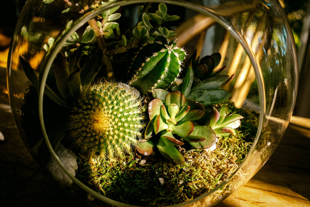
A terrarium is a glass container that contains soil and plants and may be opened to access the plants within for upkeep. Among the artificial ecosystem examples mentioned in the list, terrariums are on a rise in popularity courtesy of plant enthusiasts and botanists.

OpenTerrarium
An open terrarium containing succulents for decoration
Image: Wikipedia

Greenhouse

A greenhouse is usually referred to a glass house used for growing plants in consistent conditions where temperature and humidity and maintained. Similar to that in the atmosphere, the glass walls of the greenhouse allow heat to enter but prevent it from escaping. This maintains a constant temperature, along with added humidity making greenhouses perfect for growing tropical plants and crops in temperate or colder climates.

1024px TomateJungpflanzenAnzuchtNiederlande
Tomato plants being grown in Industrial green house Image: Wikipedia

Fish farms

Fish farms are a rising industry among the artificial ecosystem examples. These farms are similar to any farm that grows fish specifically. They can be aquarium fish, those for consumption or bait or feeders. These are usually done in small ponds dug out by the farmers and maintained to grow various fish species.

Artificial wetlands

Artificial wetlands were created for various purposes. Raw sewage, stormwater, agricultural, and industrial discharges can all be chemically cleaned in constructed wetlands. Constructed wetlands catch runoff, reduce nutrient levels, and provide diverse wildlife habitats, much like natural wetlands.

Managed forestry

Manages forests are of great importance among the artificial ecosystem examples in the field of conservation. These are forest areas created by humans, to imitate the natural forest cover that would grow in that area. These trees and vegetation and grown and transplanted, thereby helping to conserve plant species that are nearly on the brink of extinction due to deforestation and logging.

Parks

Parks are considered artificial ecosystems as they don’t sustain themselves. These can include anything from tourist parks to various national parks. Most of them contain organisms that are brought from their native habitat. They need to be fed and cared for to assure their survival. Tourist parks must be cared for by humans as well. Hence  they are among the artificial ecosystem examples

Hydroponics

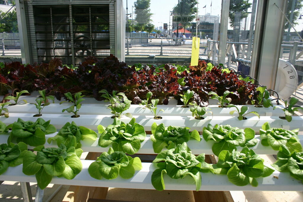
Hydroponics or aquaculture is probably the least known among the artificial ecosystem examples mentioned in the list. This method become more convenient as the amount of tillable land was not enough to grow crops. In this method farmers grow crops in water that is supplied with nutrients and inert gases, thereby reducing the need for pesticides or fertilizers and also decreasing carbon footprint.

1280px Leafy Greens Hydroponics
Another artificial ecosystem of hydroponics being used to grow salad greens
Image: Wikipedia

What is an artificial ecosystem?

 Artificial ecosystems are man-made ecosystems that cannot sustain themselves.

Thes ecosystems including biotic and abiotic factors are created by humans for specific purposes. Without them, these ecosystems can not survive.

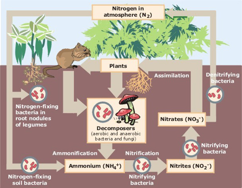
Nitrogen Cycle
Nutrient cycle of Nitrogen in a natural ecosystem
Image: Wikipedia

In these ecosystems, the setup, organisms and energy flow are all regulated by humans. These include aquariums, crop fields, terrariums and many more.

Artificial ecosystem characteristics:

Characteristics of an artificial ecosystem include:

  1. Artificial ecosystems are entirely man-made and require humans to take care of them.
  2. Most of them have very little genetic diversity in terms of the organisms-both flora and fauna.
  3. Artificial ecosystems cannot sustain the evolution of the organism residing in them.
  4. Both food chains and nutrient cycles are very short and usually incomplete to boot.

Components of an artificial ecosystem:

Like natural ecosystems, artificial ones are also composed of two main parts- biotic and abiotic components.

Biotic components: Biotic components include the flora and fauna that are part of the ecosystem. But unlike natural ecosystems, humans are always [art of the biotic components of the artificial ecosystems.

Abiotic components: These include the non-living factors including the soil, rocks, physical terrain, sunlight, gases and nutrients. Though these seem minimal they are responsible for the flow of energy, nutrients and food in the ecosystem.

Artificial ecosystem structure:

The artificial ecosystem example structures are made up of 3 parts-the biotic factors, the abiotic factors and humans.

Biotic factors include 3 main components-producers, consumers and decomposers. This is also the direction of energy flow in the ecosystem.

The abiotic factors include the soil, sunlight, nutrients, gases and everything that is non-living. These form the base ground or habitat of the ecosystem sustaining the organisms living in them.

Lastly, come the humans that actually construct these ecosystems for their own use. They are necessarily involved in the care of sustenance of these ecosystems otherwise, they will be unable to survive.

Features of an artificial ecosystem:

Features of artificial ecosystems include:

  • Artificial ecosystems are made by humans for productivity, conservation or entertainment.
  • Since humans create these ecosystems the organism count and variety are regulated, Hence the genetic diversity of these ecosystems is extremely low.
  • This also means there is no evolution due to the absence of the natural inducing factors.
  • Most times due to the absence of appropriate decomposers and bacteria the nutrient cycles and food chains are cut short. Hence they must be done manually.

Functions of an artificial ecosystem:

  1. Artificial ecosystems are goal oriented. They are planned and built for specific purposes.
  2. Some are made for aesthetic or entertainment purposes like gardens and aquariums.
  3. Artificial ecosystem examples like crop fields and dams are made for the production of food and irrigation. Dams also allow us to supply water to arid regions when required and also to produce hydroelectricity.
  4. Zoos and forests allow us to conserve flora and fauna species that are endangered, thereby helping more people to know and understand them.
  5. Lastly, wetlands were created to make sure that wastes could be chemically cleaned before they are released, thereby keeping the environment cleaner.

Also Read:

15+ Non Native Species Examples: Detailed Facts

1280px Burmese python 6887388927 300x225 1

Non native species refer to organisms that may have been transported from areas of their habitat to others by nature or by human interference.

Non native species are also called alien or exotic, but sometimes they can also become invasive causing major damage to the native flora and fauna of the area.

Non native species examples include:

  1. Burmese python
  2. Cane toad
  3. Gypsy moth
  4. Water hyacinth
  5. European wild rabbits
  6. The brown tree snake
  7. Sea lamprey
  8. Red fox
  9. Japanese beetle
  10. Nutria
  11. Comb jelly
  12. Silver pheasant
  13. Sweet clover
  14. Lionfish
  15. Cockroach
  16. Zebra mussels
  17. Kudzu plant
  18. Grass carp

Next, we will discuss the above-mentioned non native species examples in a little detail.

Points about the listed non native species examples:

Burmese python

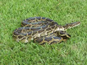
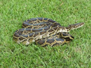
The Burmese python is technically one of the most heard of among the non native species examples listed above. Native to Southeast Asia and is one of the large snake species in the world with almost no known predator. Although the species was considered endangered, it was introduced into America via the pet trade and has now become an invasive species in the everglades of Florida.

non-native-species-examples
A Burmese python caught from the wilds of the Florida everglades
Image: Wikipedia

Cane toad

Though the cane toad is indigenous to South and Central America it is a non native species example in the Caribbean and Australia. Initially introduced into the areas to get rid of unwanted pests in the sugarcane fields, it has now become a pest itself. It considerably preyed on several native reptiles and cause a great disbalance in the natural biodiversity.

Gypsy moth

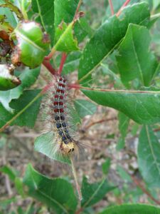
The gypsy moth Lymantria dispar dispar is native to Eurasia and is a non native species example. Originally brought to North America for scientific and commercial purposes, the most were accidentally released that causing them to be introduced into the ecosystem. It is not the moths but the larvae that are a cause of nuisance simply wiping away native vegetation in the blink of an eye.

800px Gypsy moth caterpillar eating leaves
Gypsy moth caterpillar eating leaves
Image: Wikipedia

Water hyacinth

Native to South America, the plant was introduced in Asia during colonial rule. Hence it became one of the most well known non native species examples on the list. Known for its pretty flowers, the water hyacinth started as an ornamental plant but later became an invasive species choking ponds, rivers, canals and irrigation waterways.

European wild rabbits

Originally native to Europe and the Mediterranean the European wild rabbits were introduced into Australia in the 1800s. Unlike in their native lands, in  Oceania, they had no natural predators and hence their population boomed. They are now considered pests due to the extensive damage to crops and native organisms.

The brown tree snake

Another significant animal among the non native species examples mentioned in the list is the brown tree snake that is native to Australia and parts of New Guinea. The snake was accidentally introduced into the South Pacific Island of Guam somewhere between the 1940s to 50s. The snake had no natural predators there but ended up preying on most of the native bird species and its population boomed.

Sea lamprey

The lamprey is probably the most ancient of all the non native species examples mentioned in the list. Native to the Northern and western waters of the Atlantic ocean they are the oldest of the known vertebrates, literally living fossils. These fish are parasitic feeding on other larger marine life and are non native to the Americas, where they have taken over the Great Lakes.

Red fox

 The red fox is indigenous to Europe but was introduced to the continent of Australia for recreational hunting somewhere in the mid 19th century. These mammals are voracious predators and reproduce at an alarming rate. They have caused several ground-dwelling birds, mammals and some reptiles like the green turtle populations to seriously decline.

Japanese beetle

Beetles from Japan are an invasive variety among the non native species examples. Japanese beetles eat the leaves, flowers, and fruit of over 300 different plant species. Turf grass pests include Japanese beetle grubs. They gnaw at the grassroots, turning the grass dark and causing them to die.

Nutria

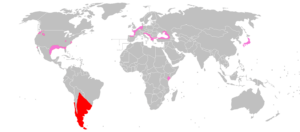
These rodents are native to South America but are considered among the non native species examples as they were introduced to the US in 1889 for fur production. But when the market collapsed in the 1940s hundreds of them were released into the wild and without any natural predators, they went about causing havoc. They not only destroy crops but also destroy embankments, ditches and canals required for natural irrigation and water flow.

Mapa Myocastor coypus
Map showing the native(red areas) and introduced regions(pink areas) of nutria
Image: Wikipedia

Comb jelly

Though the com jelly is native to the cold waters of the Atlantic in North America it has spread to the seas and oceans of Eurasia and become a threat to local wildlife. Because these jellies compete with fish and fish larvae, they can affect commercial fisheries by cascading effects on vital planktonic food webs. They also have longer feeding durations compared to their native colder counterparts.

Silver pheasant

The silver pheasant that is native to Southeast Asia and was introduced into parts of Europe and became one of the non native species examples considered exotic. Introduced to Victoria Island in 1962 mainly for ornamental purposes, the island had a pounded a substantial population by 2002.

Sweet clover

Another non-invasive variety among the non native species examples mentioned in the list the sweet clover is a plant native to Europe. It is especially good for attracting bees and butterflies and was imported to America for ground cover and to facilitate pollination of food crops. They have naturalized in the Americas and cause no disbalance in the ecosystem.

Lionfish

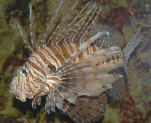
Lionfish, which are native to the Indo-Pacific maritime region, are rapidly spreading along the East Coast’s coastlines and coral reefs. Lionfish are voracious feeders, and their toxic dorsal spines have protected them from natural predators in the Atlantic thus far.

1024px Red Lionfish Pterois volitans Left Side 2460px
The lion fish is non native to the Atlantic and Caribbean Image: Wikipedia

Cockroach

As evident from their Latin name Periplaneta americana, the common cockroach was originally native to the Americas. They came to Asia among the material in the bows of ships and flourished in Asia due to the optimum temperatures and humidity.

Zebra mussels

Zebra mussels are a small, invasive gastropod that is endemic to Eurasia’s freshwaters. The black, zig-zagged stripes on each shell give them their name. In the 1980s, zebra mussels most likely entered the Great Lakes by ballast water released by huge ships from Europe. They stick to other local marine and freshwater wildlife and often end up killing them. This has put great pressure on the native riverine and freshwater fauna.

Kudzu plant

Kudzu is a Japanese and Southeast Chinese plant that is another invasive variety among the non native species examples mentioned in the list. The “Vine that Ate the South” was first brought to North America in the nineteenth century to strengthen embankments. But later they overtook the local flora often killing them.

Grass carp

Grass Carp is the last of the non native species examples mentioned in the list. The Grass Carp is a plant-eating fish that was introduced to the United States to limit the growth of plants in aquaculture ponds.

Several Grass Carp strayed into the Mississippi River basin during flooding shortly after their arrival and have subsequently spread throughout the United States. But now it has greatly increased in number and is wiping out several important marine and riverine floras along with competing with native fish and animals for food.

Also Read:

25+ Native Species Examples: Detailed Facts

1280px Rose hips 1024x768 1

Native species refers to organisms that are indigenous to a specific region or ecosystem.

Native species refer to those organisms that originated in that particular region through evolution and mutation without any sort of human interference or interaction.

Native species examples include:

  1. Kashmir Stag, Kashmir
  2. Royal Bengal Tiger, Sundarbans
  3. Rosehip
  4. Koala, Australia
  5. Nevada primrose
  6. Tarsier
  7. Nilgai
  8. Coneflower
  9. European red squirrel
  10. Walnut
  11. Bicoloured frog
  12. Large-leaved Lupine
  13. The panda
  14. European Centaury
  15. Oriental magpie robin
  16. Rafflesia
  17. Asian elephant
  18. Neem tree
  19. The proboscis monkey
  20. Snow leopard
  21. Rhododendron
  22. Trumpet vine
  23. Cougar
  24. Java sparrow
  25. Grizzly bear
  26. Eurasian lynx
  27. Platypus

Next, we will discuss the above-mentioned examples :

Discussion of the Native Species Example:

Kashmir Stag, Kashmir

The hangul or the Kashmir stag is native to India and found only in the riverine mountain regions of Jammu, Kashmir and the Himachal. The hangul is actually an Asian variety of the elk. Hence it is a native species example that is also endemic in nature.

Royal Bengal Tiger

Belonging to the Panthera tigris tigris subspecies the Bengal tiger is one of the biggest of the wild cats alive among the native species examples. Though it once roamed across the entire Indian subcontinent up to Pakistan now its range is limited to Eastern India, Bangladesh, Nepal and Bhutan.

Rosehip

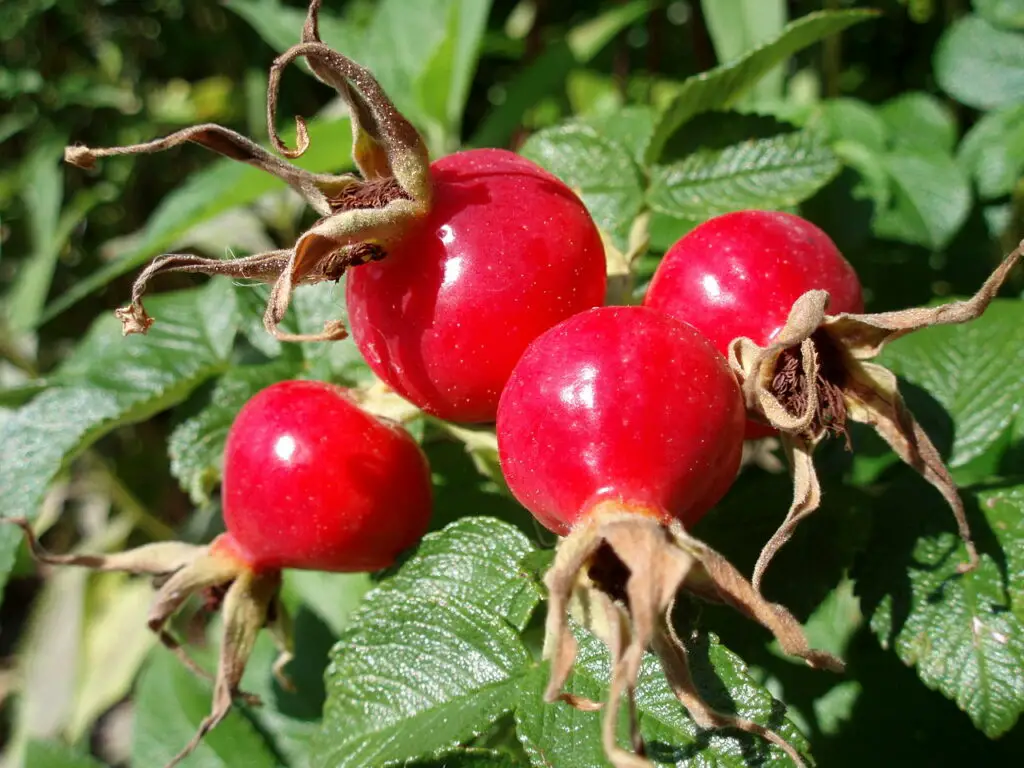
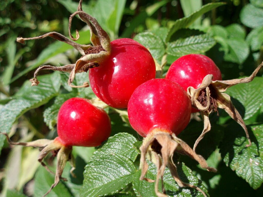
The dog-rose, sometimes known as the rose hip, is a climbing wild rose species endemic to Europe, northwest Africa, and western Asia. Rose hips form following successful flower pollination in the spring or early summer and ripen in the late summer and early fall.

1280px Rose hips
Image of the rosehip berry Image: Wikipedia

Koala

The koala bear is considered exotic among the native species examples. It is a marsupial(i.e. it carries its young in a pouch) and is native to Oceania in Queensland, Victoria, New South Wales and South Australia. Since all marsupials are endemic to Australia and New Zealand they cannot be found in any other part of the world naturally.

Nevada primrose

Primula cusickiana var. nevadensis is a variety of the Cusick’s primrose which is native to the State of Nevada in the US. These plants are known for their brightly coloured flowers that bloom in early spring.

Tarsier

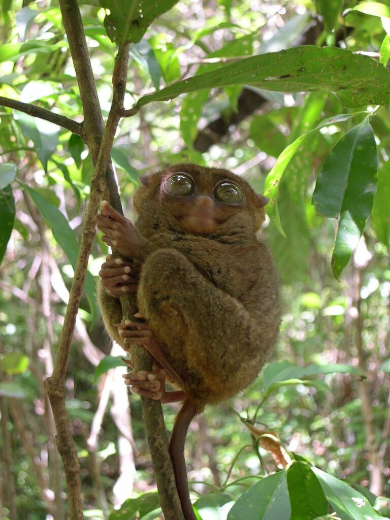
The Philippine tarsier is only found in the Philippines, as its name suggests which makes it one of the native species examples. C. syrichta populations are mostly found in the archipelago’s southeast corner. Measuring at only an average of 160 mm at max it is one of the world’s smallest primates, also known for its large eyes.

native-species-examples
Image of a Philippine Tarsier
Image: Wikipedia

Nilgai

Literally translated as the blue cow, the Nilgai which is a native species example is actually the largest among the Asian antelopes. It is native to the northern Indian subcontinent, however it can be found throughout the peninsula.

Coneflower

Belonging to the daisy family the coneflower is among the native species examples. Native only to eastern and central Nort America, the coneflower grows naturally among grows as wildflowers in prairies and the open woodlands. They are called so because the flowers actually grow to resemble something like sea urchins.

European red squirrel

The red squirrel is found throughout Europe and Asia and is among the mentioned native species examples. As the name suggests the European red squire varies in colouration from a red to an auburn not the usual brown or grey and is considered quite small in size.

Walnut

Another native species example mentioned in the list the walnut is actually native to the Americas and Southern Asia. Though considered a nut the walnut is actually a drupe fruit. Walnuts are a good source of vital lipids and a tasty snack.

Bicoloured frog

The Malabar frog or the bicoloured frog is native to the Malabar Coast of the Western Ghats. It is named so as the frog is distinctly bicoloured which makes it an excellent form of camouflage.

Large-leaved Lupine

The large-leaved lupine that is among the listed native species examples is native to the Northwest region of the Pacific. However, those introduced in other places by humans have become a threat to the native organism of those regions like the Karner blue butterfly.

Lupinus polyphyllus
Flowers of the Large- leaved lupine
Image: Wikipedia

The panda

 Distinguished by its signature black and white coat the giant panda is probably the most popular amongst the native species examples listed. Since they can only depend on bamboo for sustenance the pandas are only found in the Southwestern areas of China and nowhere else.

European Centaury

European centaury is a plant that is native to western Europe, Pakistan, and northern Africa and has been imported to North and South America, as well as Australia. It is extensively used as herbal medicine and is thought to be useful for the treatment of gastrointestinal issues.

Centaurium erythraea 220603
Flowers of the European centaury
Image: Wikipedia

Oriental magpie robin

The Oriental magpie-robin lives in open woods and cultivated areas, sometimes near human settlements in all of Southern and Southeastern Asia. They are distinctly bicoloured in their black and white plumage.

Rafflesia

Malaysia, Thailand, Indonesia, and the Philippines are home to Rafflesia. Rafflesia, often known as the corpse flower, is a parasitic plant that feeds on the wild grapes of the Tetrastigma vine genus, which give everything Rafflesia needs to exist. Botanically they are considered the largest species of flowers in the world.

Asian elephant

Smaller in size as compared to their African cousins the Asian elephant is endemic to India and Southeast Asia. They are the largest mammals across the Eurasian subcontinent.

Neem tree

Neem is thought to be indigenous to the Indian subcontinent and drier parts of South Asia among the native species examples. It has spread to areas of Africa, the Caribbean, and several South and Central American countries. The tree is mainly known for its antibacterial and anti-inflammatory properties and is greatly used in Ayurveda.

The proboscis monkey

One of the native species examples of Borneo in Southeast Asia, the proboscis monkey is an old world arboreal species. Interestinglingly it is the only species in its genus Nasalis. Currently, the proboscis monkey is considered an endangered species.

Snow leopard

Belonging to the genus Panthera the snow leopard is native to the mountains of central and South Asia. They are best known for their thick lush greyish white coat with beautiful black rosettes which allows them to camouflage effectively. Currently due to rising temperatures and poaching these magnificent predators are on the list of vulnerable animals.

Rhododendron

Rhododendrons are a bushy shrub native to temperate regions of Asia. But the reason that it is considered one of the native species examples and not an endemic species is that they have naturally spread to America as well. They produce beautiful trumpet-shaped flowers in colours like red, pink, white, purple, blue and scarlet.

Trumpet vine

The trumpet vine also called yellow trumpet vine or trumpet creeper is another native species example. Though naturalized in other parts of the continent it is actually native to the Eastern part of the US. They are named so due to the large trumpet-shaped flowers they produce that are a feeding ground for hummingbirds and sunbirds. These birds also nest in the foliage of these dense bushy creepers.

Cougar

The cougar is a puma species that is native to the American continent. Also, commonly called the mountain lion or simply the puma, the cougar has a wide habitat ranging from the Canadian Yukon range to the Andes and parts of South America. The cougar is the largest of the big cat species of the Americas following the jaguar.

Java sparrow

The java finch also called the java sparrow is another native species example found in the Indonesian peninsula. Its range extends from Java, Bali and Bawean. The java sparrow is one of the only two species in its genus Padda.

Grizzly bear

North American brown bears are more commonly known as grizzlies and are known among the native species examples. Brown bears are found in America Europe and Asia, but the grizzly is specifically native to North America. Here they range from Alaska to Mexico and then west extending up to the Hudson Bay.

Eurasian lynx

The Eurasian lynx is one of the 4 existing lynx species. It is native to the arboreal Central, Noth and Eastern Europe and extends to central Asia, Tibet, Siberia and the Himalayas. The Eurasian lynx is itself divided into six different subspecies.

Platypus

Found only in Australia, the platypus is an exemplary mammal among the native species examples. It is one among the only two existing mammals that lay eggs instead of giving birth to live young. The duck-billed platypus looks something like a beaver, an otter and a duck mixed together.

Schnabeltier 1
A duck-billed platypus swimming
Image: Wikipedia

Also Read: