Are Protists Monophyletic? A Comprehensive Exploration

are protists monophyletic

Protists, a diverse group of eukaryotic organisms, have been a subject of debate when it comes to their classification and phylogeny. The question of whether protists are monophyletic or not is a complex one, and the answer depends on the specific criteria used for classification.

Understanding Monophyly

Monophyly refers to a group of organisms that includes the most recent common ancestor and all its descendants. In other words, a monophyletic group is a natural group that shares a unique common ancestor. This is in contrast to paraphyletic and polyphyletic groups, which do not share a common ancestor or exclude some of their descendants, respectively.

The Historical Perspective on Protist Classification

are protists monophyletic

Historically, protists were grouped together in the kingdom Protista, which was a wastebasket taxon that included all eukaryotes that did not fit into the other three kingdoms (Animalia, Plantae, and Fungi). This classification was based on the shared characteristics of being eukaryotic organisms that were not animals, plants, or fungi.

The Advent of Molecular Data and Phylogenetic Analyses

However, with the advent of molecular data and phylogenetic analyses, it has become clear that the traditional classification of protists is artificial and polyphyletic, meaning that the group includes organisms that do not share a common ancestor.

The Seven Groups of Protists

Modern phylogenetic analyses based on molecular data suggest that the seven groups of protists (Amoebozoa, Excavata, Archaeplastida, Rhizaria, Chromalveolata, Hacrobia, and Incertae sedis) are monophyletic when comparing specific characteristics. These groups are defined based on their unique features, such as the presence or absence of certain organelles, the mode of nutrition, and the structure of their cells.

Measurable and Quantifiable Data Supporting Paraphyly

However, when all the diversity of protists is considered, the group as a whole is paraphyletic, meaning that it excludes some of its descendants. There are measurable and quantifiable data that support this:

  1. Freshwater Ciliated Protist Communities: A study on freshwater ciliated protist communities found measurable species-specific differences in prey ingestion, indicating that these organisms are not closely related.
  2. Reproducing Protists or Protistan Biological Species: Another study on reproducing protists or protistan biological species found that individual samples showed measurable and informative biological differences, further supporting the paraphyly of protists.

Ongoing Revisions in Protist Classification

It is important to note that the classification of protists is still evolving, and new molecular data and phylogenetic analyses may lead to further revisions in the future. As our understanding of the evolutionary relationships among protists continues to improve, the debate over their monophyly or paraphyly may be resolved with more certainty.

Key Takeaways

  • Protists are a diverse group of eukaryotic organisms, and their classification has been a subject of debate.
  • Monophyly refers to a group of organisms that includes the most recent common ancestor and all its descendants.
  • Historically, protists were grouped together in the kingdom Protista, which was a wastebasket taxon.
  • Modern phylogenetic analyses suggest that the seven groups of protists are monophyletic when comparing specific characteristics.
  • However, the group of protists as a whole is paraphyletic, as supported by measurable and quantifiable data.
  • The classification of protists is still evolving, and new molecular data and phylogenetic analyses may lead to further revisions in the future.

References

  1. Bio 2: Protists Flashcards – Quizlet. https://quizlet.com/185038087/bio-2-protists-flash-cards/
  2. Protista – an overview | ScienceDirect Topics. https://www.sciencedirect.com/topics/immunology-and-microbiology/protista
  3. Biology Practical Midterm Flashcards – Quizlet. https://quizlet.com/492290826/biology-practical-midterm-flash-cards/
  4. Correlation between fresh water ciliated protist communities andtheir micro-ecology. https://www.researchgate.net/publication/332550309_CORRELATION_BETWEEN_FRESH_WATER_CILIATED_PROTIST_COMMUNITIES_ANDTHEIR_MICRO-ECOLOGY
  5. Current practice in the approach to species – Mallet Group. https://mallet.oeb.harvard.edu/sites/hwpi.harvard.edu/files/malletlab/files/hoef-emdenbass_in_boenigk_2012.pdf?m=1450219355
Endocytosis Molecular Movement: A Comprehensive Guide

endocytosis molecular movement

Endocytosis is a fundamental process in eukaryotic cells that involves the internalization of molecules, particles, and even entire pathogens from the extracellular environment into the cell. This complex process is driven by the dynamic movement of various molecular components, orchestrating the formation, transport, and fusion of endocytic vesicles. In this comprehensive guide, we will delve into the intricate details of endocytosis molecular movement, providing a wealth of information for biology students and researchers.

Understanding the Endocytosis Process

Endocytosis can be broadly classified into two main categories: phagocytosis and pinocytosis. Phagocytosis involves the engulfment and internalization of large particles, such as bacteria or cellular debris, while pinocytosis refers to the uptake of fluid and dissolved solutes.

Pinocytosis can be further divided into two subtypes: clathrin-mediated endocytosis (CME) and clathrin-independent endocytosis (CIE). CME is the most well-studied and best-understood pathway, accounting for the majority of endocytic events in many cell types.

Clathrin-Mediated Endocytosis (CME)

CME is a highly regulated process that involves the coordinated action of various proteins, including clathrin, adaptor protein complexes, and dynamin. The key steps in CME are as follows:

  1. Cargo Selection and Clathrin Recruitment: Specific cargo molecules, such as receptors or ligands, are recognized and selected for internalization. Adaptor proteins, such as the AP-2 complex, bind to these cargo molecules and recruit clathrin to the plasma membrane.

  2. Clathrin-Coated Pit Formation: The clathrin molecules assemble into a characteristic lattice-like structure, forming a clathrin-coated pit on the plasma membrane. This pit gradually invaginates, creating a curved membrane structure.

  3. Vesicle Scission: The GTPase dynamin is recruited to the neck of the clathrin-coated pit, where it catalyzes the pinching off of the vesicle from the plasma membrane, forming a clathrin-coated vesicle.

  4. Vesicle Uncoating and Trafficking: The clathrin coat is rapidly disassembled, and the uncoated vesicle fuses with early endosomes. The internalized cargo is then sorted and either recycled back to the plasma membrane or transported to lysosomes for degradation.

The rate of CME can vary significantly between cell types, with some cells, such as macrophages, exhibiting a remarkably high rate of endocytosis, ingesting up to 25% of their own volume of fluid per hour, which equates to 3% of their plasma membrane every minute.

Clathrin-Independent Endocytosis (CIE)

CIE is a more diverse and less well-understood pathway that involves the formation of non-clathrin-coated vesicles. CIE can be further divided into several subpathways, including:

  1. Caveolae-Mediated Endocytosis: This pathway involves the formation of flask-shaped invaginations of the plasma membrane, called caveolae, which are enriched in the protein caveolin.

  2. Lipid Raft-Mediated Endocytosis: This pathway utilizes specialized membrane microdomains, known as lipid rafts, which are enriched in cholesterol and glycosphingolipids.

  3. Clathrin- and Caveolae-Independent Endocytosis: This pathway involves the formation of vesicles that do not contain clathrin or caveolin, and the molecular mechanisms are still being actively investigated.

CIE pathways are often involved in the uptake of specific cargo, such as glycosylphosphatidylinositol-anchored proteins and cholesterol.

Measuring and Quantifying Endocytosis Molecular Movement

endocytosis molecular movement

The molecular movement of endocytosis can be measured and quantified using various techniques, each with its own strengths and limitations:

Fluorescence Microscopy

Fluorescence microscopy allows for the real-time tracking of fluorescently labeled cargo and the quantification of the number and size of endocytic vesicles. This technique provides valuable insights into the dynamics of endocytic events, such as the formation, movement, and fusion of vesicles.

Electron Microscopy

Electron microscopy, particularly transmission electron microscopy (TEM), can provide high-resolution images of endocytic structures, enabling the quantification of the number and distribution of clathrin-coated pits and vesicles. This technique is crucial for visualizing the ultrastructural details of the endocytic machinery.

Biochemical Assays

Biochemical assays can be used to measure the uptake and release of specific cargo, such as ligands and receptors, and to quantify the kinetics and efficiency of endocytosis. These assays often involve the use of radioactive or fluorescently labeled molecules, which can be detected and quantified using various analytical techniques.

Regulatory Factors and Molecular Mechanisms

The endocytosis process is highly regulated by a complex network of molecular interactions and signaling pathways. Some key regulatory factors and molecular mechanisms involved in endocytosis include:

  1. Membrane Curvature and Lipid Composition: The curvature of the plasma membrane and the local lipid composition play a crucial role in the initiation and progression of endocytic events.

  2. Actin Cytoskeleton Dynamics: The actin cytoskeleton undergoes dynamic rearrangements to provide the necessary mechanical force for membrane deformation and vesicle formation.

  3. Protein-Protein Interactions: A diverse array of proteins, including clathrin, adaptor proteins, and accessory factors, coordinate their actions to facilitate the various stages of endocytosis.

  4. Signaling Cascades: Cellular signaling pathways, such as those involving phosphoinositides, small GTPases, and kinases, regulate the recruitment and activity of the endocytic machinery.

  5. Membrane Trafficking and Fusion: The transport and fusion of endocytic vesicles with various intracellular compartments, such as early endosomes and lysosomes, are mediated by a complex network of membrane trafficking proteins and regulatory factors.

Understanding the intricate molecular mechanisms and regulatory factors governing endocytosis is crucial for elucidating its role in diverse cellular processes, including signal transduction, nutrient uptake, and pathogen invasion.

Conclusion

Endocytosis is a fundamental cellular process that involves the dynamic movement of molecules and particles from the extracellular space into the cell. The process is highly complex and regulated, with multiple subtypes and diverse molecular mechanisms. By delving into the details of endocytosis molecular movement, this comprehensive guide provides a valuable resource for biology students and researchers, enabling a deeper understanding of this essential cellular function.

References:

  1. Merrifield, C. J., & Kaksonen, M. (2014). The mechanisms and regulation of endocytosis. Nature reviews Molecular cell biology, 15(1), 31-46.
  2. Farquhar, M. G., & Palade, G. E. (1981). Pinocytosis by mammalian cells. The Journal of cell biology, 91(3), 77s-103s.
  3. Conner, S. D., & Schmid, S. L. (2003). Clathrin-independent endocytosis. Nature reviews Molecular cell biology, 4(10), 771-782.
  4. Mayor, S., & Pagano, R. E. (2007). Lipid rafts and membrane domains: fluid mosaics or tight tessellations?. Nature reviews Molecular cell biology, 8(5), 361-374.
  5. Kirchhausen, T. (2000). Coat assembly and membrane deformation in clathrin-mediated endocytosis. Nature, 403(6767), 39-45.

5 Globular Protein Examples: That You Should Know

image 300x225 1

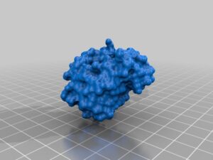
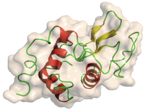
Globular protein examples comprise a group of proteins that have their polypeptide chains arranged in a spherical form.

  • Myoglobin
  • Carbonic anhydrase
  • Glyceraldehyde-3-phosphatase dehydrogenase
  • Concanavalin A
  • Cytochrome C
  • Lysozyme

Myoglobin-

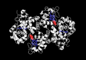
Myoglobin is the first globular protein to have its tertiary structure determined. This small, oxygen carrying protein is made of single polypeptide chain of 153 amino acids and a non-polypeptide prosthetic heme group. About 75% of the amino acid residue in myoglobin is α helical. Myoglobin lacks β sheet structures.

Carbonic anhydrase-

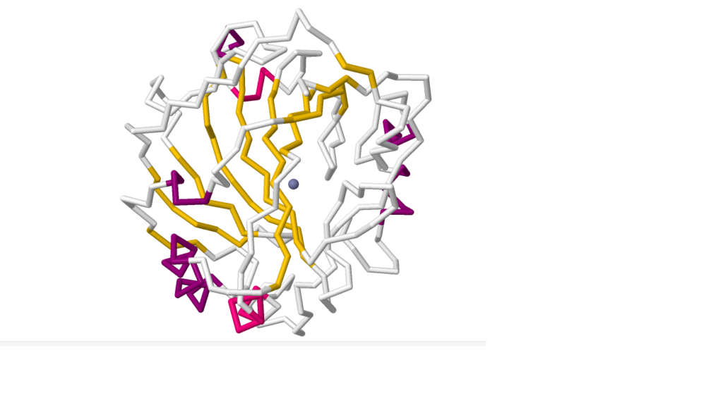
Globular protein human carbonic anhydrase is an enzyme that assist in O2 -CO2 transport. It has a single polypeptide chain of 259 amino acids with a Zn2+ ion in the center coordinated by Histidine amino acid residue in position 94,96,119. This is the only native protein in which polypeptide chain form a knot by the C-terminus going through surrounding polypeptide loop.

Glyceraldehyde-3-phosphatase dehydrogenase-

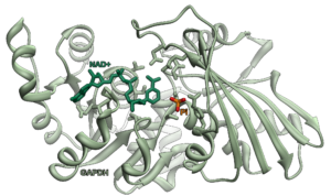
This globular protein is an important enzyme needed in glycolytic pathway and contains total of 335 amino acid residues. It is made of four identical subunits of 37 kDa, each containing a thiol(SH) group. It has two highly conserved domains, I.e., NAD+ binding domain and glyceraldehyde-3-phosphate domain. It contains a unique CREM nuclear export signal at the catalytic site. GAPDH also helps in apoptosis.

globular protein examples
Fig: “File:GAPDH with labels.png” by Vossman is licensed under CC BY-SA 3.0.

Concanavalin A-

This globular protein is a homotetramer with each subunit containing 235 amino acids and weighs 26.5 kDa. They are mostly consisting of antiparallel β sheets and non-polypeptide prosthetic Mn2+ and Ca2+.

Cytochrome C-

This small globular protein, which is made of 104 amino acid residues and heme group, is an integral part of electron transport chain. The heme group is attached covalently to the polypeptide chain. 40% of the polypeptide of this protein form α helical structure and the rest contains β turns. Except ETC this protein also helps in cellular apoptosis.

Lysozyme-

This small proteolytic enzyme found in human tears, saliva, mucus is a globular protein made of 129 amino acid residues. It contains four disulfide bonds, which provides structural stability, in-between the pairs of cysteine residues. It has five regions of α helices, about 40% of the polypeptide chain and the rest shows β sheet conformations. Lysozyme is well known for the ability to lyse or degrade microbial cell walls.

globular protein examples
Fig: “File:Lysozyme.png” by Yikrazuul is licensed under CC BY-SA 3.0.

Most globular proteins are water soluble.

While forming tertiary structure polypeptide chains fold in such a manner where the hydrophilic polar amino acids are found in the outer region whereas the core is formed by non-polar hydrophobic amino acids.

Primary and secondary structure of protein

Primary structure of protein is a polymer made of amino acids linked by peptide bonds and are called polypeptide chains. Polypeptide chains fold into different stable structures; like α helix, β pleated sheets and u-shaped β turn, depending on amino acid sequences. These structures are known as the secondary structure of protein.

Tertiary structure of globular protein-

Most proteins found inside cells; like transport and receptor proteins, enzymes and immunoglobins are globular proteins. Polypeptide chains fold back on each other forming a compact structure that varies from protein to protein. These structural diversity helps protein maintain their unique structure to perform assigned biological functions.

3-dimensional protein structure is identified using x-ray crystallography and NMR spectroscopy.

Conclusion-

Globular proteins are not only important for structural stability of tertiary protein molecules, also is imperative for specific biological function that is determined through conserved regions of tertiary structure of protein.

Also Read:

11 Lichen Examples: Detailed Facts

image 20 300x200 1

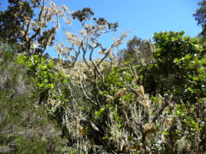
Lichen is a plantlike autotrophic existence which is the result of a symbiotic cohabitation between fungi and either green algae or cyanobacteria. They are slow growing and grow in a colonized manner; and lichen examples can be found throughout the Earth in diverse environments.

The fungal portion of the symbiosis is called mycobiont and the algal part is known as phycobiont.

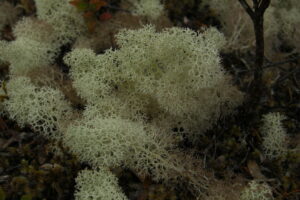
Cladonia rangiferina

They are commonly known as Reindeer moss or Caribou moss. It is a fruticose type of lichen, extensively branched and greyish in color. They are generally found in taiga pine forests and low alpine forests. Reindeer and caribou like animals use them as winter forage.

lichen examples
Fig: “Reindeer Moss” by pellaea is licensed under CC BY 2.0.

Parmelia sulcata

These corticolous,foliose lichens are commonly called Hammered shield lichen or Wax paper lichen. Due to their high tolerance of pollutants, they are found throughout the world even in metropolitan areas.

lichen examples
Fig: “Parmelia sulcata” by hedera.baltica is licensed under CC BY-SA 2.0.

Peltigera britannica

These are a type of foliose lichen, native to North America and Europe.

lichen examples
Fig:”Peltigera britannica” by Striking is licensed under CC BY-SA 2.0.

Cetraria islandica

These fruticose lichens are commonly known as Iceland moss. It can be used as food and even as traditional folk remedies.

lichen examples
Fig: “Iceland moss – Cetraria islandica” by Björn S… is licensed under CC BY-SA 2.0.

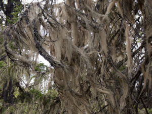
Usnea barbata

They are fruticose lichens, commonly called as Beard moss or Old man’s beard. They grow on dying trees and are very sensitive towards air pollution. They can be used as bioindicators.

Alectoria sarmentosa

These light-green, fruticose lichens are known as Witch’s hair lichen and are found in temperate zones with high rainfall.

lichen examples
Fig: “Alectoria sarmentosa” by caspar s is licensed under CC BY 2.0.

Xanthoparmelia convoluta

They are mostly saxicolous lichens attached loosely to the substratum surface. These lichens are native to Australia.

Umbilicaria grisea

They are saxicolous lichens, commonly called Velvet moss. They are native to the northern North America.

Collema crispum

These gelatinous lichens grow on moist, shady, calcareous substratum and are of foliose type.

lichen examples
Fig:”Collema crispum” by Striking is licensed under CC BY-SA 2.0.

Coenogonium implexum

These crustose type lichens are found in tropical areas throughout the world.

Lepraria lobificans

They are crustose type lichen, commonly called as Dust lichen.

Graphis scripta

These crustose type lichens are commonly called as Script lichen or secret writing lichen due to their unique growth pattern.

lichen examples
Fig: “Graphis scripta” by Odd Wellies is licensed under CC BY 2.0.

Lichen characteristic-

  • Lichens are symbiotic existence between fungi, which is the more dominant part of the symbiont, acts as a host organism for either green algae or cyanobacteria and even sometimes for both.
  •  Mycobiont forms the body of lichen, which is known as thallus and it protects the phycobionts inside.
  • A typical lichen structure can be divided into three parts, which are outer cortex or thallus, inner medulla and the basal attachment or holdfast.
    1. Outer cortex, is made of densely packed filamentous fungal cells forming a thick crusty cover.
    2. Inner medulla, is a striated structure made of alternating layers of phycobiont and mycobiont.
    3. The basal attachment, is a hair like fungal structure, known as rhizines. It acts an attachment organ that helps lichen to adhere to surface.
  • Based on the ratio of phycobionts and mycobionts, thalli of lichens are of two types;
    1. Heteromerous thalli, is where fungal cells are predominant.
    2. Homoeomerous thalli, is where algal and fungal cells are uniformly distributed.
  • Lichens reproduce vegetatively by propagating through broken thallus. The fungal part of lichens can reproduce with the help of spore formation.
  • Lichens that have cyanobacteria as their photobionts acts as a nitrogen fixator.
  • Lichens grow in various interesting shape and coloring from grey-green to bright red-orange.

Significances of symbiosis in lichen-

Phycobionts in lichen are capable of the process of photosynthesis, I.e., autotroph; because of the presence of chloroplasts. But the mycobionts cannot perform photosynthesis and thus are unable to sustain itself for long on its own. So mycobionts actively seek out the algal part through chemical reaction.

They form a mutualistic existence where the phycobionts provides the mycobionts with carbohydrate and the mycobionts offer protection from environmental elements.

Lichen types-

Based on the surface they grow on, lichens are divided into four categories,

  1. Terricolous, are lichens that grows on soil. e.g., Caldonia.
  2. Corticolous, grows on the tree barks. e.g., Parmelia.
  3. Lignicolous, are lichens found in the woods. e.g., Cyphellum.
  4. Saxicolous, lichens grow on rocks or stones. e.g., Peltigera.

Based on the growth forms of thallus lichens are classified into six main types;

  1. Crustose- they have 2-dimensional, crusty appearance and stick tightly to the substrate they are growing on. e.g., Graphis.
  2. Foliose- they have flat-leaf like appearance and grows loosely on the substratum with one or multiple points of attachment. e.g., Parmelia.
  3. Fruticose– it has filamentous, branched, 3-dimensional structure; standing erect or hanging loosely on the substratum by help of basal disc. e.g., Usnea.
  4. Leprose– they have a powdery appearance, attached superficially on the substrate. e.g., Lepraria.
  5. Squamulose- these lichens have a closely clustered leaf-like crustose attached to the surface and erect at the tip. e.g., Cladonia.
  6. Byssoid- they have a wispy, teased wool-like appearance. e.g., Coenogonium.

There are also gelatinous lichens like Collema.

Lichen classification-

Lichens are classified as their mycobiont part, as in scientific name of a lichen is same as their fungal part.

The mycobiont part consist of fungi from either phylum Ascomycota or phylum Basidiomycota.

Difference between moss and lichen-

In-spite of being called non-vascular plants, moss and lichens are very different.

Firstly, only mosses are actually plants under the phylum Bryophyta and have roots, leaves and stems likes structure with intended actions. They are considered to be the ancestor of all land plants.

Whereas, lichens are not true plants and without roots, leaves or any stems.

Ecological importance of lichen-

Lichens are of great ecological significance. Such as,

  1. They act as nitrogen fixator, entrapping atmospheric nitrogen.
  2. Mycobiont part of lichens produce chemicals that are capable of breaking down rocks and releasing the minerals in nature. This process is called biological weathering and is essential for soil formation.
  3. Terricolous lichens prevents soil erosion.
  4. Lichens can be used a food source by various animals.
  5. They can colonize barren surfaces, acting as a “Pioneer species”.
  6. They also act as bioindicators by absorbing and trapping various heavy metals, carbon or sulfur pollutants into their thallus.

Conclusion

For evolutionary advantage, the symbiosis between fungi and algae, I.e., lichens are not only important for ecosystem and wildlife, they also act as a bioindicator for anthropogenic activities that cause pollution.

Also Read:

Do Bacteria Have Peptidoglycan? A Comprehensive Guide

do bacteria have peptidoglycan

Bacteria are microscopic, single-celled organisms that are ubiquitous in our environment, playing crucial roles in various ecosystems and human health. One of the defining features of bacterial cells is the presence of a cell wall, which provides structural integrity, protection, and shape to the cell. At the core of the bacterial cell wall is a unique polymer called peptidoglycan, also known as murein.

The Importance of Peptidoglycan in Bacterial Cells

Peptidoglycan is a crucial component of the bacterial cell wall, serving as a macromolecular “exoskeleton” that stabilizes the cell and provides structural integrity. This polymer forms a mesh-like layer outside the cytoplasmic membrane of bacterial cells, giving them rigidity and shape, and protecting them from osmotic lysis and environmental stresses.

The peptidoglycan is composed of glycan strands cross-linked by short peptides, and its structure and composition can vary between different bacterial species. This variation in peptidoglycan structure is one of the key features that distinguishes Gram-positive and Gram-negative bacteria, which have different cell wall architectures.

Analyzing the Structure and Composition of Peptidoglycan

do bacteria have peptidoglycan

Researchers have developed a variety of analytical techniques to study the structure and composition of peptidoglycan in bacterial cells. These methods include:

  1. Ultraperformance Liquid Chromatography–Mass Spectrometry (UPLC-MS): This technique allows for the separation and identification of individual peptidoglycan monomers and oligomers, providing detailed information about the glycan chain length and degree of cross-linking.

  2. Atomic Force Microscopy (AFM): AFM can be used to visualize the surface topography of the peptidoglycan layer, revealing its macroscale features and changes in the cell envelope dimensions.

  3. Electron Cryotomography: This advanced imaging technique enables the visualization of the three-dimensional structure of the peptidoglycan layer, including the organization of the glycan strands and stem peptides.

  4. Genetic Screens: By identifying genes and proteins involved in peptidoglycan synthesis, modification, and degradation, researchers can gain insights into the molecular mechanisms that regulate the structure and dynamics of the bacterial cell wall.

The Structural Features of Peptidoglycan

The peptidoglycan is a porous material that lacks an ordered macromolecular structure. In Gram-negative bacteria, the pore size of the peptidoglycan layer ranges from 4 to 25 nanometers in diameter. Electron cryotomography studies have revealed that the stem peptides in Gram-negative bacteria are aligned along the long axis of the cell, while the glycan strands are wrapped circumferentially around the cell.

This unique organization of the peptidoglycan components is hypothesized to impart directional, or anisotropic, mechanical properties on the bacterial cell. The Young’s modulus, a measure of the stiffness of the material, has been measured for peptidoglycan in both Gram-negative and Gram-positive bacteria, providing insights into the mechanical properties of this crucial cell wall component.

The Diversity of Peptidoglycan Structures

The structure and composition of peptidoglycan can vary significantly between different bacterial species and even within the same species under different growth conditions. These variations can include differences in the glycan chain length, the degree of cross-linking, the amino acid composition of the stem peptides, and the presence of additional modifications, such as O-acetylation or amidation.

For example, the peptidoglycan of Escherichia coli, a common Gram-negative bacterium, is characterized by relatively short glycan chains (approximately 10-20 disaccharide units) and a moderate degree of cross-linking (40-50%). In contrast, the peptidoglycan of Bacillus subtilis, a Gram-positive bacterium, has longer glycan chains (up to 100 disaccharide units) and a higher degree of cross-linking (up to 80%).

These structural differences can have significant implications for the mechanical properties of the bacterial cell wall, as well as the susceptibility of the cell to antibiotics and other environmental stresses.

The Importance of Peptidoglycan in Bacterial Physiology and Pathogenesis

The peptidoglycan layer plays a crucial role in various aspects of bacterial physiology and pathogenesis. It is essential for maintaining the structural integrity of the cell, preventing osmotic lysis, and providing a scaffold for the attachment of other cell wall components, such as teichoic acids and lipopolysaccharides.

Moreover, the peptidoglycan layer is a target for many antibiotics, such as β-lactams and glycopeptides, which disrupt its synthesis or cross-linking, leading to cell death. The diversity of peptidoglycan structures across bacterial species is a key factor in the development of antibiotic resistance, as different bacteria may have varying susceptibilities to different classes of antibiotics.

In pathogenic bacteria, the peptidoglycan layer can also act as a pathogen-associated molecular pattern (PAMP), triggering the host’s immune response and contributing to the development of inflammatory diseases.

Conclusion

In summary, peptidoglycan is a fundamental component of the bacterial cell wall, providing structural integrity, shape, and protection to bacterial cells. The analysis of peptidoglycan structure and composition using advanced analytical techniques has revealed the diversity and complexity of this crucial polymer, with significant implications for bacterial physiology, pathogenesis, and the development of antimicrobial strategies.

As our understanding of peptidoglycan continues to evolve, researchers are exploring new ways to exploit this knowledge for the development of novel antibacterial therapies and the advancement of our understanding of the microbial world.

References:

  1. Vollmer, W., Blanot, D., & de Pedro, M. A. (2008). Peptidoglycan structure and architecture. FEMS Microbiology Reviews, 32(2), 149-167.
  2. Typas, A., Banzhaf, M., Gross, C. A., & Vollmer, W. (2012). From the regulation of peptidoglycan synthesis to bacterial growth and morphology. Nature Reviews Microbiology, 10(2), 123-136.
  3. Egan, A. J., Errington, J., & Vollmer, W. (2020). Regulation of peptidoglycan synthesis and remodelling. Nature Reviews Microbiology, 18(8), 446-460.
  4. Huang, K. H., Durand-Heredia, J., & Janakiraman, A. (2013). FtsZ ring formation in Escherichia coli. International Journal of Molecular Sciences, 14(4), 7497-7511.
  5. Bui, N. K., Eberhardt, A., Vollmer, D., Kern, T., Bougault, C., Tomasz, A., … & Vollmer, W. (2012). Isolation and analysis of cell wall components from Streptococcus pneumoniae. Analytical Biochemistry, 421(2), 657-666.

Do Bacteria Have Flagella? A Comprehensive Guide

do bacteria have flagella

Bacteria are microscopic single-celled organisms that are found in almost every environment on Earth. One of the most fascinating features of bacteria is their ability to move, and this is often facilitated by the presence of specialized structures called flagella.

The Basics of Bacterial Flagella

Flagella are long, thin, and whip-like structures that extend from the cell body of certain bacteria. These structures are composed of a protein called flagellin, which is secreted and assembled at the tip of the growing flagellum. The number of flagella that a bacterium possesses can vary, with some species having a single flagellum, while others may have multiple flagella.

The Structure of Bacterial Flagella

Bacterial flagella are complex structures that are composed of several key components:

  1. Filament: The filament is the long, thin, and whip-like structure that extends from the cell body. It is composed of thousands of flagellin subunits that are arranged in a helical pattern.
  2. Hook: The hook is a short, curved structure that connects the filament to the basal body. It acts as a universal joint, allowing the filament to rotate.
  3. Basal Body: The basal body is a complex structure that is embedded in the cell membrane and acts as the motor for the flagellum. It is composed of several rings and a central rod that extends through the cell wall.
  4. Type III Secretion System: The type III secretion system is a specialized protein export system that is responsible for transporting the flagellin subunits from the cytoplasm to the growing tip of the flagellum.

The Growth and Assembly of Bacterial Flagella

The growth and assembly of bacterial flagella is a highly regulated process that is controlled by the type III secretion system. As the flagellin subunits are exported from the cell, they self-assemble at the tip of the growing flagellum, resulting in a rapid rate of growth.

The initial rate of flagellum growth is approximately 1,700 amino acids per second, but this rate decreases as the flagellum becomes longer. Eventually, the growth of the flagellum will stop, even without any other control mechanisms in place.

The Function of Bacterial Flagella

Bacterial flagella serve a critical function in the movement and locomotion of bacteria. By rotating the flagellum, bacteria can propel themselves through their environment, allowing them to seek out nutrients, avoid predators, and colonize new habitats.

The pattern of movement exhibited by bacteria with flagella is often described as a “run-and-tumble” motion. During the “run” phase, the flagella rotate in a coordinated manner, allowing the bacterium to move in a straight line. During the “tumble” phase, the flagella rotate in a more chaotic manner, causing the bacterium to change direction.

The Diversity of Bacterial Flagella

do bacteria have flagella

While all bacterial flagella share a common basic structure, there is a significant amount of diversity in the number, arrangement, and characteristics of these structures across different species of bacteria.

Number of Flagella

The number of flagella that a bacterium possesses can vary widely, with some species having a single flagellum, while others may have multiple flagella. The arrangement of these flagella can also vary, with some bacteria having flagella located at one or both ends of the cell, while others may have flagella distributed around the cell body.

Flagellar Arrangement

The arrangement of bacterial flagella can also vary, with some species having a single, polar flagellum (located at one end of the cell), while others may have multiple, peritrichous flagella (distributed around the cell body).

Flagellar Characteristics

In addition to the number and arrangement of flagella, there are also variations in the characteristics of the flagella themselves. For example, some bacterial flagella may be longer or shorter than others, and the rotation speed of the flagella can also vary between species.

The Impact of Flagella on Bacterial Motility

While the number of flagella that a bacterium possesses can affect its overall speed and maneuverability, the relationship between the number of flagella and bacterial motility is not always straightforward.

Bacterial Speed and the Number of Flagella

Contrary to popular belief, bacteria with multiple flagella do not necessarily move faster than those with a single flagellum. The number of flagella has only a small effect on the overall speed of the bacterium, and the pattern of “run-and-tumble” motion is not significantly affected by the number of flagella.

However, the speed of a bacterial cell during the “run” phase may be affected by the number of flagella, with bacteria with more flagella potentially able to move faster. But there is a lack of reliable data on the relationship between the number of flagella and the run velocity of bacterial cells.

Flagella and Bacterial Adhesion

In addition to their role in locomotion, bacterial flagella can also play a role in adhesion to surfaces. Studies have shown that bacterial flagella can explore microscale hummocks and hollows on surfaces, which can help the bacteria to increase their adhesion and colonize new environments.

Conclusion

Bacterial flagella are fascinating structures that play a critical role in the movement and locomotion of many species of bacteria. While the basic structure of bacterial flagella is well-understood, there is a significant amount of diversity in the number, arrangement, and characteristics of these structures across different bacterial species.

Understanding the role of bacterial flagella in motility and adhesion is an important area of research, as it can have important implications for our understanding of bacterial behavior and the development of new strategies for controlling bacterial infections.

References:

  1. Renault, T. T., Abraham, A. O., Bergmiller, T., Paradis, G., Rainville, S., Charpentier, E., Guet, C. C., Tu, Y., Namba, K., & Keener, J. P. (2017). Bacterial flagella grow through an injection-diffusion mechanism. eLife, 6, e23136. doi:10.7554/eLife.23136.001
  2. “Do bacteria with multiple flagella move faster than bacteria with a single flagella? Assuming the flagella are at the same length.” Biology Stack Exchange. Retrieved from https://biology.stackexchange.com/questions/41524/do-bacteria-with-multiple-flagella-move-faster-than-bacteria-with-a-single-flagel
  3. Renault, T. T., Abraham, A. O., Bergmiller, T., Paradis, G., Rainville, S., Charpentier, E., Guet, C. C., Tu, Y., Namba, K., & Keener, J. P. (2017). Bacterial flagella grow through an injection-diffusion mechanism. eLife, 6, e23136. doi:10.7554/eLife.23136.001
  4. Friedlander, R. S., & Fenno, R. M. (2013). Bacterial flagella explore microscale hummocks and hollows to increase adhesion. Proceedings of the National Academy of Sciences, 110(14), 5460-5465. doi:10.1073/pnas.1219662110
  5. ScienceDirect. (n.d.). Retrieved from https://www.sciencedirect.com/science/article/pii/S0092867416307241

11+ Unicellular Algae Examples: Detailed Facts

33750887441 11dd6fd864 b

Unicellular algae examples can be found in various different phylum classified under kingdom Protista.

Unicellular or acellular algae are both prokaryote and eukaryote organisms containing chlorophyll. The few main phylum in which unicellular algae can be classified are; Cyanophyta, Glaucophyta, Euglenophyta, Chrysophyta, Pyrrophyta, Charophyta and a few Rhodophyta.

A few examples of unicellular algae are,

Unicellular algae are mostly phytoplankton found in both marine and freshwater habitats and some unicellular algae are found in terrestrial habitats. Many of these algae are extremophiles.

Based on the presence of either Rhizopodia or flagella, unicellular algae can be either motile or non-motile.

Cyanophyta: Blue-green algae

Commonly referred to as cyanobacteria and are prokaryotic algae. They are free-living, gram-negative bacteria, usually containing ‘chlorophyll a’ for photosynthesis. They have naked, circular fibrils of DNA in their protoplasm.

Gloeothece fuscolutea

  • Phylum: Cyanobacteria
  • Class: Cyanophyceae
  • Subclass: Oscillatoriophycideae
  • Order: Chroococcales
  • Family: Chroococcaceae
  • Genus: Gloeothece
  • Species: fuscolutea.

A rod-shaped or ovoid cyanobacterium without internal thylakoids. They are free-living, freshwater cyanobacteria with layered mucilaginous sheath. They are one of the most primitive algae based on their nucleic acid sequencing.

Prochlorococcus marinus

  • Phylum: Cyanobacteria
  • Class: Cyanophyceae
  • Subclass: Synechococcophycideae
  • Order: Synechococcales
  • Family: Synechococcaceae
  • Genus: Prochlorococcous
  • Species: marinus

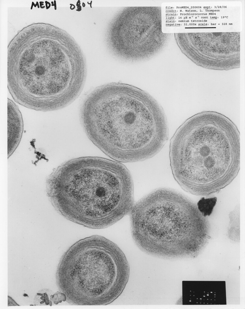
These small, marine, picoplankton are one of the most abundant and smallest photosynthetic organisms in Earth. They contain chlorophyll a and b, and lack phycobilin pigments. They are free-living, non-motile, coccoid cyanobacteria found in euphotic zones of world’s tropical oceans at a temperature range of 10-33o C.

unicellular algae examples
Fig: Prochlorococcus MED4” by Chisholm Lab is marked with CC0 1.0. Copy text

Synechococcus arcuatus

  • Phylum: Cyanobacteria
  • Class: Cyanophyceae
  • Subclass: Synechococcophycideae
  • Order: Synechococcales
  • Family: Synechococcaceae
  • Genus: Synechococcus
  • Species: arcuatus

These freshwater, motile, cylindrical coccoid picoplankton contain chlorophyll a as photosynthetic pigments and phycobilin as accessory pigments.

Glaucophyta:

They contain endosymbiotic cyanobacteria in their cytoplasm instead of chlorophyll. The endosymbiotic theory of chloroplast evolution is evident from this symbiotic association. The endosymbiotic cyanobacteria are called cyanelles and the host is called cyanome.

unicellular algae examples
Fig: “Glaucophyta” by JJT1982 is licensed under CC BY 2.0. Copy text

Cyanophora paradoxa

  • Phylum: Glaucophyta
  • Class: Glaucophyceae
  • Order: Glaucocystales
  • Family: Glaucocystaceae
  • Genus: Cyanophora
  • Species: paradoxa

Freshwater flagellate with two cyanelles present in protoplasm. Each cyanelles have a central dense body and an inner ring each, in the stroma inside the plasma membrane.

Glaucocystis duplex-

  • Phylum: Glaucophyta
  • Class: Glaucophyceae
  • Order: Glaucocystales
  • Family: Glaucocystaceae
  • Genus: Glaucocystis
  • Species: duplex

These freshwater organisms have two groups of cyanelles, one on each side of nucleus. They have reduced flagella in cell wall. These unicellular algae proliferate through asexual reproduction.

Euglenophyta:

These motile, aquatic unicellular algae have chlorophyll a &b as their respiratory pigments, and flagella with fibrillar hairs in a row. Their nucleus is mesokaryotic in nature. Their cellular body is covered by plasma membrane, underneath which lies pellicle, that is made of repeating proteinaceous units called strips and microtubules.

Peranema trichophorum-

  • Phylum: Euglenophycota
  • Class: Euglenophyceae
  • Order: Heteronematales
  • Family: Paranemataceae
  • Genus: Peranema
  • Species: trichophorum

These free-living, phagocytic organelles are mostly found in freshwaters. They have specialized ‘food vacuole’ for phagocytosis.

unicellular algae examples
Fig: “Peranema (Flagellat) – 630x” by Picturepest is licensed under CC BY 2.0. Copy text

Euglena gracilis-

  • Phylum: Euglenophycota
  • Class: Euglenophyceae
  • Order: Euglenales
  • Family: Euglenaceae
  • Genus: Euglena
  • Species: gracilis

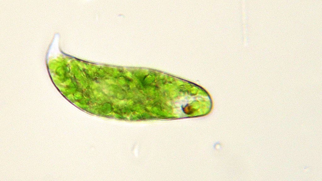
These freshwater, motile unicellular algae have two anterior flagella of which only one emerges from flagellar reservoir. They are both autotroph and heterotroph in nature and consume smaller microbes like amoeba through phagocytosis. They reproduce asexually through longitudinal mitotic cell division.

unicellular algae examples
Fig: “Euglena gracilis- Protiste (végétal)” by Philippe Garcelon is licensed under CC BY 2.0. Copy text

Chrysophyta: Golden-brown and yellow-green algae; Diatoms

They are flagellated, unicellular autotrophs with mitochondria. They have chlorophyll a and c1/c2, and carotenoid fucoxanthin. Two anteriorly attached flagella of uneven length are present.

Mallomonas elegans

  • Phylum: Chrysophyta
  • Class: Chrysophyceae
  • Order: Ochromonadales
  • Family: Synuraceae
  • Genus: Mallomonas
  • Species: elegans

These freshwater unicellular algae are covered by intricate silica scales and bristles. They are golden-brown in color.

unicellular algae examples
Fig: “Mallomonas sp.” by greenmarlin is licensed under CC BY-SA 2.0. Copy text

Cyclotella meneghiniana-

  • Phylum: Ochrophyta
  • Class: Bacillariophyceae
  • Subclass: Coscinodiscophycidae
  • Order: Thalassiosirales
  • Family: Stephanodiscaceae
  • Genus: Cyclotella
  • Species: meneghiniana

These marine water algae are found in oligotrophic environments. They have the largest genome among all the diatoms, 171 Mb in size. They proliferate through asexual reproduction which results in decreasing cellular size.

Pyrrophyta: Dinoflagellates

Unicellular, autotrophic phytoplankton which are mostly marine but some can be found in freshwater environment. They act as a primary food source for almost every aquatic food chain.

Noctiluca scintillans-

  • Phylum: Pyrrophycophyta
  • Class: Dinophyceae
  • Order: Noctilucales
  • Family: Noctilucaceae
  • Genus: Noctiluca
  • Species: scintillans

These free-living, marine dinoflagellates can be found throughout the world and have bioluminescence properties. They cause toxic algal bloom, known as red tide which causes harm to fishes and corals.

unicellular algae examples
Fig: “File:Noctiluca scintillans unica.jpg” by Maria Antónia Sampayo, Instituto de Oceanografia, Faculdade Ciências da Universidade de Lisboa is licensed under CC BY 3.0. Copy text

Pyrodinium bahamense-

  • Phylum: Pyrrophycophyta
  • Class: Dinophyceae
  • Order: Gonyaulacales
  • Family: Goniodomataceae
  • Genus: Pyrodinium
  • Species: bahamense

These autotrophic, marine dinoflagellates are generally found in Atlantic Ocean. They produce bioluminescence and also paralytic toxin. They possess chlorophyll a & c and peridinin.

Charophyta: Stoneworts or Brittleworts

These motile green algae are commonly found in fresh to brackish water. They have two flagella attached in a lateral position. They reproduce sexually by forming a dominant zygote which eventually germinates by meiotic division. The evolutionary line of charophyta later evolved into land plants.

Cylindrocystis brebissonii-

  • Phylum: Charophyta
  • Class: Conjugatophyta
  • Order: Zygnematales
  • Family: Mesotaeniaceae
  • Genus: Cylindrocystis
  • Species: brebissonii

These autotrophic green algae have a cylindrical body with asteroid shaped chlorophyll. They are generally found in freshwater or terrestrial habitats.

Euastrum humerosum-

  • Phylum: Charophyta
  • Class: Conjugatophyta
  • Order: Desmidiales
  • Family: Desmidiaceae
  • Genus: Euastrum
  • Species: humerosum

These are freshwater algae. They have a pyramidal outline of their cell, with deep invagination of the median apical incurvation, dividing the cell into apical, lateral and basal lobes.

unicellular algae examples
Fig: “Euastrum sp – Algue verte” by Philippe Garcelon is licensed under CC BY 2.0. Copy text

Also Read:

15+ Methanogenic Bacteria Examples: Detailed Explanations and Images

With a few methanogenic bacteria examples we will know about methanogens which are a group of bacteria that produce methane as a byproduct of their anaerobic metabolism.

Methanogenic bacteria example as prokaryotic organisms that belong to the phylum Euryarchaeota under domain Archaea. They are obligatory anaerobes and obtain energy by breaking down CO2, hydrogen, acetate, formate, methanol etc. into methane and carbon dioxide.

A few Methanogenic bacteria examples,

They are a diverse group of organism which can survive in various extreme habitats; like hydrothermal vents, geothermal vents, anaerobic digesters, bogs etc. They are also found in decomposing organic matters, rumen of herbivorous animals, intestine of various mammals, guts of insects etc.

Process of methane formation:

These bacteria are very sensitive to oxygen even at a trace level and susceptible to oxygen stress by prolonged exposure. They break down CO2 into Methane in presence of hydrogen,

CO2 + 4H2 —> CH4 + 2H2O

Methanobacterium bryantii

  • Phylum: Euryarchaeota
  • Class: Methanobacteria
  • Order: Methanobacteriales
  • Family: Methanobacteriaceae
  • Genus: Methanobacterium
  • Species: bryantii

This non-motile, rod-shaped bacterium has an optimal growth temperature ranging from 37-45oC. These gram positive bacteria are mostly found in aquatic sediments and  have a 3.46 Mb long genome sequence with 3300 different proteins.

Methanobacterium oryzae

  • Phylum: Euryarchaeota
  • Class: Methanobacteria
  • Order: Methanobacteriales
  • Family: Methanobacteriaceae
  • Genus: Methanobacterium
  • Species: oryzae

This rod-shaped, non-motile bacterium was first isolated from paddy field of Philippines. They grow at a temperature of 40oC and a neutral pH of 7.

Methanobrevibacter acididurans

  • Phylum: Euryarchaeota
  • Class: Methanobacteria
  • Order: Methanobacteriales
  • Family: Methanobacteriaceae
  • Genus: Methanobrevibacter
  • Species: acididurans

These hydrogenotrophic methanogens are acid tolerant and are usually found in gut of larger animals such as mice, termites etc. These organisms can grow optimally at a temperature of 35oC to 37oC and 6 pH. They don’t have peptidoglycan as their cell wall component, rather are made of pseudomurein. Under acidic condition these bacteria shows better Methanogenic capacity than other bacteria of same order and decrease accumulation of volatile fat causing obesity.

Methanosphaera cuniculi

  • Phylum: Euryarchaeota
  • Class: Methanobacteria
  • Order: Methanobacteriales
  • Family: Methanobacteriaceae
  • Genus: Methanosphaera
  • Species: cuniculi

These gram-positive, non-motile, spherical or oval methanogens are commonly found in ruminant gut because they prefer low hydrogen or methane producing host. They were first isolated from gut of rabbit and have an optimal growth temperature of 30-40oC and optimum pH is 6.8.

Methanothermobacter thermoflexus

  • Phylum: Euryarchaeota
  • Class: Methanobacteria
  • Order: Methanobacteriales
  • Family: Methanobacteriaceae
  • Genus: Methanothermobacter
  • Species: thermoflexus

These rod-shaped, filamentous, gram positive methanogens are moderate thermophiles and have an optimal temperature of 55-60oC and a neutral pH value of 7. It was first isolated from digester sludge of methacrylate wastewater in Russia.

Methanothermus sociabilis

  • Phylum: Euryarchaeota
  • Class: Methanobacteria
  • Order: Methanobacteriales
  • Family: Methanothermaceae
  • Genus: Methanothermus
  • Species: sociabilis

Small, rod-shaped, gram positive, motile bacteria generally occur singly or form a short chain or cluster. Their cell wall is made of pseudomurein with an outer layer of protein S. They are highly thermophilic and can survive up to 97oC.

Methanococcus aeolicus

  • Phylum: Euryarchaeota
  • Class: Methanococci
  • Order: Methanococcales
  • Family: Methanococcaceae
  • Genus: Methanococcus
  • Species: aeolicus

These small cocci-shaped methanogens are isolated from marine sediment and is the first archaeal genome to be sequenced. They are gram negative, non-motile bacteria that have their cell wall made of protein.

Methanothermococcus okinawensis

  • Phylum: Euryarchaeota
  • Class: Methanococci
  • Order: Methanococcales
  • Family: Methanococcaceae
  • Genus: Methanothermococcus
  • Species: okinawensis

These motile, irregular cocci, thermophilic archaeon was first isolated from a deep sea vent chimney at Iheya ridge in Okinawa, Japan. They have an optimum pH of 6.7 and optimum temperature of 60-65oC.

Methanocaldococcus indicus

  • Phylum: Euryarchaeota
  • Class: Methanococci
  • Order: Methanococcales
  • Family: Methanocaldococcaceae
  • Genus: Methanocaldococcus
  • Species: indicus

They are motile, coccoid methanogens; first found in deep sea hydrothermal vent in Central Indian Ridge in international waters. It can grow from 50oC to 85oC temperature and around 6.5 pH.

Methanotorris igneus

  • Phylum: Euryarchaeota
  • Class: Methanococci
  • Order: Methanococcales
  • Family: Methanocaldococcaceae
  • Genus: Methanotorris
  • Species: igneus

Small, free living, hyperthermophilic methanogens first found in shallow submarine hydrothermal vent at Kolbeinsey ridge in Iceland.

Methanopyrus kandleri

  • Phylum: Euryarchaeota
  • Class: Methanopyri
  • Order: Methanopyrales
  • Family: Methanopyraceae
  • Genus: Methanopyrus
  • Species: kandleri.

This rod shaped, hyperthermophilic methanogen is the only species of methanopyraceae family. They were first found surviving at a 2000m depth and 84-110oC temperature at Gulf of California. They have their cell wall made of terpenoid lipids.

methanogenic bacteria examples
Fig: “Methanopyrus kandleri Kurr et al. 1992” by Matt Crook is marked with CC BY-SA 3.0.

Methanocella arvoryzae

  • Phylum: Euryarchaeota
  • Class: Methanomicrobia
  • Order: Methanocellales
  • Family: Methanocellaceae
  • Genus: Methanocella
  • Species: arvoryzae

These non-motile, rod shaped bacteria was first isolated from Italian rice field soil. They grow at an optimal 45oC temperature and 7.1 pH. They mainly use acetate or formate as their primary energy source.

Methanogenium cariaci

  • Phylum: Euryarchaeota
  • Class: Methanomicrobia
  • Order: Methanomicrobiales
  • Family: Methanomicrobiaceae
  • Genus: Methanogenium
  • Species: cariaci

These non-motile, gram negative, rod shaped methanogens are mesophilic, having an optimal temperature of 25oC. First found in marine sediment of Cariaco trench in Venezuela.

Methanomicrobium mobile

  • Phylum: Euryarchaeota
  • Class: Methanomicrobia
  • Order: Methanomicrobiales
  • Family: Methanomicrobiaceae
  • Genus: Methanomicrobium
  • Species: mobile.

Non-motile, slightly curved rod-shaped, gram negative methanogens are commonly found in ruminant stomach. They have an optimum growth temperature of 40oC and 6.1 – 7.0 is optimal pH value.

Methanospirillum lacunae

  • Phylum: Euryarchaeota
  • Class: Methanomicrobia
  • Order: Methanomicrobiales
  • Family: Methanospirillaceae
  • Genus: Methanospirillum
  • Species: lacunae

These motile, gram negative, curved rod or spiral shaped hydrogenotrophic methanogen are naturally found in wetland sediments and anaerobic sewage digesters.

Methanothrix soehngenii

  • Phylum: Euryarchaeota
  • Class: Methanomicrobia
  • Order: Methanosarcinales
  • Genus: Methanothrix
  • Species: soehngenii

They are non-motile, rod-shaped, mesophilic methanogens with an optimum growth temperature of 35oC. These organisms were first isolated from anaerobic sewage sludge in Switzerland. They cannot process carbon dioxide like other methanogens and solely dependent on acetate as an energy source.

Methanosarcina barkeri

  • Phylum: Euryarchaeota
  • Class: Methanomicrobia
  • Order: Methanosarcinales
  • Family: Methanosarcinaceae
  • Genus: Methanosarcina
  • Species: barkeri

These non-motile, gram positive, irregular coccoid methanogens grow into different sizes based on salt concentration of the medium. They were first isolated from aquatic sediments, digesters and in rumen of herbivores. They have a 4.8Mbp long nucleotides sequence and 3606 different proteins; which make the bacterium a model organism to study biotechnology and cell biology.

methanogenic bacteria examples
Fig: “Methanosarcina barkeri” by Matt Crook is marked with CC BY-SA 3.0.

Also Read:

15+ Red Algae Examples: Detailed Facts

With a few red algae examples we will discuss what red algae are and how they differ from other algae.

Red algae or Rhodophytes are eukaryotic algae with over 6000 species, of which most are marine except a few freshwater species like Batrachospermum etc. They are autotrophs and have a red color due to the presence of phycobilin pigments, I.e., phycoerythrin and phycocyanin. They are mostly autotrophic except a few like Harveyella, which are parasitic to other algae. Unlike other algae red-algae are sessile because they lack flagella.

A few common red algae examples are,

Bangia atropurpurea

Division: Rhodophyta, Subdivision: Eurhodophytina, Class: Bangiophyceae, Order: Bangiales, Family: Bangiaceae, Genus: Bangia, Species: atropurpurea. They grow in the littoral intertidal zones of both freshwater and marine water. Filamentous body which is uniseriate at base and multiseriate above, is formed from unbranched, erect thalli.

Porphyra umbilicalis

Division: Rhodophyta, Subdivision: Eurhodophytina, Class: Bangiophyceae, Order: Bangiales, Family: Bangiaceae, Genus: Porphyra, Species: umbilicalis. Growing in cold, shallow marine water; these edible algae are a type of Nori. It can reproduce both asexually, with help of spores or sexually using male and female gametes produced by single thallus. They are a rich source of vitamin B12.

Lemanea fluviatilis

Division: Rhodophyta, Subdivision: Eurhodophytina, Class: Florideophyceae, Order: Batrachospermales, Family: Lemaneaceae, Genus: Lemanea, Species: fluviatilis. Found in temporal streams and rivers, these algae are native to Europe and North America.

Thorea ramosissima

Division: Rhodophyta, Subdivision: Eurhodophytina, Class: Florideophyceae, Order: Thoreales, Family: Thoreaceae, Genus: Thorea, Species: ramosissima. These freshwater algae are distributed through temperate and tropical regions. They are small, filamentous and dark green in color and reproduces asexually.

Gelidium amansii

Division: Rhodophyta, Subdivision: Eurhodophytina, Class: Florideophyceae, Order: Gelidiales, Family: Gelidiaceae, Genus: Gelidium, Species: amansii. Stiff, cartilaginous algae with compound lobed, pinnate leaves on each branch. This seawater alga is usually found in the East Asian shallow coasts and is an important part of Asian diet.

Ethelia hawaiiensis

Division: Rhodophyta, Subdivision: Eurhodophytina, Class: Florideophyceae, Order: Gigartinales, Family: Cruoriaceae, Genus: Ethelia, Species: hawaiiensis. These mesophotic marine algae have large filament cells and their thallus cavity is often inhibited by microalgae.

Corallina officinalis

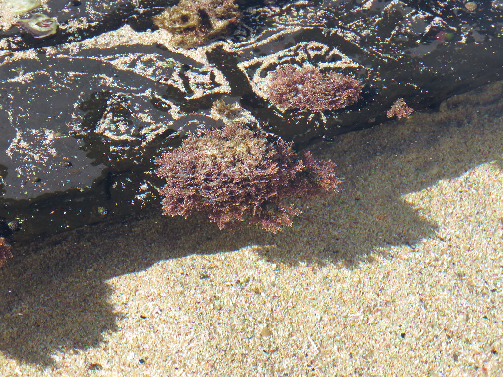
Division: Rhodophyta, Subdivision: Eurhodophytina, Class: Florideophyceae, Order: Corallinales, Family: Corallinaceae, Genus: Corallina, Species: officinalis. These marine algae are found in lower or mid littoral zones on the rocky shores. They have a calcareous deposition within its cells and strengthen the thallus. It is used as a skincare product.

red algae examples
Fig: “Corallina officinalis” by dhobern is marked with CC BY 2.0.

Platoma cyclocolpa

Division: Rhodophyta, Subdivision: Eurhodophytina, Class: Florideophyceae, Order: Nemastomatales, Family: Schizymeniaceae, Genus: Platoma, Species: cyclocolpa. Thallus is gelatinous, slippery and pale red and are usually found in Western Pacific.

Gracilaria arcuata

Division: Rhodophyta, Subdivision: Eurhodophytina, Class: Florideophyceae, Order: Garciliariales, Family: Garciliariaceae, Genus: Gracilaria, Species: arcuata. These agar yielding, marine algae are native to Indo-Pacific Ocean & Atlantic Ocean and grow in shallow sub-tidal zones attached to corals. Thallus is branched and cartilaginous and the plant is unisexual. They are used for human consumption and as wastewater purifier.

Rhabdonia spp

Division: Rhodophyta, Subdivision: Eurhodophytina, Class: Florideophyceae, Order: Gigartinales, Family: Areschougiaceae, Genus: Rhabdonia. Thalli are erect, cylindrical or segmented into clavate of these marine algae. Each individual alga can grow up to 25 cm.

Champia lumbricalis

Division: Rhodophyta, Subdivision: Eurhodophytina, Class: Florideophyceae, Order: Rhodymeniales, Family: Champiaceae, Genus: Champia, Species: lumbricalis. These marine, worm like algae are native to South Africa.

Ceramium affine

Division: Rhodophyta, Subdivision: Eurhodophytina, Class: Florideophyceae, Order: eramiles, Family: ceramiaceae, Genus: Ceramium, Species: affine. They are small, dioecious, marine algae that are consisted of a terete of single row of cells surrounded by smaller cells forming cortex.

Chondrus crispus

Division: Rhodophyta, Subdivision: Eurhodophytina, Class: Florideophyceae, Order: Gigartinales, Family: Gigartinaceae, Genus: Chondrus, Species: crispus. These soft and cartilaginous marine algae is commonly known as Irish moss. They grow on the surface of rocks, from intertidal zone to all the way to the sea floor and can survive in minimal light. They are found in Northern Atlantic and are commonly edible, also used as medicine.

Cyanidioschyzon merolae

Division: Rhodophyta, Subdivision: Eurhodophytina, Class: Bangiophyceae, Order: Cyanidiales, Family: Cyanidiaceae, Genus: Cyanidioschyzon, Species: merolae. A primitive, unicellular alga, commonly found in hot springs. This eukaryotic cell is a model cell to study cell & molecular biology and bio-technology because of having an extremely simple, compact genome of 16.5 Mbp containing 5,331 genes.

Palmaria palmata

Division: Rhodophyta, Subdivision: Eurhodophytina, Class: Florideophyceae, Order: Palmariales, family: Palmariaceae, Genus: Palmaria, Species: palmata. These sessile, marine cold-water algae have a leathery texture and is commonly called Dulse or Irish seaweed. They are usually found in different shores throughout Europe and North Atlantic shore of America. Dulse is consumed as food due to its rich mineral content.

Mastocarpus stellatus

Division: Rhodophyta, Subdivision: Eurhodophytina, Class: Florideophyceae, Order: Gigartinales, Family: Phyllophoraceae, Genus: Mastocarpus, species: stellatus. These red algae are also known as carrageenan moss or false Irish moss and generally found in coast of Ireland and Britain. They can tolerate high environmental stress. It is used as a food item because of its high antioxidant content and also being used as a plastic substitute.

Also Read:

9+ Mesophilic Bacteria Examples: Detailed Explanations and Images

1486848966 c23fc91a5c b

With a few mesophilic bacteria examples we will discuss their optimum growth temperature and other details.

Different bacteria grow at a different temperature and the temperature in which bacteria colony growth rate is maximum is called optimum growth temperature. Bacteria with the optimum growth temperature of 20o to 45o Celsius or approximately 65o – 113o Fahrenheit, are called mesophilic bacteria and clostridium and E. coli are mesophilic bacteria examples.

The average optimal temperature for reproductive activity of mesophilic bacteria is around 37oC. That is why most bacterial infections in human is caused by mesophilic bacteria. Mesophilic bacteria can be isolated from decomposing organic matters, sewage, soil etc.

A few common mesophilic bacteria examples are;

Listeria monocytogenes

Phylum: Firmicutes, Class: Bacilli, Order: Bacillales, Family: Listeriaceae, Genus: Listeria Species; monocytogenes. Rod shaped, commonly foodborne bacteria. Optimum temperature for these bacteria is 25o– 35o C. This bacterium has a 2.94 M bp long genome with 2853 ORFs and a total 13 different serotypes based on surface antigens. This bacterium causes listeriosis human and infects almost 1600 people every year.

Escherichia coli

Phylum: Pseudomonadota, Class: Gammaproteobacteria, Order: Enterobacteriales, Family: Enterobacteriaceae, Genus: Escherichia, Species: coli. This rod-shaped bacterium is mostly transmitted through fecal-oral transmission and commonly found at lower intestine of warm-blooded animals. They are mostly harmless but a few strains cause food-poisoning. Optimum growth temperature for E. coli is 37oC. Its circular DNA is 4.6 M bp in length with 4,288 protein coding genes. Origin of replication in prokaryotic bacteria was first found in E. coli; and is called Ori-C.

mesophilic bacteria examples
Fig: “Escherichia coli” by mrmin123 is marked with CC BY-SA 2.0.

Staphylococcus aureus

Phylum: Firmicutes, Class: Bacilli, Order: Bacillales, Family: Staphylococcaceae, Genus: Staphylococcus, Species: aureus. Cocci shaped, facultative aerobe bacterium and forms cluster in culture media. Their optimal growth is observed in 30o – 37o C temperature. It has a 2.86 M bp long genome and about 2677 different proteins. This opportunistic pathogen is generally transmitted through direct contact and causes various skin and soft tissue inflammations, food poisoning etc.

mesophilic bacteria examples
Fig: “Staphylococcus aureus Bacteria” by NIAID is marked with CC BY 2.0.

Streptococcus pneumoniae

Phylum: Firmicutes, Class: Bacilli, Order: Bacillales, Family: Streptococcaceae, Genus: Streptococcus, Species: pneumoniae. These airborne, pathogenic, lancet-shaped bacteria is the main reason behind human pneumonia. The optimal growth temperature for these bacteria is around 37oC. It has a 2.1 M bp long genome, about 1553 genes and 154 genes containing virulome. It colonizes in the nasopharyngeal tract and causes ailments like fever, cough, breathing troubles etc. In case of severe inflammations, it breaks down hemoglobin causing respiratory failure.

Streptococcus pyogenes

Phylum: Firmicutes, Class: Bacilli, Order: Bacillales, Family: Streptococcaceae, Genus: Streptococcus, Species: pyogenes. Optimal growth temperature is 37oC. These small, round human-specific bacteria is also known as group A streptococcus. It releases various toxins like streptolysin, Spe A, Spe B etc. and cause impetigo, pharyngitis, peritonsillar cellulitis, brain & liver abscess, various autoimmune disorders etc. It has roughly 1.9 M bp long genome with 1700 different proteins.

mesophilic bacteria examples
Fig: “Streptococcus pyogenes” by VeeDunn is marked with CC BY 2.0.

Stenotrophomonas maltophilia

Phylum: Pseudomonadota, Class: Gammaproteobacteria, Order: Xanthomonadales, Family: Xanthomonadaceae, Genus: Strenotrophomonas, Species: maltophilia. These aerobic, rod-shaped bacteria found in wet environments are highly antibiotic resistance and is a newly emerging concern. They thrive in a temperature of 35oC. Their genome sequence consists of 159,365 bp and produces 271 predicted proteins. It causes chronic respiratory illness, endocarditis, peritonitis, UTIs etc.

Clostridium kluyveri

Phylum: Firmicutes, Class: Clostridia, Order: Clostridiales, Family: Clostridiaceae, Genus: Clostridium, Species: kluyveri. These rod-shaped, obligatory anaerobe bacteria have around 3.96 M bp long genome sequence. These bacteria can use ethanol or acetate as energy source and grow. They are mostly aquatic, free living and form spores. They are not known for any pathogenicity.

Neisseria gonorrhoeae

Phylum: Pseudomonadota, Class: Betaproteobacteria, Order: Neisseriales, Family: Neisseriaceae, Genus: Neisseria, Species: gonorrhoeae. These host associated diplococcus is sexually transmitted, I.e., transmitted through body fluid contamination. It has a circular DNA of 2,153,922 nucleotides and 2069 genes. Their optimal growth temperature is 35oC. It causes gonorrhea in human. Purulent discharge from urethra and dysuria are common symptoms of gonorrhea. Almost 95% of infected men are symptomatic whereas, only 10% – 20% infected women are symptomatic.

Lactobacillus plantarum

Phylum: Firmicutes, Class: Bacilli, Order: Lactobacillales, Family: Lactobacillaceae, Genus: Lactobacillus, Species: plantarum. These rod-shaped, free living, facultative anaerobes grow in an optimal temperature of 25oC and is one of the most studied species of probiotic microbes in food industries. They produce lactic acid which is been used for fermentation for centuries. They have a 3,308,274 nucleotides long genome sequence with 3,052 ORFs. These bacteria have been used for producing various fermented products like pickles, Kimchi, sauerkraut or fermented cabbage, Nigerian Ogi and also various fermented milk products. It also has antioxidant activities and also increases intestinal permeability.

Please click to learn on Decomposer Bacteria Examples.

Also Read: