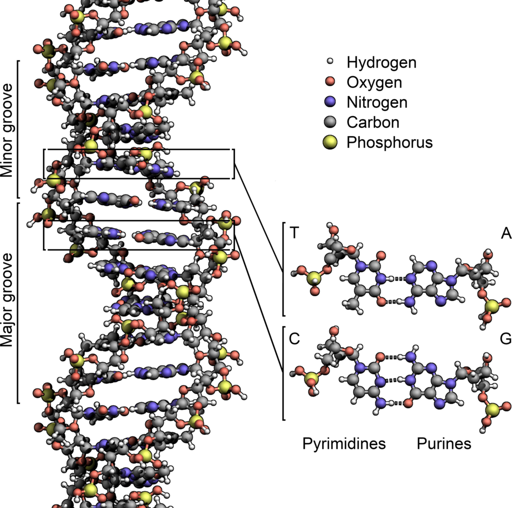
Is Adenine a Nucleotide: Detailed Facts

bb 1

The very base for making of a nucleic acid is said to be a nucleotide. A nucleotide has a sugar molecule linked with phosphate.

With concern to is adenine a nucleotide, it said to be a nucleotide in the RNA and DNA. It is said to be a part of adenosine triphosphate and is linked with the thymine in DNA and uracil in RNA and even has a base of sugar, phosphate in its structure with adenine being classified as a nucleotide.

This complex is said to be an ATP base and helps in getting the molecule phosphorylated. Adenine is said to be purine with lots of roles to offer in biochemistry. Adenine is one of the four bases in the nucleic acid that are in the DNA and RNA.

The rest of the bases in them are the guanine, cytosine, thymine in DNA and uracil in RNA. Adenine helps in protein synthesis and also is considered a chemical component in DNA and RNA. The adenine has its shape complementary to either uracil or thymine in both the strands.

They serve as monomeric units of the nucleic acid polymers – deoxyribonucleic acid and ribonucleic acid. The monomer units of DNA are nucleotides, and the polymer is known as a polynucleotide. Each nucleotide consists of a 5-carbon sugar (deoxyribose), a nitrogen containing base attached to the sugar, and a phosphate group

Is adenine a nucleotide?

The nucleotides are the molecules that have a phosphate and also a nucleoside and it is organic in nature.

The question for is adenine a nucleotide, it’s a Yes, adenine is said to be a nucleotide with having many of the uses in the field of biochemistry and can have many tautomer with shape opposite to the pyrimidine bases.

Adenine is a nucleotide serves as a unit of monomer in the polymer of nucleic acid and for the DNA and RNA. These both are vital biomolecules needed for every living organism. Nucleotides can be generally taken from diet and can also be synthesized from the genera, nutrients by the organ called liver.

Any of the nucleotide that is seen is made of subunits that are counted to be three in number. They are the nucleobase, a group of phosphate that has one to three attached phosphates to it and a sugar having five carbon chains namely can be the deoxyribose or ribose.

Nucleotide seems to play a good role in as a fundamental for metabolism in the cell level. They help in getting the chemical energy needed to make a nucleoside triphsophates, guanosine triphosphate, uridine triphosphate and adenosine triphosphate. A nucleotide is the basic structural unit and building block for DNA. These building blocks are hooked together to form a chain of DNA.

This helps in many functions for the cell and demand power for the synthesis of cell membrane, amino acids and proteins and also functions for cell to cell transfer both external and internal. They also help in cell signaling and help in the reaction involving certain enzymes.
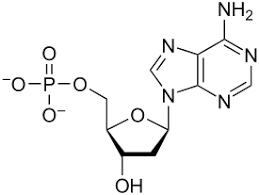
Is adenine A nucleotide
Image credit-
NucleotideWikipedia

Why is adenine a nucleotide?

Adenine is one among the four bases in both the stands of nucleic acids and differs just in one base for DNA and RNA.

The adenine as a nucleotide has a structure typical to it and consists of the purine base linked up with the five chain of carbon sugar and one to three of the phosphate groups. The sugar and phosphate group make up the backbone of the DNA double helix, while the bases are located in the middle.

In order to get the energy stored back, there is an endergonic reaction where the ADP and the free phosphate get to be used. They have the energy as their base and are common for be adenine triphosphate and gets the molecules phsophoralized.

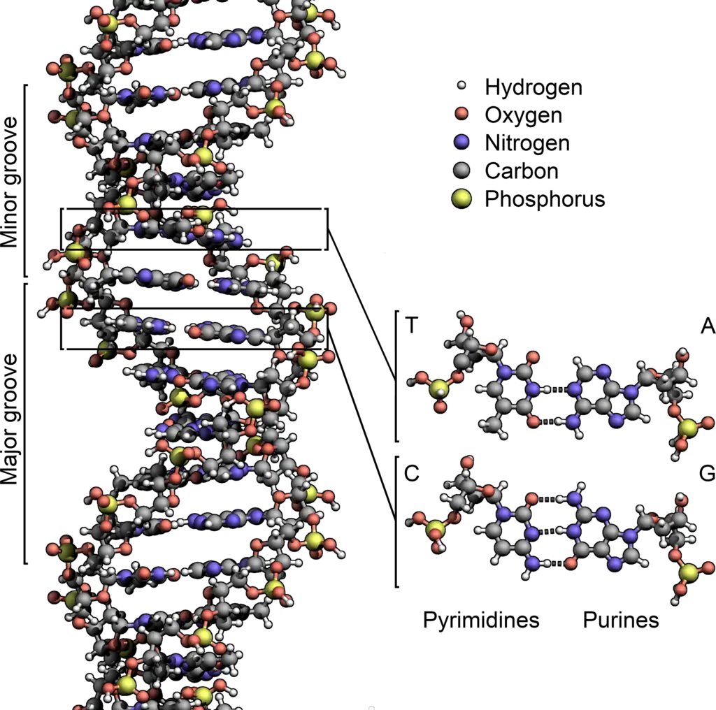
Adenosine is a greater molecule which is made of adenine, a sugar being either deoxyribose or ribose and more or one of the groups of phosphate. There are bases of both purines and pyrimidine in the nucleotide. Adenine is said to be a purine base. These are structures composed of a 5-sided and 6-sided ring. Cytosine and thymine are pyrimidines which are structures composed of a single six-sided ring. Adenine always binds to thymine, while cytosine and guanine always bind to one another.

Adenine being used is used up in many parts of the cell and just not in both of the both of acids strands being DNA and RNA. It is a part of the adenosine triphosphate molecule and is seemed to be the energy place for the cells. So it gets to play two roles inside the cell which are helping to build the nucleic acid and storing up cell energy.
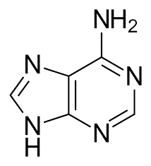
mm 1
Image credit-
AdenineWikipedia

How is adenine a nucleotide?

There are two groups of nucleotides in the nucleic acid. They are the purines and pyrimidine. Adenine is a purine.

Adenine is surely the one base among four and stands first in all description. It is also one of the two purine bases ad is sued in for making nucleotides with bonding to thymine in DNA. The  molar mass is 120 g/mol.

A molecule which is made up of nitrogen many of the hydrogen atoms and carbon is called to be adenine with the chemical formula of C5H5N5. The time when a base just like adenine attaches itself to phosphate and the ribose, it gets to form a nucleotide. The Nucleotide database is a collection of sequences from several sources, including GenBank, RefSeq, TPA and PDB.

This base gets to interlink with thymine in DNA while guanine and cytosine binds up together always with one another. This linking of bonds is referred to as complementary base pairing. These bases get to complement each other and are liked via the hydrogen bonds that can be broken down easily while the DNA replicates itself.

The base of adenine seems to be fused with guanine and form an attached ring skeletal like formation which is made from purine and thus it is said to be a base of purine. The nitrogenous base of purine is a character by the group of amino acids that are single and the C6 carbon in the adenine and the C2 for the guanine.

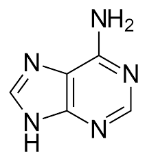
The adenine being a purine is an aromatic compound being heterocyclic with having the chemical formula of C5H4N4. The chemical structure of purine is made of the pyrimidine rings that have an imidazole ring attached to it and thus have the two of the carbons ring and a close of four nitrogen atoms.

Pyridopyrimidine - Wikipedia
Image credit-
Pyrimidine ringWikipedia

Function of adenine as nucleotide

The nucleotide of adenine is mostly referring to as the adenylates or the adenosines. They are molecules being organic and consist of ADP, AMP and the ATP.

Adenines along with rest of the bases are said to perform different in both of the strands. Adenine helps in protein synthesis inside the RNA and binds up with the base of uracil.

Inside the DNA adenine gets to attach itself with thymine which is a base of pyrimidine and uses up the nucleotide that helps in making of nucleic acid. They bind up with the other with the help of the hydrogen bonds which can be broken down without any efforts and helps in getting the structure stable.

The molecules that are in the adenine making it a nucleotide are used in playing a vital role for having the energy transfer and stored. The base of purine is guanine and adenine and adenine is said to be a major product for the ATP. ATP is made when adenine links with a molecule of ribose and the three chain of phosphate.

Nucleoside serves to be work even out of the cell and outside of storing the genetic data. They work as passing on messages and get the energy moved from cell to cell. Despite these they also help in many of the cell functions like metabolism, signal transfer ad power up the enzymatic reactions.

The basic function of the bases in the strands of nucleic acids namely adenine, guanine, cytosine, thymine or uracil are that they are the primary nucleobase. They function in having the space for the basic unit in the genetic coding and having initials to be A,C,G, T or U. In DNA and RNA, nucleotides are covalently linked by the phosphate group; the negative charge of the phosphate group at neutral pH is essential for stabilizing nucleotides.

Also Read:

Adenine Structure In RNA: Detailed Facts

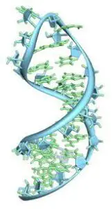
hj 1 162x300 1

RNA is commonly called to be ribonucleic acid and is a vital molecule being polymeric.

The base adenine structure in RNA is to get itself linked up with uracil. Adenine gets itself form adenosine which links to the ribose sugar and the deocyadenosine while getting connected to deoxyribose.

A polymer is a material that has many great sized molecules or even macromolecules made up of many repeating units. For this diverse property in spectrum, both the natural and synthetic polymers do play a vital role in all day. 

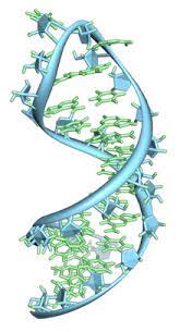
RNA is concerned to be a chain of nucleotides but is a bit different from DNA in its structure as DNA is said to be a double helix while RNA has a single stand folded on top of itself in spite being double. The bases that RNA holds are adenine, guanine, uracil and cytosine.

There are also many molecules of RNA play a good role inside the cells by catalyzing the reactions in biology along with having to control the expression of the gene or sensing the signals by the cells. It also takes part in the universal process being the protein synthesis where RNA directly synthesized protein on ribosomes.

Structure of RNA

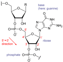
Each of the nucleotide in the RNA is made of a sugar called the ribose with having the carbons numbered 1’ via 5’.

In general the base in connected to 1’ state with adenine, cytosine, guanine and uracil. Adenines are the guanines are the purine while uracil and cytosine are the pyrimidine.

The 3’ is the position for one of the ribose which also has a phosphate group linked and then having the 5’ stage as its next. There seems to be a negative charge on the phosphate in each of them that makes RNS have a molecule that is charged which is called to be polyanion.

Cytosine and guanine have bonds of hydrogen in between them and also same goes for uracil and adenine. These are the pairs that link up during any process in RNA. There is also possibility of different linkage like the adenine in bulk getting connected with each other of the GNRA tetra loop also can be made which is a base pair of guanine and adenine.

RNA has a good component that helps itself separate from DNA which is the presence of the hydroxyl group at the second base position of the ribose sugar. As there is seen the function group of hydroxyl the helix of RNA is caused to generally take almost the form of A-form geometry.

RNA also has the ability to take up the B-form mostly used in DNA. The geometry that outcomes from the A firm is quite deep and narrow with major groove and the minor groove is shallow and length stays wide. There is also another possibility of the presence of hydroxyl chain which is the flexible conformational area of the RNA molecule.

There are found to be more than 100 types of nucleosides that can take place naturally with the largest diverse modifications only seen in tRNA. The roles in specific for many of the modifications are not known yet discovered but it is noted that the RNA linked with ribosome take part in post transcription.

adenine structure in rna
Image credit- RNAWikipedia

Adenine Structure in RNA

It is one of the four nucleotide bases found in RNA and DNA both and have their letters in as C-A-G-U in RNA.

The adenine structure in RNA gets to make tautomer which are compounds that can grow fast and can be easily inter converted and are generally said to be similar. In conditions that are isolated, 9H adenine tautomer is seen.

The workings of the purine consist of making of two of the bases which are guanine and adenine. Both of the nucleic acid bases are taken from the nucleotide called the inosine monophosphate termed also as IMP. The IMP in turn gets synthesized by the already present ribose sugar phosphate via a complex phase or pathway.

The complex pathway that uses up atoms from glycine, which is also amino acids along with aspartic acid and glutamine, is also used up here with the help of a coenzyme called tetrahydrofrolate. The process of making adenine is not surely known but a present method for preparing it in large scale if to use formamide way.

Adenine is considered to be one of the purine bases with the other said to be guanine and is used in making of the nucleotides of the nucleic acids. In the DNA, the adenine tends to link with thymine through two hydrogen bonds that helps in balancing the structure of the nucleic acid.

hh 2
Image credit- Adenine StructureWikipedia

Function of adenine in RNA

The strands of RNA are mostly single but also exists few RNA microbes that are double stranded. There can be variety in RNA.

Translation is the way when RNA is made from protein synthesis and transcription is said to while RNA makes DNA.  Thus, the function of RNA differs along with the type of cell they are in being in eukaryotic and prokaryotic.

There are particular molecules of RNA that make gene expression and then have the capability to serve as a therapeutic agent for diseases in human. There are three main type of RNA in for protein synthesis. They are messenger RNA, transfer RNA, and ribosomal RNA. Mutation in RNA can lead to many human diseases.

The function of the base adenine is-

  • Used in protein synthesis and binds with uracil
  • It helps in providing energy and form ATP
  • It helps in cellular functions
  • Helps in converting chemical energy to chemical reaction.

Messenger RNA

This is seen to transcribe from the DNA and has the blueprint of genetic that makes the protein.

The mRNA in the prokaryotes is not intended to be processes and thus can directive used fir protein synthesis at a go. In the eukaryotes, there is sued a fresh RNA that is transcribed along with a pre mRNA and needs to go for maturation to make mRNA.

Any pre mRNA has all the non-coding and coding areas called the exons and introns. At the time of processing of the pre mRNA, the intron split up while the exons are connected together. There is a 5’ cap and a 3’cap. The 5’ cap protects this from getting degraded and helps in balancing of mRNA. Messenger ribonucleic acid is a single-stranded molecule of RNA that corresponds to the genetic sequence of a gene, and is read by a ribosome in the process of synthesizing a protein.

mm
Image credit-Messenger RNAWikipedia

Transfer RNA

These are the molecule that has a structure of cloverleaf and is of RNA that helps in translating the mRNA into protein.

The basic function of tRNA is to carry the amino acids on the 3’ reception area to the complex of ribosome with the support of aminoacyl tRNA synthetize. There can be defects seen in their own RNA. Transfer RNA does this by carrying an amino acid to the protein synthesizing machinery of a cell called the ribosome.

Aminoacyl tRNA synthetize is actually an enzyme that binds with the perfect amino onto the free tRNA to get the protein synthesized. The amino acid type is based on the mRNA codon, which acts as the sequence for three nucleotide coding for all amino acids. There is also an anticodon arm for the tRNA that is complementary to mRNA.

Ribosomal RNA

These are the most vital from of RNA needed in protein synthesis. Any ribosome has a large but small unit of ribosome.

Inside the prokaryotes there is a tiny 30s and a big 50s unit of ribosome and makes up 70s in together. While in for the eukaryotes, there is a 60s and an 40s subunit of ribosome and together makes 80s. It is the primary component of ribosomes, essential to all cells. 

The ribosomes can have peptidyl, an acceptor and exit that shall help them in binding of aminoacyl-tRNAs and then connect the amino acids together to make the desired polypeptides. Still, there is a major issue for the RNA mutation that can hamper the normal function of the RNA. The error in RNA can be an outcome for the defects that had been overlooking in RNA.

Also Read:

Adenine Function in RNA: Detailed Facts

ij

RNA is a nucleic acid which is same as DNA but RNA is just single stranded. They are used for much purpose.

Adenine is one of the double bases that are used in forming of the nucleotides for the nucleic acids structures being the DNA and RNA. The adenine function in RNA is to bind uracil through the two molecules of hydrogen and is a pyrimidine.

Along with the above mentioned adenine function in RNA, adenine is also used up in places within the cell with just not in DNA or RNA, it is a part of the adenosine triphosphate molecule and plays as a source of energy in the cell. It plays a dual role in cell with building RNA and DNA.

Ribonucleic acid is commonly short termed as RNA. It is a molecule which is polymeric and is vital for different biological roles in regulating, coding, expression of genes and decoding. RNA also is vital for synthesis of protein and cellular respiration.

Taking in all the function of adenine in RNA or any other, along with the proteins, the lipids, the carbohydrates, the nucleic acid seems to be of much importance with respect to the four great macromolecules needed for life forms.

The most basic adenine functions in RNA are-

  • It is one of the two based of purines that is used to make nucleotides of the nucleic acids. It helps in bonding with the thiamine in the structure of DNA and also in RNA binds with uracil.
  • They have their role in for cellular respiration in form of NAD, ATP, FAD and synthesis of protein as a chemical for in RNA and DNA.
  • It is an organic and pre base of purine. Thus, have many roles to play in biochemistry mostly in cell to cell respiration. It is also a chemical compounder of RNA.
  • It provides with formation of the bonds by supporting it with energy needed.
  • They link with the phosphate in order to keep energy. When 3P is linked with adenine, it is ATP while in DNA adenine interacts with in the form of AMP. So, in general in DNA the formation of adenine is used as a substrate.
ij
Image Credit- Structure of RNA moleculeWikipedia

Adenine function in RNA as a base

The bases of adenine take up one of the two forms. The purines have a double ring in where a 5 ring atom links to a 6 atom ring.

Pyrimidines are usually a 6 atom ring. The purines in the acids are the adenine and guanine. The pyrimidine refers to uracil, cytosine and the thymine. The chemical formula for adenine is C5H5N5. Most DNA is found inside the nucleus of a cell, where it forms the chromosomes.

The base of adenine binds with thymine in DNA and uracil in RNA. It is a vital base as it is not only supportive in both the nucleic acids strands bit also acts as a source of energy and a carrier for the molecule of ATP, the cofactor for flavin adenine dinucleotide and the factor for nicotinamide adenine dinucleotide. . Chromosomes have proteins called histones that bind to DNA.

The metabolism purines consist of the formation of guanine and adenine. Both of the bases are made for the monophosphate and the present method is still being recognized for getting adenine made in large scale. DNA has two strands that twist into the shape of a spiral ladder called a helix. DNA is made up of four building blocks called nucleotides: adenine (A), thymine (T), guanine (G), and cytosine (C). The nucleotides attach to each other (A with T, and G with C) to form chemical bonds called base pairs.

There are many that tend to confuse both the names of bases which are adenosine and adenine. Both however are not the same. Adenine is the name which is popular to hat if the purine base. Adenine is the great molecule of nucleotide that is made up of pure adenine and the sugar being deoxyribose or ribose and more or one of the phosphate groups.

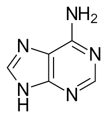
adenine function in rna
Image Credit- AdenineWikipedia

What is adenine in RNA?

The adenine in RNA is a derivative of purine and is a nucleobase. It is on among the rest three bases.

The rest of the bases except adenine are cytosine, guanine and thymine in DNA. In RNA, the four bases are adenine, cytosine, uracil and guanine. Adenine is represented as A and binds with uracil.

The derivatives of the bases have a good role in biochemistry which consists of getting the protein synthesized and then taking pat is cell respiration. The adenine has its shape complementary to that of wither thymine in DNA or uracil in RNA.

Pure adenine is always said to be a molecule that is independent. When it is connected with DNA there is a formation of covalent bond in between the sugar called the deoxyribose and in the left side bottom nitrogen that gets a help in removing the atom of hydrogen. Genes are short pieces of DNA that carry specific genetic information.

After it the rest of the structures is said to be adenine residue and forms great part of the entire molecule. Adenosine is the adenine that reacts with ribose sugar which is used in RNA and the adenosine triphosphate. The deoxyadenosine is the adenine linked with the deoxyribose and then used to make DNA.

Any of the hard structures are made of smaller blocks, just like a house is made of smaller parts. Living beings are also made in the same way with combining the smaller parts being the molecules made of atoms. Adenine is vital life building block. It is among the major four bases. So adenine plays a dual role in the cell: it’s used for building DNA and RNA, but it’s also used at storing energy in the cell.

The RNA and DNA have the genetic code for all living organism that are the plants, animals, the fungi, humans and other microbes. Adenine helps them to get a stabilized nucleic acid composition for the molecules. It is found in adenosine triphosphate which is a molecule that helps in carrying the enemy needed for life.

hj
Image Credit- RNAWikipedia

Structure of the Adenine RNA molecule

Adenine is one of the rest there molecules found in both of the stands of nucleic acids that is needed for sustaining any of the living being.

Adenines gets top make many types of tautomer and compounds that can fast be inter changed and are generally in common referred to be as same. Yet, in condition being isolated, the adenine tautomer is seen in inert or gas phase.

The metabolism of the purines involves the making of guanine and adenine. Both of the components being guanine and adenine are taken from the nucleotide called the inosine monophosphate that in turn is made to synthesize from the already kept ribose phosphate. Adenine is one of nitrogenous bases utilized in the synthesis of nucleic acids.

This complex is taken as a tunnel and via this pathway of the complex with using the atoms from the amino acids like the aspartic acid, glutamic and glycine atoms and also used up is the coenzyme called the tetrahydrofolate. It also involves the transfer of protein molecules form amino to the ribosomes. Adenine also bonds with Thymine in the DNA structure. Adenine is likewise utilized somewhere else in the cell, in DNA and RNA, however it’s essential for the atom adenosine triphosphate, which is the energy hotspot for the cell.

Some of the molecules of RNA do play a good role in getting the reactions in biology catalyzed by letting the control of gens or communicating with sensing responses and sending back cell signals. One of the vital adenine functions in RNA is getting the synthesis of protein done. In DNA, adenine binds to thymine via two hydrogen bonds to assist in stabilizing the nucleic acid structures. In RNA, which is used in the cytoplasm for protein synthesis, adenine binds to uracil.

Also Read:

Types of Nitrogenous Bases In RNA: Detailed Facts

nn 1

RNA is also termed as ribonucleic acid and is a same molecular to that of the DNA with RNA just being single stranded.

The types of nitrogenous bases in RNA are-

Any molecule that has nitrogenous has quite of a chemical characteristic at the base.

The nitrogenous bases in DNA are adenine, guanine, thymine and cytosine. RNA also has same types of nitrogenous bases yet with one only exception which is the uracil is there instead of thymine. The nitrogen have biological related material with complements of nitrogenous.

Any RNA has a backbone which is made up of several sugars in alternating form namely the ribose and also having a group called the phosphate groups. The RNA is actually a linear molecule that has several other four small molecules.

As said already with the types of nitrogenous bases in RNA, RNA is an acronym to the word ribonucleic acid. It is an important molecule that is seen in the cells and is needed for life. The pieces of the RNA are actually used up to make the proteins in the body for the growth of the cell and make them stay in place.

Adenine

Adenine is in general a nucleobase with a derivative of purine. It is the one of the four seen bases in the nucleic acid.

The one in the DNA is represented as G-C-A-T and in RNA is as G-C-A-U. Apart from adenine, the others are guanine, cytosine, and thymine in DNA or uracil in RNA. They are derives from biochemistry.

The derivatives of the bases are much rich in energy with ATP and has coenzyme A. It is also need in synthesis of proteins and also is a chemical component for the DNA and RNA. The shape of it is complementary to that of wither uracil or thymine.

The time when adenine is linked with DNA, there is a formation of covalent bond and is made in between ribose sugar and then at the base of left is nitrogen. The rest of the residue is called to be adenine residue. It is made to react with ribose and used in RNA.

nn 1
Image credit- AdenineWikipedia

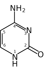
Cytosine

The nucleoside of cytosine is cytosine. In the base paring of Watson Crick model, cytosine forms a hydrogen bond along with guanine.

It is one of the rest nitrogenous bases in DNA and RNA. It is a derivative of pyrimidine along with the heterocyclic ring being aromatic and two of the substitutes that have been attached. 

It was discovered by Albrecht Kossel in 1894. A structure was synthesized in lab was proposed in 1903. Cytosine is much part of DNA AND RNA. It acts as a co factor of the enzyme and then can transfer to the phosphate and get t converted from ADP to ATP.

Cytosine is one of the four building blocks of DNA and RNA. So it’s one of the four nucleotides that’s present both in DNA, RNA, and each cytosine makes up part of the code

hn
Image credit- CytosineWikipedia

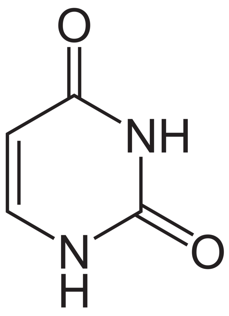
Uracil

The base of uracil is replaced by thymine in DNA. Uracil is seen to be demethylated from the thymine.

In the RNA, Uracil is actually much common and has a natural occurrence. It can be found in herring sperm, spleen and the thymus. It is an unsaturated compound which can absorb light.

Uracil base replaces thymine and links with adenine during DNA transcription in RNA. There is an increase in thymine substitution and thus DNA replicated well. When the phosphate combines with uredines, it makes urine 5-monphsphare.

kj
Image credit- UracilWikipedia

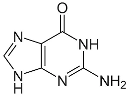
Guanine

RNA as a part in its role is the actual photocopy of the DNA in the cell with just a gap of one difference in the base.

It is made to pair with cytosine. The nucleotide of cytosine is called guanosine. It is a purine derivative and forms a ring system and is a unsaturated molecule being planar.

It is an organic compound with two ring face made of carbon and atoms of nitrogen. It is free in its occurrence and is combined with many diverse sources that are natural like the guano excrement and the dead body of the bats seals and birds.

The composition of guanine in human is about 28% with adenine being 22, thymine being 22. T helps in binding with the proteins that activates the receptors and used up as ion channel. The word guanine derives from the Spanish loanword guano which itself is from the Quechua word wanu, meaning “dung”.

nn
Image credit- GuanineWikipedia

How are the types of nitrogenous bases in RNA and DNA different?

RNA is said to be one of the three major molecules in biology which is vital for all the living forms with the rest being the DNA and the proteins.

Uracil is seen in RNA and combines with adenine while thymine is seen in DNA that binds with adenine. The DNA makes RNA makes proteins. The other adenine, cytosine and guanine are same in both DNA and RNA.

The cells have their own workhorses and those are the proteins and they its role inside the cell as the enzymes with helping in cell signaling, getting the components structures. DNA is said to be the short of deoxyribonucleic acid.

The DNA is the blueprint of cell with carrying all the genetic related information for the growth of the cell and the stakes in nutrients to help it propagate. There is a central tenet for the molecular biology that states that the genetic flow if data in the cell is from the DNA to the proteins via the RNA.

At the time while the cells need to produce a specific protein, the gets to activate the gene where the part of the DNA that codes the proteins and then makes multiple copies if the specific part of DNA in the form of messenger RNA. The several copies of the messenger RNA are then made to translate the genetic coding in protein via cell action making ribosomes.

With this the RNA then expands the amount of protein which is specifically given  and is made one at a time with having vital control points for regulation and managing the protein that has been made. All the protein is made at one time and is sure to have coded the gene one by one.

types of nitrogenous bases in rna
Image credit- Nitrogenous basesWikipedia

There are three major roles of the RNA in the cell which are-

  • RNA acts as a copy of DNA and as a coupler for the genetic code and the amino acids as a component of structure fir the ribosomes. The uses of RNA are much broader and vital with being interesting.
  • The RNA also can act as the enzymes that shall help up in getting the reaction work faster. In many of clinics it has been seen that, the microbes RNA carry the sprayed genetic data.
  • It also helps I getting the function of cell regulated. Its tarts from the process of cell division and the goes to differentiation and the cell growth and along with its aging and the end with cell death. There are certain defects that are seen in RNA or while having it regulated and is faced as diseases in humans.

Also Read:

Function of plasmid in bacteria: Detailed Facts

aa

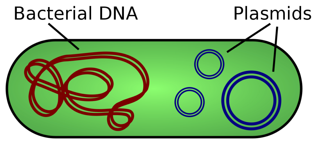
Plasmids are the tiny molecule inside the cell which is also an extrachoromosomal DNA and can be separated.

The Function of plasmid in bacteria can differ and serves quite a bit of usage. They help in gene carrying that helps in organism to survive, some are sensory organelle, some help replication.

The genes that they have are for having the organism to survive wither by getting to kill the rest of microbes or also can be by getting to defeat the host cell that is done by generating of toxins.

Some of the bacteria get to work via this process by helping the bacteria in replication of its DNA. As the plasmids are nothing they actually tend to have less genes with some functions. There are provisions for multiple plasmids to exist in the same cell with having different functions.

The plasmid can be made to part ways from the chromosomal DNA and can replicate on its own. They are mostly found in as small, being double stranded and in circular shape inside the bacteria with sometimes even showing its presence to archaea and eukaryotes.

The main function of the plasmid in bacteria are-

  • They carry in genes that are antibiotic resistant and then spread them in entire human or the body of animal. This helps the eukaryotes get them treated.  
  • The plasmids on the DNA are also able to produce proteins that are antibacterial.
  • They also are capable of carrying out those genes that are involves in all the metabolic process and let themselves be used u for having the pollutants digest from the outer space.
  • They are also able to let carry genes that are connected with getting the pathogenicity of the bacteria increased that can help cause the diseases like that of tetanus and anthrax.

Function of plasmid in bacteria as Gene therapy

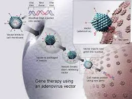
Plasmids play a good role in gene therapy. They are most in common to be used for getting the therapeutic genes inserted.

They are also easy to be manipulated and also are easy for the replication in the cells of the bacteria. They have the ability to be efficient to target the cells that shall be defeating and triggering the genes inside them.

They are also no plasmids that are harmful or have any bad effect like that of viral spreaders. The genes is inside the human system for getting them fights against the disease. Gene therapy is the introduction of genes into existing cells to prevent or cure a wide range of diseases.

aa
Image credit- Gene therapyWikipedia

Function of plasmid in bacteria as Recombinant DNA technology

The recombinant DNA technology makes a good use of the plasmids for a lot of purpose.

For the part of the drug delivery, the technology makes a good use of the plasmids to have the likely drugged get inserted in the body. The function of plasmid in Bacteria is much likely and targeted here.

The DNA technology being recombinant which is applied in plasmids for being resistance and can be used up to get the bacteria killed for being harmful to the human cells.

The very first rime when the plasmid was applied inside the human was while insulin was getting inserted inside the human cells. It did give good result and the other application for it was inserting it in the hormone for human growth in the animal’s mammalian cells.

aa 1
Image credit- Recombinant DNA technologyWikipedia

Application of plasmid in bacteria

The humans have been able to develop a lot of uses that can be obtained from plasmid and thus have created software.

The Function of plasmid in bacteria is so diverse that a software has been made that shall be able to record the sequence of DNA of the plasmids for getting it used in many techniques.

The plasmids are sued up in genetic engineering for amplifying or making of many copies of specific genes. In the molecular cloning, a plasmid is considered to be a variable. It is a sequence in DNA that shall help in transferring any foreign genetic particle form one cell to other.

This is done so that the genes can be made to express and also replicated. Plasmids are also used in cloning the tiny segments of the DNA. They can also be made to replicate proteins like that of the protein that are used up to code for insulin in big spaces.

On addition, the plasmids are seen to transfer genes into the human cells as part of the gene therapy. There is investigation or research going on this term. The cells may be lacking few of the proteins if the person has a hereditary disorder that has gene mutation. A plasmid in the DNA shall allow the cells to express a protein that they lack.

Function of plasmid in bacteria
Image credit- PlasmidWikipedia

Characteristic of plasmids

The plasmids can be classifies in many ways with mostly being conjugative and non-conjugative ones.

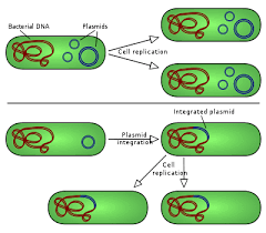
For the purpose of the plasmids to get into the process of replication on its own, they have to stretch the DNA inside the cell. The DNA that is trenched acts as a self-replicating unit here.

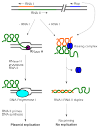
The bacteria replicon is quite typical and may have a number of elements like that of the gene plasmid specific to replication, initiation and protein and the repeating units is called introns along with the DNA boxes and with the adjacent AT-rich area. The ones that integrate are called the episomes.

There are also many small plasmids that make the use of the host replicative enzymes to make themselves copies while the bigger plasmids can carry out genes that is specific to replication with respect to plasmids. There are a few of them that can get inserted into the chromosome which are the host.

The plasmids carry out only one gene. Mostly the genes that they carry on are useful to the host cells like that of the cells that enable them to live in a surrounding that should or else be lethal or restrict their growth. Some genes tend to encode trait for being antibiotic resistance and are unlike toward heavy metals.

Some of the other characteristics of plasmids in bacteria are-

  • They have size ranging from small to being less that 1 kilobase pairs.
  • They are generally circular but can also be linear sometimes.
  • Plasmids vary in different individual with being in range from one to many.
  • The plasmids that are larger in size tend to have less number of copies.
  • The plasmids can be carried on from one cell to other.
tt
Image credit- Self replicating PlasmidWikipedia

Plasmid in bacterial transformation

The bacteria can take in any of the foreign DNA and this is called transformation. It is the key step towards cloning of DNA.

After the very step of transformation, the bacteria are already made to get selected on the plates of antibiotic. The bacteria along with the plasmids are then said to be antibiotic resistant with each making a colony.

The colonies that have the correct plasmids shall be able to grow to make cultures that are bigger if that of the identical bacteria that shall be sued up to make plasmid or making of proteins. The key steps to get the DNA cloned is first transforming and selecting the bacteria.

In any method of general cloning method, the scientist at the basic insert a DNA piece like that of the gene, inside the circle shape DNA and is called the plasmid. The step that is used is restriction enzymes and the DNA ligase and is called the ligation.

After the method of ligation is done, the next way if to get the DNA of travelled inside the bacteria in a way called the transformation. Thus, one can use the process of antibiotic selection and then analyses DNA ways to make sure the bacteria is correct and then contain it in the plasmid.

There are many steps that should be kept in mind while plasmids are being utilized in bacteria transformation. They are-

  • The bacteria that are made special are mixed long with the DNA just like ligation
  • The bacteria are then treated with heat shock that results in some of them to take up on as a plasmid.
  • The plasmids that are used in cloning have a gene that is antibiotic resistance. Thus all the bacteria that are placed on any of the antibiotic plate are selected for the ones that are able to make up on a plasmid.
  • The bacteria that are not able to take on any plasmid are dead. Each of the bacterium having the plasmid produce a cluster that is identical and have plasmid have those bacteria called the colony.
  • There are many colonies that are formed and are checked upon to get them recognized with the correct one like that of PCR or the restriction digestion
  • A colony is made that has all the right plasmids grown in large quantity and is then used to make plasmid or produces proteins.

There always need to be an eye kept on the colonies for a good use of plasmid for transformation. The cause’s are-

  • All the colonies should have plasmids taken up and would be antibiotic resistance.
  • It is however not needed that the plasmid that have the colonies need to be of the same plasmid.
  • When any of the DNA is made to cut and paste, it is possible that the side materials of the products can are formed which was not made to build.
  • The bacteria can be lysed up to release the protein along with gene releasing to express itself having chemical sign.
  • For this reason sometimes the gene can back up by without taking any of the gens and sometimes it can move backwards. It can be complex process depending on the hands.
  • If the gene seems to move backwards, the wrong strand of the DNA shall be transcribed and no protein can be made out of it.
  • In some of the cases, the bacteria can be sued up for building of proteins by using a lot of plasmid DNA. This is also one of the functions of plasmid in bacteria.

Also Read:

What Is Plasmid DNA In Bacteria: Types, Structure And Detailed Facts

h 300x292 1

Bacteria are referred to as prokaryotic organism that have single cell and have existence in million in the environment.

With regards to what is plasmid DNA in bacteria, it is a small yet circular in shape and doubles stranded molecule of DNA that is places quite distinct from the chromosomal of the cell DNA.

The word plasmid was first kept in place by Joshua Lederberg in 1952. The term was forts observed in paper and he was the one to get it published with also explain the experiments that were taken in consideration. It was conducted on the species of salmonella bacteria.

The plasmids in the cell of prokaryotes concerned with the bacteria exist in its natural form and also occur in some of the eukaryotes. Most often the genes that are carried out in the plasmids actually do provide the bacteria with some of the advantages over genetics like that of being antibiotic resistance.

The very own function of the plasmid DNA in bacteria is to help the organism in its survival process. It helps in getting the other host cells killed by generating proteins that are toxic and also gives a cover to its own cell by making resistance or also by killing the unknown organism. It also deals with DNA replication in harder state.

Plasmids not just in bacterial cells but also in general play a good role in getting the bacteria to evolve and getting itself used to the surrounding that keeps on changing. They also carry on genes that have vital traits for the usage of the bacterial cell. The bacteria having plasmids are resistant towards antibiotic and each also get to form colony.

The bacterium is held responsible in getting to take the foreign DNA in the process and is termed as transformation. It takes place after restriction ligation and digestion and converts the newly generated plasmid to the bacteria. After this, the bacteria are selected and kept on antibiotic plate.

Types of plasmid DNA in bacteria

Then bacteria having plasmids are important for them helps in getting the bacteria adapt and evolve via the change on surrounding.

Concerned with what is plasmid DNA in bacteria, The plasmids are bacteria are defined by their function and are four in their types. Plasmids can be found in both prokaryotes and few of the eukaryotes.

The types of plasmids are

Resistance plasmids

These plasmids buy their definition carry many or only one of the genes that are antibiotic resistance.

They are mainly connected by the gens that encode the harmful determinants, many of the specific enzymes or also are resistance to the heavy materials that are toxic. The resistance genes are located on plasmids which have the ability to transfer in vitro.

Plasmids add up to being antibiotic resistance solving two problems. It allows the scientist to easily get to known about plasmids that have the bacteria when the cells that grows on specific media and then gets to provide with a pressure to keep the plasmid. There is also pressure on bacteria to keep in the plasmids. This implies that a bacterium can always be said to be resistant for multiple antibiotics at one time by picking up a single plasmid.

A resistance plasmid is actually small element which is placed out of the chromosome and also carries the information of DNA that helps in fighting against the antibiotic drugs.  An example of it can be the pBR322 that helps in transferring of the gens for the ampicillin and tetracycline resistance.

what is plasmid dna in bacteria
Image credit- Plasmid-mediated resistanceWikipedia

Virulence plastids

They are in general not small but are large and of size more than 40kb and are actually low copy stuff.

They helps in encoding the genes that help I promoting the ease to bacteria in many determine situations which they often impose the cost of its fitness to the host. Plastids are said to be organelles that are plant-specific and arise from the olden endosymbiosis of a bacterium that is photosynthetic.

The plasmids do actually carry the genes that are resistance and virulent ones that shall disseminate via the S.aureaus gang by the transfer of horizontal gene process. They are also termed to be self-replicating with having extra chromosome. Some example of it is the phagocytosis, the receptors binding the cell of the host.

The elements that are considered to be extra in the chromosomes are actually the keep agents that help in changing of the microbial organism. They helps in spreading of many traits which also includes the resistive, improving fitness and also is resistance to the microbes and helps in metabolism of the rate product.

jj
Image credit- Virulence plastidsWikipedia

Degradative plasmids

These are the ones that carry the genes that get to confer above the host bacteria the boon of degrading recalcitrant compounds that are organic.

They are mostly not seen much in the nature. The genes on the TOL plasmids, which consist of the pWWO and the nab related to t along with the dmp genes are actually enabled in comparative studies.

It has been seen in decades that there are few usage of the temperate phages which are seen in the host genome as a part of the extra chromosome that helps in getting them replicate within the cycle of cell. the phages are also termed to be plasmids for this every purpose.

These degradative plasmids tend to aid mostly the host microbe in getting to work out the unknown molecules like that of salicylic acid and toluene. These are somehow able to go for horizontal coverage and the find any bacteria which are isolated naturally.

Col plasmids

The col plasmids are the antibiotic types of plasmid that helps in coding for the colicining the proteins that gets to kill the bacteria.

There are also col factors which determine the making of the proteins called the colicins. They are actually helpful in antibiotic use and can also help in killing bacteria and also are resistance to them.

They make up the bacitracin that are said to be the proteins that get to kill the rest of the bacteria and thus are also let to defend the host bacteria. Bacteriocins are seen in most type of bacteria which consists of the E.coli, the common one and it gets this from the plasmid ColE1.

They are made of circular molecules of the DNA and are on occasions the plasmids are seen to be linear or may be made of RNA. They are also mall in size and some are large with less number of copy. The copy number varies in the all the cells.

hh
Image credit- Col plasmidsWikipedia

Structure of plasmid in bacteria

Plasmids were first experimented to be seen in the salmonella bacteria in 1952 with tons of experiments being performed in hand.

The plasmid is circular in shape and has replicons that have an origin of replication in them and thus are said to be self-replicating. They also have point for being resistance towards antibiotic and helps in bacterial survival.

The plasmid has a vital role in the technological breakage. They have made a steal part in developing the molecular biotechnology; they act as a transport system that introduces the foreign DNA inside of the bacteria.

Along with the above mentioned usage of the plasmids and with considering the question of what is plasmids DNA in bacteria, plasmids help in getting the DNA delivered that consist of genes for getting it antibiotic resistance and is also a therapeutic stage for infections, and genetic caused disease.

The plasmids can have variety of functions just like they can help facilitate the process of DNA replication, get to improve the survival rate of the organism and then also carry on with at least single gene and mostly the genes are useful to the host species.

The plasmids do actually carry a minimum of a single gene with also providing the bacteria its ability to help fixing of nitrogen. The plasmids can be present each of the cells in many numbers varying from each type and then also ranging from one to numerous.

The larger the plasmids the lower is the number of copies in them. There are also some presences of plasmid which have no effect and are zero observed on the phenotype of the host. Such type of plasmids is called to be the cryptic plasmids.

Also Read:

The Multifaceted Functions of Bacterial Flagella: A Comprehensive Exploration

function of flagella in bacteria

Bacterial flagella are complex, multifunctional organelles that play a crucial role in various aspects of bacterial physiology, including motility, adhesion, biofilm formation, pathogenesis, and flagellar assembly. This comprehensive blog post delves into the intricate details of these functions, providing a wealth of biological and advanced information to serve as a valuable resource for biology students and enthusiasts.

Motility and Chemotaxis: The Driving Force of Bacterial Navigation

Flagella are the primary means of locomotion for many bacterial species, enabling them to navigate their environments and respond to chemical cues. Studies have shown that the number and arrangement of flagella can significantly impact a bacterium’s swimming speed and behavior.

  • Swimming Speed: Escherichia coli, a well-studied model organism, can swim at speeds of up to 30 μm/s, with the number of flagella influencing their speed. Peritrichous bacteria, which have flagella distributed over their entire cell surface, can swim faster than monotrichous bacteria, which have a single flagellum.
  • Chemotaxis: Flagella-driven motility allows bacteria to sense and respond to chemical gradients in their environment, a process known as chemotaxis. This enables them to move towards favorable conditions, such as nutrient-rich areas, and away from harmful substances or environments.
  • Flagellar Structure and Function: The flagellar apparatus is a complex structure composed of numerous proteins, including the flagellar motor, hook, and filament. The motor, powered by the proton motive force, generates the rotational force that propels the bacterium forward. The hook acts as a universal joint, allowing the filament to change direction, while the filament itself acts as a propeller, driving the bacterium through the surrounding medium.

Adhesion and Colonization: Flagella as Anchors and Bridges

function of flagella in bacteria

In addition to their role in motility, flagella can also contribute to bacterial adhesion and colonization of surfaces, a crucial step in the establishment of bacterial communities and biofilms.

  • Penetration of Subsurface Features: Studies have shown that flagellar filaments can penetrate into microscale hummocks and hollows on surfaces, accessing areas that the bacterial cell bodies cannot reach. This allows the bacteria to establish a more secure attachment to the surface.
  • Bridging Gaps and Weaving Webs: Flagellar filaments can also bridge gaps between surface features, creating a web-like structure that facilitates the attachment of additional bacterial cells, further enhancing the colonization process.
  • Adhesion Mechanisms: The flagellar filament itself can act as an adhesive structure, with specific proteins or glycans on the surface of the filament interacting with receptors on the host cell or surface. This direct adhesion can complement other adhesion mechanisms, such as the production of extracellular polymeric substances (EPS) or the expression of adhesins.

Biofilm Formation: Flagella as Pioneers and Architects

Flagella play a crucial role in the initial stages of biofilm formation, enabling bacteria to attach to surfaces and initiate the development of the complex three-dimensional structures that characterize mature biofilms.

  • Initial Attachment: Studies on Pseudomonas aeruginosa have shown that flagellar motility is required for the initial attachment of bacterial cells to surfaces, a critical step in biofilm formation.
  • Biofilm Architecture: Flagella-driven motility also contributes to the spatial organization and architecture of the biofilm, as bacteria use their flagella to navigate and position themselves within the growing community.
  • Biofilm Dispersal: Interestingly, flagella can also play a role in the dispersal of biofilms, as some bacteria may use their flagella to actively swim away from the biofilm in search of new colonization sites.

Pathogenesis: Flagella as Virulence Factors

Flagella can also contribute to the pathogenesis of bacterial infections, enabling bacteria to reach the site of infection and evade the host’s immune response.

  • Reaching the Site of Infection: Flagella-driven motility allows pathogenic bacteria to navigate through the host’s tissues and reach the site of infection, where they can establish a foothold and initiate the infection process.
  • Immune Evasion: Studies have shown that the flagellin protein, the main structural component of the flagellar filament, can be injected into the host cell cytosol by some pathogens, such as Salmonella enterica serovar Typhimurium. This triggers an immune response, but also allows the bacteria to evade certain host defense mechanisms.
  • Adhesion to Host Cells: Flagella can also contribute to the adhesion of pathogenic bacteria to host cells, facilitating the initial stages of infection and the subsequent colonization of the host.

Flagellar Assembly: The Intricate Dance of Protein Localization

The assembly of the flagellar apparatus is a complex process that involves the coordinated expression and localization of numerous proteins, forming a highly organized and functional structure.

  • Flagellar Rotor Structure: Studies on the flagellar rotor of Salmonella enterica serovar Typhimurium have revealed a three-dimensional structure consisting of 34 pegs fitting into 26 + 8 holes, demonstrating the intricate and precise nature of flagellar assembly.
  • Protein Localization: The localization of specific proteins to the appropriate sites within the flagellar structure is crucial for its proper assembly and function. This process is tightly regulated and involves complex signaling pathways and protein trafficking mechanisms.
  • Flagellar Biogenesis: The biogenesis of the flagellar apparatus is a hierarchical process, with the expression and assembly of different flagellar components occurring in a specific order to ensure the proper construction of this intricate molecular machine.

In conclusion, bacterial flagella are remarkable organelles that play a multifaceted role in the physiology and behavior of bacteria. From motility and chemotaxis to adhesion, biofilm formation, pathogenesis, and flagellar assembly, these structures demonstrate the remarkable adaptability and complexity of bacterial systems. The wealth of biological and advanced information presented in this blog post provides a comprehensive understanding of the diverse functions of bacterial flagella, serving as a valuable resource for students, researchers, and anyone interested in the fascinating world of microbiology.

References:

  1. Bacterial flagella explore microscale hummocks and hollows: implications for surface adhesion. https://www.ncbi.nlm.nih.gov/pmc/articles/PMC3619351/
  2. Bringing order to a complex molecular machine: The assembly of the bacterial flagella. https://www.sciencedirect.com/science/article/pii/S0005273607002556
  3. The flagellum in bacterial pathogens: For motility and a whole lot more. https://www.sciencedirect.com/science/article/abs/pii/S108495211500230X
  4. Flagellar motility is required for the formation of bacterial biofilms. https://www.ncbi.nlm.nih.gov/pmc/articles/PMC98952/
  5. Flagellin, a major proinflammatory agent, is a key component of the type III secretion system of Salmonella. https://www.ncbi.nlm.nih.gov/pmc/articles/PMC98952/

Is Prokaryotic DNA Circular Or Linear: Why, How And Detailed Insights

Prokaryote cell.svg

The prokaryotes are the one that consist of the domains of age Archaea and the Eubacteria. Some examples of them are cyanobacteria, the bacteria and archaea.

With concern to the question for is prokaryotic DNA circular or linear, most of the cells of the prokaryotes have chromosome which is singular and is actually circular. Thus, they have circular DNA.

The cells of the eukaryotes have the chromosomes which are linear in shape and have terminal ends. In most of these, the DNA is seen to be arranged in many linear chromosomes. Also, the linear is just not always concerned with the eukaryotes. Eukaryotes have their DNA in the nucleus having a nuclear membrane.

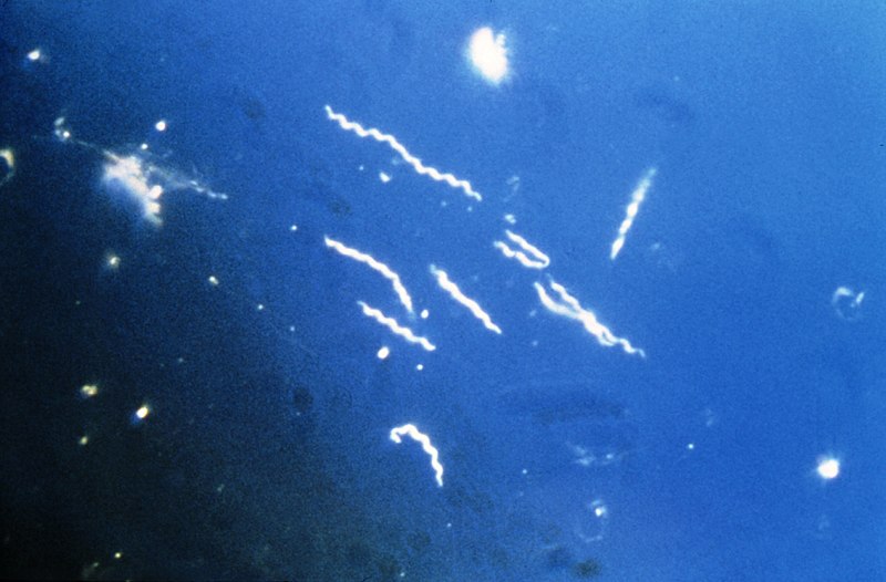
Some of the organism which are termed to be prokaryotic also have linear chromosomes leading them to also have DNA being linear in shape. One such example of it is Borrelia Burgirferi. There are experiments that show that some of the prokaryotes can contain to maintain viability with linear ones.

There are quite a lot factors that determine the linear DNA getting evolved with one of the best suited explanation to be the selective pressure which is in favor of the ones that related the length of genome of an organism. There can be clarity for mutation with concern with the type of DNA.

The prokaryotic ones that have linear DNA can face up problem like the one with the end replication issue. This is the time when the issue is raised by the directionality of the DNA enzymes getting replicated which ends up in much loss of the genetic substances in the end of the linear cycle of cell and replication.

Borrelia burgdorferi

They are the species of bacteria and of the class spirochete in the genus of Borrelia which caused the Lyme disease.

The concerned query for is the prokaryotic DNA circular or linear, the answer quite contradicts to the actual. The prokaryotes have circular DNA but however this species of bacteria have linear chromosome.

They were termed to be the third genome of microbe that was sequenced after the Mycoplasma in 1995. It has linear chromosome which has 853 genes and 910,725 base pairs. The method that was used up to get it sequenced was the whole genome shotgun.

There are quite a lot variation in genomes of it which plays to contribute in various dissemination and disease. The specific group of genome has much variety of genomes on the membrane receptors that are specific to the host infection.

bo
Image credit- Borrelia burgdorferiWikipedia

Why is prokaryotic DNA circular?

The cells of the prokaryotes do have chromosome which is single and is circular in shape and is seen in nucleoid.

The molecules in the circular DNA is made to get extracted from the bacteria that are mesospheric and have a lacking of duplex that turns out to be relative compared to the linear DNA of the equal size. This lacking of the duplex causes an exertion of strain on the DNA and then making it to coil up.

There are many of the organism that are prokaryotes have a typical cell with also linear chromosome but is mostly circular which is cause they trend to replicate faster than the cells of the eukaryotes making DNA replications take place during division of cell.

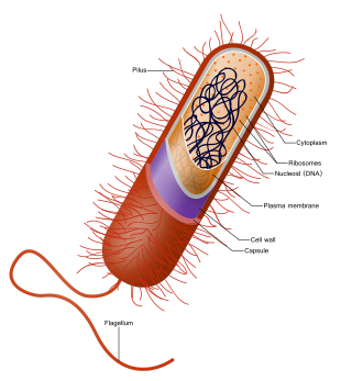
Mostly the prokaryotic cell carry a little amount of the genetic substance in the form of the chromosome or molecules in single quantity of the DNA that is circular. The prokaryotic DNA is placed in the center of the cell and is called the nucleoid and does not have any nuclear membrane.

The DNA has in general a charge which is negative inside the cells of bacteria and do lack the helical turns. The DNA is not nor overwound or under overwound but takes up the confirmation of being relaxed.

is prokaryotic dna circular or linear
Image credit- ProkaryoteWikipedia

How is DNA organized in prokaryotic cells?

The cells of prokaryotes lack a nucleus and have no nuclear membrane as well which leaves it to having a nucleoid.

The genetic substances in the cells of the prokaryotes are carried by a single piece of DNA that is circular and is linked to the membrane of cell and comes in direct touch with the cytoplasm.

The prokaryotic DNA is circular and a double helix structure and is a single chromosome. The structure of double helix is enclosed around the proteins to make the nucleosomes. The DNA is packed up quote tight to let in fit inside.

The gene of the prokaryotic microbes is organized all together in a form of cluster and is called the lac operon. The organization form of the DNA in the prokaryotes thus is different from that of the eukaryotes in many separate ways. The most obvious difference is the process of condensation that the prokaryotic molecules of DNA undergo to for in the cells.

Inside the eukaryotes the material of genes is a home to the nucleus and is kept packed as the linear chromosome. The chromosome is made up of the complex of DNA protein and is called chromatin which islet to kept organized into the subunits called the nucleosomes.

Do prokaryotes have non coding DNA?

The genomes of the prokaryotes seem to be much compact and gets transcript often and covers one or more genes.

It was in original advice that over the 98% of the human genomes have not been encoded with the sequences of protein that consists of most of the one in the introns while 20% of the prokaryotic genome stays non-coded.

In almost all the eukaryotes, the large mass of the genome stays up for the non-coded in form of proteins. The part of the eukaryotic genomes that is non-coded is likely to have been expanded through the various mechanisms via evolution like that of the deletion and insertion of the DNA.

The non-coding sequence of the DNA is the one that do not tend to get coded for the amino acids. Most of the non-coded DNA lies in between the genes of the chromosome and has been found to give zero function. There is other DNA which is non-coded and is called the introns and is found inside the genes.

The eukaryotic organism seem to be much complex than the prokaryotic ones which leads to the cells of the eukaryotes to have more number of DNA than the other. The cells of the eukaryotic organism tend to be more complex and are made of the DNA and the protein histones.

non
Image credit- Non-coded DNAWikipedia

DNA in extant prokaryotic cell is circular?

There is formation of two copies as the outcome after replication with one getting separated from another cause of the cell growth.

There is evidence to proof that the DNA of the prokaryotes are circular and are single chromosome. The cells of the prokaryotes do not tend to go for mitosis and thus the chromosome is seemed to replicate and result in two copies.

The prokaryotes do not have any nucleus and thus yet the DNA of them are not scattered all over the cell. The DNA is kept inside the nucleoids of the prokaryotes and is circular with being double stranded and is coiled up tight. As for the structure being a circle and coiled tight, the DNA is not scattered in the cell.

In many of the organism like that of the humans, the chromosomes are linear but in the organism like in that of bacteria or the archaea they are typical being circular. The DNA of the prokaryotes is concerned in the nucleoid which is in the center of the cell.

Also Read:

Is Prokaryotic DNA a double Helix: Why, How and Detailed Insights

dna

The prokaryotes are referred to the organism that has no specific nucleus and no rest number of organelles with inside membranes.

With concern to the response for is prokaryotic DNA a double helix, the reply to it is yes. The DNA of all the prokaryotes that includes the bacteria and the archaea is circular in their structure and is double stranded.

Just not similar to that of the eukaryotes, these are the structures having no specific region or are boundary less and have a double stranded DNA being circular and are smaller than that of the DNA of the eukaryotes in their cell. There is a formation of a loop in the prokaryotes which is the plasmids and is of no specific use in the cell growth.

The molecule of the DNA can be long and get stretched where the DNA in the cell of a human may be of length of 2m. Thus, the cell DNA need to be packed in such a way that it can be fitted in the cell and function inside its boundary and shall not be seen with naked eyes.

There is also a presence of a second type of nucleic acid called the ribonucleic acid. Just like the DNA, RNA is also a polymer of the nucleotides. Each of these is made up of the nitrogenous base with a phosphate group and also a five carbon one called the ribose.

Cell arrangement to prove is prokaryotic DNA a double helix

DNA is referred to as the deoxyribose nucleic acid and is the concerned molecule that works and gets replicated.

The model for the DNA suggests that prokaryotic DNA is a double helix which is double helix was given by Watson and Crick where the DNA is a polymer of the nucleotides. Each of the nucleotides includes the nitrogen bases and is made of two strands.

There are two of the purines which are adenine and guanine, the two of the pyrimidine which are the thymine and cytosine. Along with the composition of nitrogenous bases they also have a phosphate group and a five carbon sugar called the deoxyribose.

Each of the DNA stand is made up of nucleotide bonds attached together which are covalent in between the phosphate group of the one and the deoxyribose of the next sugar seen. This is the actual backbone for the DNA and here is the base extension point. The prokaryotes have cytoplasm, ribosomes, cell wall, flagella and cell membrane.

The strand having one of the bases does get to bond with the base of the strand that comes second along with the hydrogen bonds. Adenine always gets to bond with thymine then with cytosine and then gets to bond with guanine. Most prokaryotes have circular, single chromosome placed beside nucleoid.

It gets into the part of duplication when the cell is ready to get divided and shall read in order to generate the molecules like that of proteins to let the functions of the cell be carried out smooth. This is the absolute reason for the DNA getting to be packed and let protected in many ways. The genome is made of double stranded DNA ns is single in form of a circle.

is prokaryotic dna a double helix
Image credit- DNAWikipedia

Why is prokaryotic DNA a double helix?

The prokaryotes are seen with a single chromosome which is circular and is double stranded. The DNA of the prokaryotes are free floating and not in the nucleus.

With response to is prokaryotic DNA a double helix, The adenine always gets to bond with thymine and cytosine gets to pair with guanine. This is the cause that makes two of the strands to become spiral around each of them in a shape that forms the double helix.

With the reply to is prokaryotic DNA a double helix they have a chromosome that covers the area of the cytoplasm known as the nucleoid and is single being circular. They are also seen to be made up of tiny rings that look like the double stranded chromosomal DNA that are extra and are called plasmids. 

The eukaryotes have the one with being linear packed up in the chromosomes. The helix of DNA is enclosed around the proteins to make the nucleosomes. The coils of proteins and made to coil up and during division the chromosomes are seen to be coiled up more to help in movement.

The chromosomes of the prokaryotes are likely to have two specific areas that shall separate them by reflecting certain degrees of packing, staining, and then getting to determine if the DNA in any of the area is being euchromatin (expressed) or heterochromatin (not expressed). Some of the prokaryotes make loops called plasmids which are not useful for any normal growth of cell.

The DNA of the prokaryotes seems to be circular for the circular DNA did evolve at the first even before the linear eukaryotic DNA. This is also for the prokaryotes were one among the common ancestors that descended with circle polymerase and getting to replicate DNA in circle. Thus, the part of answer for is prokaryotic DNA a double helix or not, its an yes.

Also Read:
Do Prokaryotes Have Golgi: Why, How and Detailed Insights

dnaa 1 1024x1016 1

Prokaryotes are said to be the organism having a single cell and have no nucleus present along with organelles having a membrane.

Along with the tagged with job for do prokaryotes have Golgi, the prokaryotes are just one compartment cell and have no Golgi apparatus or any nucleus to store DNA or a mitochondria.

The cells of the prokaryotes also have no cytoskeleton. There is no presence of any of the complex materials seen in the cells of the prokaryotes with also no membrane enclosed nucleus. The Golgi apparatus are special to just the cells of the eukaryotes and being absent to the prokaryotes.

The Golgi apparatus are also called the Golgi body or the Golgi complex. It is a membrane covered organelle seen in the cells that have a good bounded nucleus and is referred to as the cells of the eukaryotes. It is made up of many chains of pouched stack that are flat and is called the cisternae.

The Golgi body is present in both the cells of the animals and plant and it is the term which is attached to the groups of the disc like features situated quite close to the place of the endoplasmic reticulum. The presence of the Golgi bodies in the cells can vary and can be variable as well.

Do all cells have a Golgi apparatus?

The Golgi apparatus in its presence in any type of cell us used for getting the proteins packed up along with the lipids as well.

The Golgi apparatus which is also the Golgi body or complex is seem to show their presence and use only in the cells of the eukaryotes which concerns both the animals and plants cells.

The response to do prokaryotes have Golgi is a No and with support to that there is no organelle in the cells of the prokaryotes that do have a membrane and thus they have no Golgi body.

If there is no presence of any Golgi bodies in the cells, there will be any generation of lysosomes which is a cell organelle having within them certain enzymes for digestion. They help in getting the parts of the cell or the cells that are worn out get broken down. Golgi is placed near nucleus.

With a lot of Golgi bodies being there in the eukaryotes do there is no or minimum cell death. For if there is no Golgi complex then there will be no accumulation of the organelles that shall be damaged or dead and the cell having the molecules will result to being death called as cell death. There will also be a stop to transfer and packaging of materials in cell.

Do prokaryotes have Golgi
Image credit- Golgi apparatusWikipedia

Do prokaryotes have Golgi plasma?

The term prokaryotes can be addressed to the microbes that lack organelles having no nucleus and the ones that have only single cell.

With reference to the question for do prokaryotes have Golgi, the cells of the prokaryotes lack any membrane bounded organelles and thus have no Golgi’s as well. They are just unique to eukaryotes.

The cells of the prokaryotes have plasma membrane that surrounds them yet have no presence of any organelles that have no membrane bound within the region of the cytoplasm. The difference from the eukaryotes and prokaryotes lies in the presence of the membrane enclosed organelles.

The Golgi plasma is nothing much different from the Golgi body or the Golgi apparatus. They help in getting the proteins modify and transport them in the cells of the plant and the animals which are the cells of eukaryotes only. It is the central part organelles that mediate the lipid and the protein transfer inside the eukaryotes.

pro
Image credit- ProkaryotesWikipedia

Do prokaryotic cells have Golgi body?

There are many organelles that are found in the eukaryotes and do have a definite regions for all the cells.

Golgi apparatus is found only in the cells of the eukaryotes which concerns with the membrane enclosed around the cell and in seen in both the eukaryotes which are the animals and plants. So, prokaryotes have no Golgis.

The Golgi complexes are the part of the cells that are made up of the membranes and have several membrane types. Some of them are the tubules and some are in similarity with the vesicles. The Golgi is right beside the nucleus and is also called the perinuclear body.

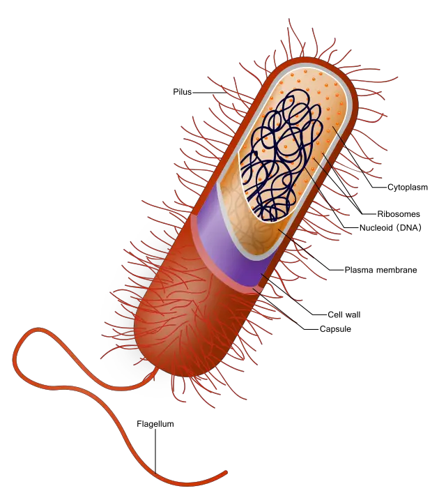
With regards to the prokaryotic cells having no organelles that are membrane bound they consist of a plasma membrane which is simple, the ribosomes and the DNA structure of nucleoid. The cells of the prokaryotes are actually differentiated. The prokaryotes can include the archaea and the bacteria.

The cells of the entire prokaryote category are almost tiny, with being cells that are simple and have a measurement of about 0.1 to 5 micron in the diameter. The cells of prokaryotes have no specific regions for its cells has they have no boundary to guard or mark the cell regions.

dnaa 1
Image credit- DNAWikipedia

Do prokaryotes have Golgi complex?

The cells of the prokaryotes like the archaea and the bacteria have a plasma membrane but have no organelles inside cell that are membrane bounded.

Prokaryotes have no Golgi complex. The complex for the Golgi’s are has been given a lot of names. The several names given to them are the Golgi bodies, the Golgi complexes or the Golgi apparatus.

In the unicellular or the organism that are primitive, the cell type is simple and has more than one or few of many chromosomes. It also has membrane that is double bounded and includes the hereditary material which is naked. The Golgi can appear as sac, in compartment or like vesicles and found just near the nucleus.

The Golgi complex is not useful much for the unicellular organism as there here is ultimately no need for the proteins to get itself processed and packaged and to also get the enzyme and protein sorted out giving them direction to the rest of the cells as it shall make up to only one of the cell.

Also Read: