What Happens to a cell in a Hypotonic Solution: Detailed Insights

image 22

Any cell is said to be a mass of the cytoplasm that is guarded well by a cell membrane. They are all microscopic in size.

A cell in a hypotonic solution, has a net movement for water from the part of the solution in the body. A cell that is placed into a hypotonic solution shall seem to swell and then expand till it shall burst via a process called cytolysis.

A solution that is hypotonic or a cell in a hypotonic solution has a concentration that is lower than that of the solutes of the other solution. In the term of biology, the solution that is out of cell is said to be hypotonic only if it has less concentration for solutes considering to that of cytosol. There is a diffusion of water in the cell, for osmotic pressure and then the cell leads to often being turgid or also bloated.

In chemistry, a solution is a special type of homogeneous mixture composed of two or more substances. In such a mixture, a solute is a substance dissolved in another substance, known as a solvent. Some examples of solutions are salt water, rubbing alcohol, and sugar dissolved in water. When you look closely, upon mixing salt with water, you can’t see the salt particles anymore, making this a homogeneous mixture
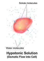
 cell in a hypotonic solution
Image credit-Hypotonic solutionWikipedia

Unlike osmotic pressure, tonicity is influenced only by solutes that cannot cross the membrane, as only these exert an effective osmotic pressure. Solutes able to freely cross the membrane do not affect tonicity because they will always equilibrate with equal concentrations on both sides of the membrane without net solvent movement. It is also a factor affecting imbibition. A hypotonic solution has a lower concentration of solutes than another solution.

Solution are said to be a mixture of a compound made of a solvent and a solute. The substance that is the one that is present in more concentration is called as solvent and the one that is in less level is said to be solute. A salt water is a good example of hypotonic solution. Thus, cell in a hypotonic solution shall have less solute concentration than the cell. An iso-osmolar solution can be hypotonic if the solute is able to penetrate the cell membrane.

What happens to a red blood cell in a hypotonic solution?

The very simple definition of tension or more tone means to have a large osmotic pressure that the rest medium or fluid.

The time when cell in a hypotonic solution shall have lower concentration, the red blood cell kept in this solution shall have a free water net movement in the cell. This means less water than the cell.

On having a cell in a hypotonic solution or rather a red blood cell in it shall have it bloated up and might explode. If it is kept in a hypertonic mode, it shall shrivel that shall make the cytoplasm dense and the components shall remain concentrated and also can die. Thus, for this reason a plant cell seems to be ideal. To prevent the process of burst, the cell needs isotonic mode.
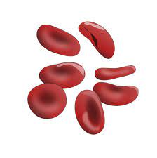
image 22
Image credit-Red blood cellWikipedia

After having the red blood cell in a hypotonic solution there shall be seen a net movement for free water inside the cell. This phase shall outcome in an increase volume in the cell with having less concertation of solute. The solution shall end up with finally high level of concentration. It would eventually lead to it swelling up and then bursting in the method called hemolysis.

To avoid situation like this, cell in a hypotonic solution can never be the red blood cell. Thus to level this up or avoid the process of hemolysis, on needs to place the cell in a solution called to be isotonic solution. This shall have 0.9% m/v of NaCl and glucose shall be in a concentration of 5% m/v. A solution is isotonic when there is equal amount if solutes on both side of membrane and no swelling or shrinking is seen.

Any example of isotonic solution can be the normal saline water that is 0.9% in concentration and the lactated ringers. These fluids are said to be useful at the time when there is a huge loss of body fluids from any trauma, dehydration, vomiting, diarrhea, blood loss or nausea. This term is used both in biology and in chemistry for transport across the semipermeable membrane.

What happens to an animal cell in a hypotonic solution?

Any cell in a hypotonic solution shall, have less amount if solute concentration and more solvent in it. Osmosis is the process involved in it.

The solutions that are hypotonic shall have less water than the cell. Thus with animal cell in a hypotonic solution shall be filled up with water completely and then shall burst. Pure water and tap water are hypotonic.

An animal cell is a eukaryotic cell type that do not have a cell all yet seems to have a nucleus that has a membrane and is true. It also has several other cellular organelles. The contents that it has are cytoskeleton, centrosome, lysosome, mitochondria, Golgi apparatus. Any typical cell that is of an animal shall have cytosol, a cell membrane, the organelles and cytoplasmic structure.
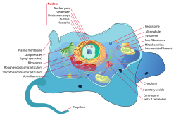
Wikipedia:Featured picture candidates/Typical animal cell - Wikipedia
Image credit-Animal cell-Wikipedia

The process of osmosis takes place at the time when animal cell in a hypotonic solution. Thus for this, the animal and plant cell seem to both appear a lot plump while placed in a hypotonic solution. After this has been seen under the microscope, the vacuoles seem to appear a lot larger considering only the plant cell. It has more of solute and net movement. It is all a result of the method called as osmosis.

The swelling of a cell in a hypotonic solution is for the less amount of solute in it and the net movement of water in the cell causes a breaking or swelling of the cell. There is a movement of water at a place where there is low outside fluid or osmolality to an area that has more of it. The cell shall also want to expand. Just like the animal cell, the plant cell shall mot burst. The increase in solute level leads to it being broken finally.

What happens to a plant cell when placed in a hypotonic solution?

Both the plants and animal ell are said to be different and thus perform and show different types of property when placed to conditions.

When a plant cell in a hypotonic solution is exposed it shows up osmosis that takes up water and then begins to swell up. There is a rigid wall in the plants that help prevent the cell from getting it busted making it turgid.

Osmosis is said to be a process having the spontaneous path or water diffusion system in rest of the solvents via a semipermeable membrane. It is the simple movement concerned to water from the place of high concentration for water to the place of low level water. It helps in getting the two of the solutions separated by having the solute concentrated.
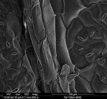
image 23
Image credit-Plant cellWikipedia

If there is a link made between the two areas via a membrane, then the one that has more of the solute that the rest then the eater shall flow from the second point to the first. The ability of the solution to have the water move is said to be tonicity. The tonicity of any of the solution shall be called as osmolality. Concentration and solubility is directly related to each other.

Water seems to enter the cell for the plant and the also the plasma membrane that tends to swell up and the push up against the walls of cell. If the plant cell in a hypotonic solution has a concentration of solute less than the within if the cell the water shall tend to enter the cell via osmosis. The cell shall swell but with zero lyse as the cell wall gives the structure to expand. Thus, the cell membrane shall press up the wall and make a turgor force that shall give the support to plant.

The cell after attaining the osmolality in the hypotonic solution, shall not burst but expand. But the animal cell in a hypotonic solution shall explode. This is so as the plant cell have a wall for the cell that is rigid all the way via the plasma membrane. On getting to swell with the water they shall turn turgid. Hypotonic solutions also help in making the vegetables like bell pepper turn crisp.

What happens to a bacterial cell in a hypotonic solution?

Usually if there is a placement of cell in a hypotonic solution it is quite likely to get ruptured or swell up due to its tonicity.

If a bacterium cell is kept in a hypotonic solution, it shall seem to rupture by having the cell swell due to the gradient of osmotic pressure within the cell of the bacteria.

The bacteria are microorganism that have a cell boundary and is simple that that of the rest of the organism, they have their own center of control that have genetic data and is kept in a single loop for the DNA. Some of the bacteria do have an extra genetic circle of cell material and is called to be plasmid rather than being a nucleus. They are the prokaryotic cell type.
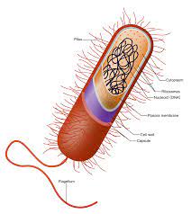
image 24
Image credit-Bacterial cellWikipedia

For the instance of osmotic pressure and the gradient that is made the relative presence of the hypertonic solution in the cell makes it swell and in the method it becomes much slow and some of them are finally resistance to each other in action by all means of the cell wall. If there is the control of water, the cell might lead to damage and burst. The plasma membrane helps in getting the pressure kept normal with bacteria having straight characters.

Most of the cell wall of the bacteria, fungi and algae have a cell wall that is rigid and are able to tolerate the osmolality and then enjoy the surrounding being hypotonic. If the solution seems to be hypertonic then the water that is in, then cell shall be able to leave it and then the bacteria will be shrinking. The cell movement outside the water is called to be osmosis. There is a prevention of the cell from expansion and thus have lysis.

Can a Hyperosmotic solution be hypotonic?

Osmosis is said to be the solute number that gets dissolve while tonicity leads to having no units. Osmolality is said to be comparison of two solutions.

Yes, it can be so. If the cell is placed in a hyperosmotic solution but the hypotonic part is like dextran of 10% concentration, there shall be a water movement. Thus, a hyperosmotic solution can be hypotonic.

With also have this question of can hyperosmotic solution can be hypotonic, it is not always needed to be so. Yet, the hyperosmotic solutions shall always remain hypotonic. There is a good response for the widespread presentation of tonicity and osmolality that had a good result. It also needs bit of an additional arrangement.
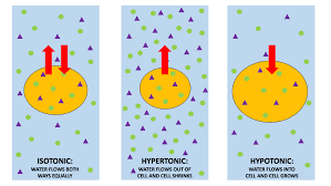
image 71
Image credit-OsmoticWikipedia

Hyperosmotic stress in most mammalian cells causes cell shrinkage due to osmotic efflux of water leading to increases in intracellular ionic strength. Hyperosmotic solution has been shown to disrupt mating pairs at two different time points. First, as described earlier, osmotic solution will erase the preparative changes brought about by “initiation”. The increase of intracellular ions and the accompanying influx of water cause RVI. 

A solution can be either isotonic or hyperosmotic and both. If a cell needs to be placed in a hypertonic solution, there will be a net flow of water out the cell and then the cell shall loose volume. A solution shall be hypertonic in a cell and thus its solute shall be having a high concentration. There is always a swelling due to influx and the cell shrinks.

Can a Hyperosmotic solution be isotonic?

A solution can be either be isotonic or also be hyperosmotic. Hyperosmotic refers to the capacity of achieving more than the normal osmosis.

The solutes that are hard to penetrate or actually have zero penetration via the cell membrane, thus the water movement across the membrane of the cell shall be able to take place to reach stability. Solutions of equal solute concentration are isotonic.

Tonicity is a measure of the effective osmotic pressure gradient; the water potential of two solutions separated by a partially permeable cell membrane. Isotonic is a term used to describe solutions and chemistry and, sometimes, muscles in human biology. In chemistry, a solution is said to be isotonic when it has the same concentration of solutes as another solution across a semipermeable membrane. The use of isotonic in human anatomy is used more rarely.

The very term hyperosmotic means the property of having a pressure with less osmosis. This means that the molecules in solute shall number in a single side of the membrane that shall allow only specific molecules to pass via the low side than the one that has on the rest side considering the number of solute. The water molecules shall be travelling fast and via the cell membrane that shall have the particles to solute in.

Hyperosmotic vs hypertonic

The solutions that tend to have less number of solute is called to be hypotonic. The word hypo means less and is same to hyperosmotic.

The solutions that are hyperosmotic are never always hypertonic. But the hypsometric ones are always said to be hypotonic. This depends on the tonicity and the osmolality of the solutions.

In the solutions that are hyperosmotic the solutes tend to move from the place where the surrounding has a high osmotic pressure that the rest part of the solution. On the other part, the hypotonic have the solutes that travel from the rea that have high part of concertation and move to the less area concentration or the surrounding.

In simple way, hyperosmotic refers to the character that has a pressure high is so osmolality while hypotonic refers to the feature of having an osmotic pressure that is less. Hyperosmotic also have pressure that is high in the rest of the area concerned to the cell while a cell in a hypotonic solution shall have less pressure out. Solution in hyperosmotic tend to move from the solution to rest while opposite in hypotonic.
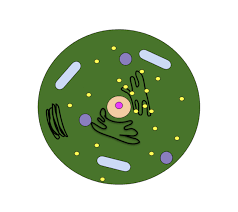
image 72
Image credit-Cell shrinkageWikipedia

Hyperosmotic stress results from an extracellular osmolyte or solute concentration in the serum or medium that is higher than physiological, and high in comparison to the intracellular environment.  Hyperosmolality is classified as hypertonic or isotonic according to whether cell shrinkage occurs. A hypertonic medium contains solutes that are relatively membrane impermeable, such as peptides, metabolites, and small ions.

Also Read:

Bacterial Cell Type: Detailed Analysis on Every Types, Events And Facts.

types of bacterial cell 300x141 1

In nature there are several types of bacteria are present. Bacteria are generally classified basing on their structure and shape. Usually bacterial cell is prokaryotic cell.

Bacteria have several classifications according to their shape, structure and number. There are many subclasses in bacteria. Bacterial cell basically contains cytoplasm, genetic material, nucleoid and cell wall. In this topic we shall discuss more on bacterial cell type.

Dormant bacterial cell type

The dormant bacterial cell is commonly known as an endosperm. The endospore is usually referred as a spore.

An endospore is likely to be tough and non-reproductive cells. Endosperms are formed from bacteria. In this stage the bacterial is in non-reproductive stage for days or even for several months too.

The temperature can also determine the reproduction stage of bacteria. For example at -18 degrees centigrade the bacteria cannot undergo reproduction and will be in dormant stage. At 0-5 degrees the bacteria will be sleeping stage and the reproduction will be very slow. But at 5-63 degrees the bacteria can reproduce at faster rate.

What is the most common type of bacterial cell?

The most common type of bacteria are cocci, spirilla, bacilli, and comma forms.In this section we shall discuss bacterial cell type.

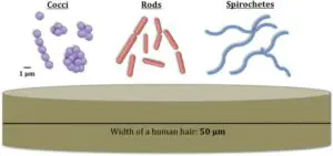
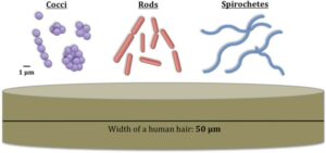
Bacteria are typically classified in to five types basing on their shape. They are cocci, bacilli, spirilla, vibrios and spirocheates.

The rod shaped is commonly known as bacilli. The spherical shaped bacteria is known as cocci. The common examples of cocci are Streptococcus pneumoniae and Neisseria gonorrhoeae. The spiral shaped bacteria is known as spirilla.  Campylobacter jejuni is the example for spiral bacteria. The comma shaped bacteria is known as vibrios. Vibrio cholera is example for vibrio bacteria it causes cholera in humans. It leads to diarrhea.

bacterial cell type
Image credit: Bacterial Cell Type – Wikimedia

The cocci bacteria are sub classified depending on the arrangement of cells. They are diplococcus, strepto cocci, staphylo cocci, tetrads and sarcina.Diplococcus bacteria means a pair cocci are present. For example streptococcus pneumoniae and Neisseria gonorrhoeae).

Streptococci means that the cocci are present in string like structure. Example for this type is streptococcus pyogenes.Staphylo coccus bacteria means that the cocci are present in colonies in irregular manner.Example for staphylo coccus bacteria is Staphylococcus aureus.

If we found four cocci together then it is said to be tetrad. Example for tetrad is Micrococcus species.If eight cocci are found together it is said to be sarcina bacteria.An example for this type is Sarcina ventriculi.

The spiral bacteria can be sub classified into two types basing on the cell flexibility, cell mobility, number twists present in a cell. There are two types of spiral bacteria. One is spirillum and the an other one is spirochetes. The spirillum and spirochete is differentiated basing on the presence of flagella. Usually the spirilla are short and rigid cells whereas spirochetes are relatively long and flexible cells. The spirilla have external rigid flagella whereas spirochetes have internal flexible flagella.

Camphylobacter species are the best examples of spirilla. They are pathogenic and may cause food borne camphylobacteriosis.

Borrelia species are the best examples of spirochetes. These are also pathogenic in nature. For example Borrelia recurrentis may cause relapsing fever in humans. These are the common bacterial cell type.

What bacterial cell type is a flexible spring-like structure?

The spirochetes are the flexible spring like structure bacteria having internal flagella.

Among all the species of bacteria the spirochetes are the more flexible bacteria. They contain internal periplasmic flagella which help the bacteria in locomotion. Flagella are the locomotors in bacterial cells. They stick the bacterial cells in liquids or to the surface so that a cell can move freely in the environment.

Read more on: Eukaryotic Cells Vs Bacterial Cells: Detailed Insights

Frequently Asked Questions

Are cocci bacteria  gram positive or gram negative?

The cocci bacterial cells are gram positive in nature. Because they contain peptidoglycan in their cell membrane.

Cocci bacteria like streptococci, staphylococci, enterococci are gram positive bacteria.

How can we indicate whether the bacteria is gram positive or gram negative?

By using gram staining we can differentiate the bacteria.

In gram staining the gram positive bacteria turns purple and gram negative bacteria turns red or pink.

Why the gram positive bacteria turns purple in gram staining?

The gram positive bacteria turns purple in gram staining because of the presence of peptidoglycan in the cell membrane.

The gram negative bacteria do not contain peptidoglycan layer.

What are common infectious bacteria in humans?

  • Coliform bacteria, bacterial vaginosis, gonorrhoea, chlamydia, syphilis, E. coli, Salmonella are the common infectious bacteria.
  • For example Coliform bacteria can cause urinary track infections. E.coli and salmonella are common food poising bacteria.

Also Read:

Is Cyanobacteria Unicellular Or Multicellular: Why, How And Detailed Insights

cy

Cyanobacteria are of the phylum from Gram negative bacteria and are also called Cyanophyta.

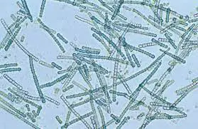
With the question for is cyanobacteria unicellular or multicellular, cyanobacteria are the most diverse type that can range from being unicellular to multicellular and do have filaments.

They do take up energy from the process of photosynthesis. The name of this means the colour blue in ancient Greek. The name thus given to these is also blue green algae. The botanist does not agree on giving the term algae to the eukaryotes but cyanobacteria are exception.

They are seen in the fresh water or even on the terrestrial lands. The cyanobacteria generally use the photosynthetic pigments, like the carotenoids and the phycobilins and other form of the chlorophyll that helps in absorbing the energy form the sun.

Cyanobacteria do mark good variety of morphology from the unicellular colonial to the filaments. The filamentous forms do show up cell differentiation like the heterocyst, the akinetes and the hormogonia. All of these together have connections and are termed to be the first mark of multicellularity.

Where is Cyanobacteria found?

They are much diverse and large in their phylum and are the photoautotrophic prokaryotes.

They are seen in almost all places. The sea spray consists of the marine microbes that includes the cyanobacteria and can be swept high inside the atmosphere which turns them into aeroplankton and can move anywhere. 

They do often inhabit in colonial aggregates that can take up thousands of forms. They do have filaments dominating the upper area and is found in much of the extreme surrounding conditions like in the hot springs, the hyper saline water in polar areas and the dessert.

They are microscopic in nature and are in general in all types of aquatic form being their natural habitat. While in the fresh water, marine or in the mixed up ratio of marine and fresh, these do have one cell and also use up sunlight to make their own food.

They are actually optimized for all the next to be place conditions of having less oxygen. Some of them are used for nitrogen fixing and some live inside the water and the moist soils with being free or having a symbiotic equation with the fungi or the plants.

is cyanobacteria unicellular or multicellular
Image credit- CyanobacteriaWikipedia

Are all cyanobacteria unicellular?

The cyanobacteria may have many sheaths to het the filaments or the cells from the colonies to bind to themselves.

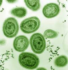
Some of the cyanobacteria can be cells that do free live and can be unicellular like the Prochlorococcus, the Synechococcus, the Crocosphaera but also some of them do show symbiotic links with many haptophyte alage like the Coccolithophores.

The marine life cyanobacteria are quite extensive and do have high blooms with having their appearance in blue green or scum. The blooms can be toxic and can quite lead to the closure of the waters with many of the marine one being vital parasites of the unicellular range.

With the fact of is cyanobacteria unicellular or multicellular, the symbiotic state clears it all while getting itself linked up with the rest organism for its survival.

Many of the species of this can also glide up in the way as the cell moves differing from the movement of swimming or crawling relying on any outside organelle change and take place in the presence of substrate. The ones that do glide through the plants do possess filaments that help them move.

The gliding of the filamentous bacteria seems to be energized by a mechanism called the slime jet where the cell do release a gel that gets itself expanded fast and helps the, in getting itself hydrated also giving them a force of propulsion with some of the unicellular ones also using it.

The bacteria can enter into the plants via the stomata and then get to colonize the inter space that shall form loops and many inner coils. Some of them colonize the roots of plants while some target the root system.

phl
Image credit- ProchlorococcusWikipedia

Why are cyanobacteria multicellular?

They are on among the unicellular ones that are the oldest and did make it up gradually into many forms consisting of colonies as well.

The cyanobacteria are the largest with being the diverse prokaryotic phylum, along with the morphophytes that are of all range and varies from being unicellular to the multicellular one and also being filamentous.

 Some of the cyanobacteria are able to get the nitrogen from the surrounding fixed and get them converted into the forms that can be used by the animals and plants. They also have seemed to raise the level of oxygen in the atmosphere by 2.45 billion and give the base of aerobic evolution.

With is cyanobacteria unicellular or multicellular, the species of the cyanobacteria are all filamentous or form colonies. The ones that are multicellular have developed many cell having separate functions. Some of the cells do carry on photosynthesis while other respiration and other absorb nitrogen.  

The cyanobacteria get their energy from the process of photosynthesis while getting to absorb the light as the source of energy and use the nitrogen form atmosphere to make amino acids which are the protein building blocks. Yet, this can also lead to make trouble for getting to exchange and communicate materials.

Filamentation
Image credit- FilamentsWikipedia

FAQs-(Frequently Asked Questions)

Why are cyanobacteria not said to be multicellular?

This is because as the bacteria do not have any of the cell compartments thus they are said to be prokaryotes even after not having the same functions as the organism being multicellular.

Are the protists unicellular or multicellular?

Any group members of the protists that are eukaryotes are basically termed as the unicellular microscopic ones.

They can share any physical attributes or morphology same to that of the plants or the animals or even both yet they are unicellular. The very vast part of the protists is unicellular having many cells and does from up colonies.

How to cyanobacteria respire?

The process of respiration in these can take place inside the membrane of thylakoids along with photosynthesis sharing the same place for respiration.

The class of cyanobacteria uses up several protein pairs to perform aerobic respiration which marks the trait for being independently getting acquired in each of the class. It is thus consistent with the rise of the oxygen that takes place after the divergence.

Also Read:

5 Facts On Is Fungi Multicellular Or Unicellular? Why & How

1280px Mouldy Clementine 300x200 1

Fungi are found both as unicellular and multicellular organisms in nature.

Fungi are a kingdom of eukaryotic organisms ranging from single-celled to multi celled. Single cell fungi like yeast have a more simple structure in comparison to those of mushrooms.

To answer the question “is fungi multicellular or unicellular” it is both. Moving up the hierarchy as fungi go from unicellular to multicellular they grow more complex, with distinguishing features and characteristics. They also have developed specialized cells and structures to facilitate their mode of living.

How fungi are multicellular?

Fungi can form multicellular filamentous systems that can be both microscopic and macroscopic.

In the case of moulds, they are made of very fine threads called hyphae produced by repeating cells in lines and branches. Mushrooms on the other have their cells forming large and visible fruiting bodies that hold the spores.

In molds, the hyphae can grow into the air and forms spores at the end of themselves. The fruiting body of mushrooms is made up of densely packed hyphae that divide to generate the various elements of the fungal structure, such as the cap and stem.

Are all fungi multicellular?

All fungi are not multicellular.

Fungi are composed of both unicellular and multicellular organisms. Then some are dimorphic i.e. they can have both unicellular or multicellular forms based on their requirements(can alternate between yeast and hyphal forms).

Kingdom fungi consists of 3 main types of organisms- yeast, mold and mushrooms. But have a broader classification that also includes – rusts, stinkhorns, puffballs, truffle(yes the insanely expensive truffle used in gourmet food) and mildews.

But based on cellular organization they are of 3 types mainly:

Single-celled yeasts. These are the most simple of those found in kingdom Fungi. Next in complexity are molds. These are multicellular and filamentous in nature. The most complex in the organization is the mushrooms.

Simple diagram of yeast cell en.svg 2
A typical unicellular yeast structure Image: Wikipedia

Mushrooms are Macroscopic filamentous fungi that make up large and visible fruiting bodies. The fruiting body is the top of the mushroom that we see and often consume.

Fungi multicellular characteristics:

  • Multicellular fungi mainly include molds, mushrooms and toadstools.
  • In the case of molds, the body structure is simply made of hyphae, formed by repeated dividing cells both linearly and branching.
  • The hyphae can extend and form spores at the end of the hyphae.
  • All multicellular fungi reproduce via spore formation and dispersal.
  • They continue to be microscopic.
  • In mushrooms, the cells are more specialized. They form mycelium under the ground, that act as roots.
  • The body extends above the ground to form a fruiting body with specified parts called the cap and gills.
  • The gills are under the cap and contain millions of spores. The gills help in protecting the spores from environmental conditions.
  • The gills also facilitate in allowing better spore dispersion. They resemble fish gills and may open to release the spores contained within them into the air and forest floor.
  • Toadstools are mushrooms that simply look like toads can sit on them. They can cause anything from nausea, vomiting to even death in case of consumption.

Multicellular fungi examples:

Multicellular fungi include

MOLDS:

Molds are tiny(microscopic) filamentous fungus that feed on plant and animal materials and produce spores. They may be found both indoors and outside and are an important element of our natural environment.

is-fungi-multicellular-or-unicellular
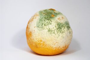
Mold growing on a clementine Image: Wikipedia

Molds in nature come in a variety of colours, including white, black, green blue and black. “Toxic mould” isn’t a species or a kind of mould, and “black mould” isn’t either. The words “toxic mould” and “black mould” are sometimes used in the news to refer to moulds that generate mycotoxins or to a specific mould, Stachybotrys chartarum. Toxigenic fungus is a term used to describe moulds that create mycotoxins.

Spinellus fusiger 51504
Mold growing on a mushroom Image: Wikipedia

The green or white furry things that grow on old bread, fruits and even on the damp walls are all molds.

MUSHROOMS:

A mushroom is the spore-bearing fruiting body of a fungus that grows above the earth, on soil, or any other media that it obtains its nutrition from -including plants, buildings or other mushrooms.

The word “mushroom” and its variants may have been derived from the French word “mousseron”, which literally translates to “moss” (mousse). Because it is nearly impossible to tell the difference and distinguish between edible and toxic fungi, a “mushroom” might be edible, poisonous, or unpalatable(tastes terrible).

The most commonly known ones include- Button mushroom(Agaricus bisporus), Shiitake mushrooms (Lentinula edodes), King Oyster mushrooms(Pleurotus eryngii) and many more including the Truffle that is more expensive than a gold bar.

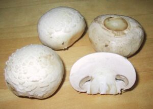
ChampignonMushroom 1
Commonly consume Button mushroom
Image: Wikipedia

Toadstools are the mushroom species that are poisonous by nature. Structurally they are in no way different from mushrooms and they look very similar as well, apart from the fact that they are poisonous. Some toadstools are often mistaken as edible mushrooms that can cause severe medical issues even death.

Some mushrooms use this fact and have adapted to look like their poisonous counterparts to keep predators like us away. Hence new forest foragers are always cautioned to pick mushrooms only if they are absolutely sure of their identity.

Technically mushrooms and toadstools are the same and also not all toadstools are poisonous and not all mushrooms are safe to consume. Hence the distinction between them is not really as clear as we would like it to be.

800px Amanita phalloides 1
Poisonous Death Cap mushroom Image: Wikipedia

One of the world’s most poisonous mushrooms is called the Death Cap (Amanita phalloides) found throughout Europe. Extreme stomach discomfort, vomiting, and bloody diarrhoea can begin within 6 to 12 hours after ingestion, causing fast fluid loss from the tissues and severe thirst.

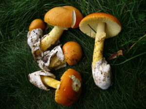
1024px Oronges
Edible Caesar’s mushroom
Image: Wikipedia

Early indicators of substantial involvement of the liver, kidneys, and central nervous system include a decrease in urine production and a dip in blood sugar. If the patient is not treated it can lead to coma and even death. But the worst part is that this deadly mushroom looks very similar to edible ones like the straw mushroom and Caesar’s mushroom.

Also Read:

Are Yeast Multicellular :Why,How And Detailed Insights And Facts

Yeast S cerevisiae secretory vesicles 300x300 1

Are yeast multicellular or not is a very tricky question.Yeast are one of the most economically important fungus having more than 1,500 species all over the world. Let’s try to find out if yeast are multicellular or not?

Yeast are mostly unicellular, evolved from multicellular ancestors. In some cases they can adapt multicellular characteristics or depending on the environmental factors they can switch between unicellular and multicellular life cycle, so we can say that yeast are facultatively multicellular.

In the case of Baker’s yeast or Saccharomyces cerevisiae, it can adapt multicellular characteristics in it by forming colonies or by other mechanisms. Hence we can say that yeast are mostly unicellular but in some cases they are facultatively multicellular.

To know more about Eukaryotes read the article Eukaryotic Cells Examples: Detailed Insights

What is facultative multicellularity?

Facultative means optional so in simple words facultative multicellularity is a condition in which unicellular organisms can be able to achieve multicellular characteristics depending on external factors.

Facultative multicellularity is a condition in which a species which is naturally unicellular, can be able to adapt multicellular characteristics. Some specific environmental factors trigger the unicellular organism for adaptation. In response that unicellular individual becomes a part of the multicellular body. After overcoming that particular obstacle it can revert to its previous unicellular form.

Facultative multicellularity can be present in two different forms, the first one is the simplest way to achieve multicellularity, that is forming a clump by sticking to each other (unicellular cells).The second one is more complex than the first one, cell differentiation.

Examples of facultative multicellularity:

Baker’s Yeast ( saccharomyces cerevisiae) is the most common facultative multicellular.

Are yeast multicellular

Saccharomyces cerevisiae from Wikimedia commons

Causes of facultative multicellularity

When we talk about the causes of facultative multicellularity, then there are some specific environmental factors that trigger an unicellular organism for adaptation. 

Depending on all these factors, an individual unicellular organism changes its gene expressions and adapts multicellular lifestyle for its evolutionary survival. 

  • Structured environment: Structured environment highly influences the facultative multicellularity. Presence of this factor stimulates multicellular metabolic activity by changing the gene expression. Due to that the individual cells start to differentiate into non-dividing forms and make colonies or biofilms.
  • Starvation : Starvation is also an important factor that can trigger facultative multicellularity in unicellular organisms. Starvation or lack of glucose in cells stimulates the cell differentiation process. In this case the diploid organism starts to produce haploid spores (sporulation process). The environmental stress resistance power of these haploid spores are much better than vagitative forms that give the evolutionary benefits to that organism. Due to starvation some organisms like saccharomyces cerevisiae form “pseudohyphal growth” by which they can produce special Q cells (quiescent cells) that give evolutionary benefits to it.
  • Aging: Aging is also a factor for facultative multicellularity. In case of yeast (saccharomyces cerevisiae) after dividing a defined number of times (replicative lifespan or RLS) the mother cells are getting old. Then the metabolic activity and gene expressions are slightly changed. In colonies the cells change from acidic phases to alkaline phases to synchronize better metabolism and communication with each other. Hence the older colonies or biofilms are more resistant than others. 

Importance of facultative multicellularity

Facultative multicellularity gives various evolutionary benefits to those organisms. It makes their lifestyle easier for natural selection. 

  • In some cases where a single celled organism fails, a multicellular group or colonies easily performs all those tasks. 
  • Facultative multicellularity give an organism fitness advantages. Bigger biofilm allows more effective nutrient intake and affects the growth factors positively.
  • Facultative multicellularity also prevents food deficiency related problems. From multicellular yeast cells an enzyme invertase secretes polysaccharide (sucrose) into monosaccharide (glucose and fructose). That’s how the cellular sugar levels maintain and they thrive successfully.
  • The stress resistance power increases. The developing spores or Q cells are more stress resistant than the vagitative forms. In exposure to environmental factors such as starvation or mechanical stress these have more stress tolerance to survive.
  • By achieving multicellularity, the organism also develops an extra protection mechanism. In biofilm colonies the inner cells remain protected by extracellular matrix and surface cells from environmental toxins and external forces. 

Facultative multicellularity in saccharomyces cerevisiae 

The bakers Yeast or Saccharomyces cerevisiae is the best example of facultative multicellular organisms. They achieve multicellular characteristics by both simple cell adhesion to more complex cell differentiation developments. 

  • Cell adhesion : In the most simple way the S. Cerevisiae sticks with each other and performs cellular interactions between them by forming a clump. In other ways through budding a mother forming a daughter cell. After DNA and cytoplasmic division devoid separation they both stay attached with each other and perform a stay together strategy to achieve multicellular characteristics. 
  • Cell differentiation: In complex format the yeast cells interact and perform cell differentiation processes. In this model division of labour is seen. By cell differentiation process they form non-adhesive colonies and biofilms. They achieve multiple evolutionary benefits from it. In this method the yeast cells perform multicellular activities to avoid nutritional deficiencies, develop protection mechanisms and many more. Stress resistance power increases through making biofilms.

To know about eukaryotic cells and bacterial cells read the article on Eukaryotic Cells Vs Bacterial Cells: Detailed Insights

Yeast unicellular characteristics

Yeast are mostly unicellular organisms so they possess several unicellular characteristics in its lifestyle.

  • Simple eukaryotic cell organization. Typically smaller in size approximately 3-4 µm in diameter. Depending on the species variation it can grow upto 40 µm in diameter. 
  • As they are having only a single-cell, the total body is directly exposed to the environment (except colonies).
  • As an unicellular organism, yeast usually reproduce by asexual methods (by budding). A small bud forms in the mother cell, after completing DNA and cytoplasmic division, the bud (daughter cell) separates from the mother cell.
  • Due to absence of proper movement mechanisms yeast cells are normally non-motile ( unable to move).
  • They are usually saprophytic, which means they don’t eat ( metabolism by eating needs multiple cellular mechanisms); they absorb nourishment (unicellularity friendly nutritional method) from other compounds.
  • As an unicellular organism in presence of oxygen yeast undergo cellular respiration convert sugar into carbon dioxide and ATP. 

Are yeast multicellular?

Yeast are naturally unicellular but can be able to adapt multicellular characteristics in presence of advarce situations. They make colonies or biofilms to achieve evolutionary benefits. This condition is called facultative multicellularity.

Through achieving facultative multicellularity they are able to avoid nutritional deficiencies. They develop more complex protection mechanisms and all.

How is yeast unicellular?

Yeast belongs to kingdom fungi. They are normally single-celled organisms. That means it possesses a single cell and performs all its Metabolic activities through it. So, that is why we can say that yeast are unicellular organisms. 

According to a study, yeast are single-celled organisms which evolved from multicellular ancestors. They also possess facultative multicellularity that means they are able to achieve multicellular characteristics.

Are all yeast multicellular?

The answer of this question is very tricky because as we previously discussed all yeasts have only a single eukaryotic cell and possesses it’s Metabolic activities through it. But when the situation is adverse some yeast cells adapt multicellular characteristics, becoming a part of the multicellular body. They form colonies. 

So the answer is most of the yeasts are unicellular and some are facultatively multicellular. 

Baker’s Yeast or Saccharomyces cerevisiae is the best example of facultative multicellular organisms. 

As a whole we can say that yeast are unicellular organisms, having the ability to adapt multicellular characteristics in adverse situations. Which means they are facultatively multicellular. We briefly describe all the possible aspects of facilitative multicellularity. We describe how yeast are multicellular? We also discuss unicellular characteristics of yeast. Hope this article will be helpful to you.

Also Read:

Does Fungi Have A Cell Wall: Why, How And Detailed Insights And Facts

1024px Fungi collage 2 300x261 1

Fungi are a separate classified kingdom that has a cell wall.

Fungi, like all other Kingdom Plantae species, have a cell wall that surrounds the cell membrane. Because of this before the five-kingdom classification, they were considered as a branch of kingdom Plantae.

So the answer to the question “Does fungi have a cell wall?” the answer would simply be yes they do. But later on careful investigation, it was found that they had a lot of differences from plants are hence was classified into a resolutely separate kingdom from plants. Fungi have characters that share similarities to both plants and animals.

This is because unlike plants fungi are heterotrophic or “other-eaters” by nature. This means that they solely depend on other organisms as a food source. This is different from insectivorous plants that require certain compounds absent in their natural growing medium, that they obtain from other animals.

Why do fungi have cell walls?

The cell wall in fungi is mainly a structural feature.

All organisms apart from those in the kingdom Animalia have a cell wall. Fungi were once confused as plants due to the presence of cell walls in them to give structure and shape to the cell.

Fungi appeared on the earth even before plants. So they have several similarities, but an equal number of dissimilarities. Before the Five Kingdom classification came into play, Fungi were considered a sum class of plants. But unlike plants fungi are not autotrophic organisms.

 Also, they reproduce only by asexual means using spores or vegetative propagation as means. These major properties helped to distinguish Fungi into a separate kingdom of their own.

1024px Fungi collage 2
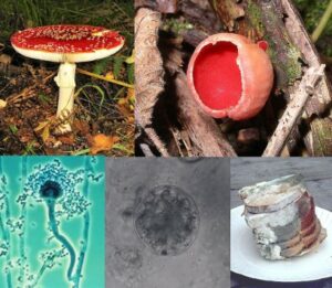
All types of fungi found in nature- mushrooms, toadstools, yeast and mold (clockwise from top left)
Image: Wikipedia

What is unique in the cell wall of fungi?

The uniqueness of the fungal cell wall lies in its composition.

Unlike plants, fungal cell walls are made of a heteropolysaccharide called chitin. Chitosan found in crustacean and insect exoskeletons is actually a derivative of this very chitin.

Fungal cell walls are made up of matrix components embedded in and attached to fibrous load-bearing polysaccharide scaffolds. The polysaccharide, in this case, is chitin. Chitin is a linear homopolymer of N-acetyl-d-glucosamines linked through β-(1,4)-glycosidic bonds.

This is different from plant cell walls that are composed of cellulose a linear polymer of β-1,4 linked d-glucose molecules. N-acetyl-d-glucosamine is a chemical derivative of glucose found naturally.

How do fungi have cell walls?

Fungi are primitive organisms and have a cell wall naturally.

All eukaryotic organisms apart from animals have a cell wall, so Fungi have a cell wall as well. It is merely that their cell walls have different compositions.

The plant cell wall is structural and made of cellulose. This cellulose can harden and form rigid structures like bark by accumulating lignin in themselves. On the other hand, chitin cannot produce such a structure but is strong enough to support larger structures snd also protect smalled unicellular fungi.

does-fungi-have-a-cell-wall
Haworth projection molecular image of Chitin Image: Wikipedia

Chitin is a natural amino-acid sugar derivative that is found to occur naturally. Chitin in nature is more stable and strong when compared to cellulose normally as the hydroxyl group in the sugar is replaced with an acetyl amine group. This results in stronger intermolecular bonds.

Fungi cell wall characteristics:

  • Specific to kingdom Fungi the cell wall lies outside of the cell membrane.
  • The fungal cell wall is made of a mixture of glucans and glycoproteins instead of conventional cellulose.
  • The cell wall’s function is to supply structure and support thus giving the cell stability.
  • The fungal cell wall is also responsible for the cell’s ability to interact with its surroundings.
  • Though chitin is the main component the fungal cell wall is a mixture of 3 main components- glucans, chitin, chitosan and glycosylated proteins.
  • Several pathogen-associated molecular patterns (PAMPs) and epitopes for the immune response in a fungus are found in its cell wall.
  • Toadstools are brightly coloured mushrooms found across the globe. These mushrooms are so brightly coloured to ward off their predators.
  •  This is because toadstools are highly poisonous and can cause severe health issues including death in some cases.

Fungi cell wall composition:

As discussed before the fungal cell wall is polymeric and arranged in various layers. It has 4 main components- chitin, chitosan, glucan and glycoproteins.

The major portion of the cell wall is made of glucans-around 50-60% in dry weight. Β-1,3-D-glucan is the most abundantly present glucan (60-95%). It is the main structural polysaccharide in the fungal cell wall.

Next comes- Chitin. The quantity of chitin in the cell wall varies depending on the species. In yeast, it constitutes only 1-2% of the total dry weight. But if we consider filamentous fungal species that proportion can reach from 10-20%. Chitin is a linear polymer composed of N-acetyl glucosamine subunits. This is a derivative of glucose produced by removing its hydroxyl group to add an acetyl amine group in its place.

The next major component are glycosylated proteins or to simply put glycoproteins. They constitute a hefty 30-50% of the dry weight in most filamentous fungal species. Glycoproteins are formed when most proteins are linked to carbohydrates via O or N links. Cell wall proteins play a variety of roles, including maintaining cellular form, adhesion activities, cellular protection against various chemicals, molecule absorption, signal transmission, and the production and rearrangement of wall components.

Chitosan chair
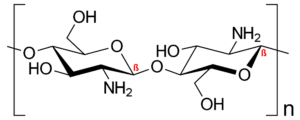
Chitosan molecule
Image: Wikipedia

Chitosan is not considered a major factor as it is just a derivative of chitin.

800px Eumelanine.svg
Melanin molecule structure
Image: Wikipedia

The last and most overlooked component is melanin. This is a pigment that is present in our skin and hair and weight. Melanin is a high-molecular-weight pigment that shields fungi from stress-causing factors and allows them to persist in the host. It is negatively charged, hydrophobic, and insoluble in an aqueous solution. It is also responsible for the most commonly coloured mushrooms.

Also Read:
Eukaryotic Cells Vs Bacterial Cells: Detailed Insights

Prokaryote cell.svg 268x300 1

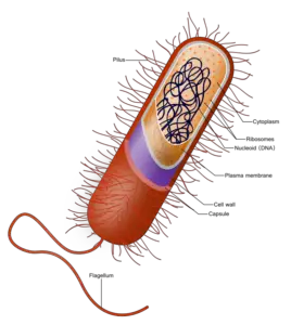
The main difference between eukaryotic and prokaryotic (bacterial cells) lies in the assembly and presentation of the genomic material in the cells.

A Comparison Of Eukaryotic Cells vs Bacterial Cells:

POINTS OF COMPARISON BACTERIAL CELLS EUKARYOTIC CELLS
Presence of Nucleus Absent. They have something called a nucleoid instead. Eukaryotic cells have a membrane-bound nucleus.
DNA arrangement DNA or genetic material is usually double-stranded and arranged circularly. DNA is made up of multiple double-stranded linear DNA.
Cellular complexity Always unicellular Can range from unicellular to multicellular.
Reproduction Mitosis, Meiosis and fusion of gametes Unidirectional DNA transfer and also cloning.
Chloroplasts Absent. Chlorophyll is found scattered in the cytoplasm. Present in plant and algal cells.
Cell wall Present but made of peptidoglycans. Present only in plant and algal cells.
Vacuoles Present Present
Cell size 1-10 µM 10-100 µM
Ribosomes Present but smaller in size and fewer in number Similarly present but greater in size and greater in number.
Other organelles Bacterial cells do not have any other membrane-bound organelles. The characteristic of eukaryotic cells is the presence of various cell organelles like – Endoplasmic Reticulum, Mitochondria and suchA table comparing Eukaryotic cells vs Bacterial Cells

Characteristics of bacterial cells:

  • Bacteria are small unicellular microscopic organisms that make up a major portion of prokaryotes.
  •  They can be free-living or host dependant. The ones that are host dependant may be parasitic or symbiotic.
  • Bacteria belong to prokaryotic cell organization, which means that they do not have an organized and membrane-bound genetic material.
  • Instead, they have a nucleoid – a mass of genetic material floating about in the cell cytoplasm.
  • They can survive in the harshest environmental conditions including the gut of various other organisms.
  • Bacterial cells also do not have any form of membrane-bound organelles in their system.
  • Since a lot of them are free-living they have structures like flagella which help them to locomote.
  • Others that depend on hosts have structures like pilli or fimbriae that allow DNNA transfer(in case of infection) or cellular attachment respectively
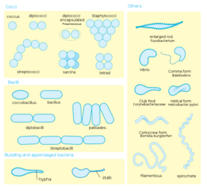
  • They are mainly differentiated on their shapes- spherical, rod-shaped or spiral.
  • They are respectively named coccus, bacillus or spirullus.
  • They have a peptidoglycan cell wall that can be stained using some specific chemicals.
  • Based on the type of stain that the cell wall catches they can bed distinguished into Gram-positive and Gram-negative bacterial cells.
eukaryotic-cells-vs-bacterial-cells
Typical bacterial representation Image :Wikipedia

Some examples of Bacteria:

Some bacteria have found the ability to love in the most adverse environments, while some have beneficial effects on natural flora and fauna. They can also cause various diseases that may even be fatal. Here we will look at some of them:

800px Bacterial morphology diagram.svg
Bacterial classification based on morphology Image: Wikipedia
  • Halophiles: These bacteria live in extremely salty environments where most other organisms cannot survive.
  • Acidophiles: These bacteria like in highly acidic environments.
  • Alkaliphiles: These bacteria on the other hand live in extremely alkaline environments with extremely high pH.
  • Psychrophile: These bacteria live in cryogenic temperatures below 0 degrees Celcius in places like glaciers and polar snow caps.
  • Nitrogen-fixing bacteria: Nitrogen-fixing bacteria live in the soil or the root nodules of leguminous and fix atmospheric nitrogen either into the plant root or into the soil directly.
  • Beneficial bacteria: These bacteria can live in the gut(intestine) of larger animals. They help break down nutrients for better absorption. Some of them also synthesize Vitamins that keep the intestinal flora and fauna(beneficial organisms in the gut) healthy.
  • Commercially useful bacteria: Some bacteria like Lactobacillus is used commercially and also at home for the preparation of curd and cheese.
  • Pathogenic bacteria: Not all bacteria are beneficial. We mostly associate bacteria as disease-causing pathogens. They can cause diseases in both plants and animals, that can also be life-threatening. Some bacterial diseases can wipe out a year’s worth of crops. In humans, they can cause typhoid, pneumonia, tuberculosis and many more. Before the discovery of vaccines and antibiotics these diseases had caused massive numbers of deaths.

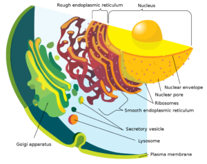
Characteristics of Eukaryotic Cells:

Endomembrane system diagram en.svg
Parts of a eukaryotic cell
Image: Wikipedia

What can be found in eukaryotic cells but not in bacteria?

The most important distinguishing factor between eukaryotic and bacterial cells is the presence and absence of the nucleus respectively. In bacterial cells, the genetic material is arranged in a circular material called a nucleoid. There is no membrane covering the genetic material so we can say that it practically floats in the cytoplasm of the bacteria.

Another important characteristic of eukaryotic cells is the presence of other membrane-bound organelles. This is also absent in bacterial cells as they are nearly devoid of any extra organelles. Bacteria are structurally very simple organisms.

What do bacteria and eukaryotes have in common?

On closer look bacterial cells are in some ways similar to plant cells in general:

Conclusion:

Bacteria arrived on Earth millions of years before the advent of simple unicellular eukaryotic organisms. Even then they have managed to survive to this day without having to undergo any major adaptations or evolutions. This is probably why they could survive over the harsh atmospheric changes. Hence bacteria have acquired the ability to live in all circumstances and environments where no other organism can dream of surviving. Though they seem lacking in comparison to eukaryotes in terms of size or complexity they are still much more individually capable

Also Read:

Fuel Cell Aircraft: A Comprehensive Guide for Science Students

fuel cell aircraft

Fuel cell aircraft have the potential to significantly reduce greenhouse gas emissions in the aviation industry. The design and analysis of fuel cell systems for aviation is a complex process that involves the generation of flight mission profile data, the development of fuel cell system models, and the use of stochastic models to predict mission profiles under uncertainty. This comprehensive guide will provide science students with a detailed understanding of the technical specifications, design considerations, and practical implementation of fuel cell aircraft.

Understanding the Fundamentals of Fuel Cell Aircraft

Fuel cell aircraft utilize hydrogen-powered fuel cells to generate electricity, which is then used to power the aircraft’s electric motors. This technology offers several advantages over traditional jet engines, including:

  1. Reduced Emissions: Fuel cell aircraft produce zero direct emissions, making them a more environmentally friendly option for air travel.
  2. Improved Efficiency: Fuel cells have a higher energy conversion efficiency compared to internal combustion engines, leading to better fuel economy and longer flight times.
  3. Quiet Operation: Fuel cell aircraft are significantly quieter than their jet-powered counterparts, reducing noise pollution.

To understand the design and operation of fuel cell aircraft, it is essential to delve into the underlying principles of fuel cell technology.

Fuel Cell Fundamentals

A fuel cell is an electrochemical device that converts the chemical energy of a fuel, such as hydrogen, directly into electrical energy. The basic structure of a fuel cell consists of an anode, a cathode, and an electrolyte membrane. The electrochemical reactions that occur within the fuel cell can be described by the following equations:

Anode reaction: $2H_2 \rightarrow 4H^+ + 4e^-$
Cathode reaction: $O_2 + 4H^+ + 4e^- \rightarrow 2H_2O$
Overall reaction: $2H_2 + O_2 \rightarrow 2H_2O$

The specific power and energy density of fuel cells are crucial parameters in the design of fuel cell aircraft. The specific power, measured in kW/kg, determines the power-to-weight ratio of the fuel cell system, while the specific energy, measured in kWh/kg, determines the energy-to-weight ratio.

Fuel Cell System Design for Aircraft

The design of fuel cell systems for aircraft involves several key considerations, including:

  1. Flight Mission Profile: The energy system design process starts with the generation of flight mission profile data, which includes parameters such as flight duration, altitude, and power requirements.
  2. Fuel Cell System Modeling: Detailed models of the fuel cell system, including the electrochemical, thermal, and mechanical aspects, are developed to accurately predict the system’s performance.
  3. Stochastic Modeling: Stochastic models are used to predict mission profiles under uncertainty, accounting for factors such as weather conditions and air traffic.
  4. Energy System Design Optimization: The energy system design process explores the design range and evaluates design options using Monte Carlo-based sampling of mission profiles.

Fuel Cell Aircraft Design and Analysis

fuel cell aircraft

The design and analysis of fuel cell aircraft involves several key components, each with its own technical specifications and design considerations.

Fuel Cell Stack Design

The fuel cell stack is the core component of the fuel cell system, responsible for generating the electrical power. The design of the fuel cell stack involves the following considerations:

  • Proton Exchange Membrane Fuel Cells (PEMFCs): PEMFCs are commonly used in fuel cell aircraft due to their high power density, fast start-up, and low operating temperature.
  • Stack Configuration: The fuel cell stack can be designed with a specific number of individual cells, depending on the power requirements of the aircraft.
  • Cooling System: An effective cooling system is essential to maintain the optimal operating temperature of the fuel cell stack.

Hydrogen Storage and Delivery

The storage and delivery of hydrogen fuel is a critical aspect of fuel cell aircraft design. Factors to consider include:

  • Hydrogen Storage: Hydrogen can be stored in various forms, such as compressed gas or cryogenic liquid, each with its own advantages and challenges.
  • Hydrogen Delivery: The fuel delivery system must ensure a reliable and efficient supply of hydrogen to the fuel cell stack.
  • Safety Considerations: Proper safety measures must be implemented to mitigate the risks associated with handling and storing hydrogen.

Power Conversion and Distribution

The electrical power generated by the fuel cell stack must be converted and distributed to the aircraft’s various systems, including:

  • Power Conversion: Power conversion components, such as DC-DC converters and inverters, are used to transform the fuel cell’s output to the appropriate voltage and current levels.
  • Power Distribution: The power distribution system ensures that the electrical power is delivered to the aircraft’s motors, avionics, and other systems.
  • Energy Storage: Batteries or other energy storage devices may be integrated into the system to provide additional power during peak demand or to store excess energy.

Aircraft Integration and Optimization

The integration of the fuel cell system into the aircraft design is a complex process that involves the following considerations:

  • Weight and Balance: The fuel cell system’s weight and placement must be carefully considered to maintain the aircraft’s overall weight and balance.
  • Aerodynamic Integration: The fuel cell system components must be integrated into the aircraft’s design in a way that minimizes aerodynamic drag and maximizes efficiency.
  • System Optimization: The overall fuel cell aircraft system must be optimized to achieve the desired performance, range, and efficiency.

Practical Implementation and Validation

The practical implementation and validation of fuel cell aircraft systems involve several key steps, including:

  1. Prototype Development: Building and testing fuel cell aircraft prototypes is essential to validate the design and performance of the system.
  2. Flight Testing: Rigorous flight testing is necessary to evaluate the fuel cell aircraft’s performance, safety, and reliability under real-world conditions.
  3. Simulation and Modeling: Computational fluid dynamics (CFD) simulations and other modeling techniques can be used to further refine the aircraft’s design and optimize its performance.
  4. Certification and Regulation: Fuel cell aircraft must comply with strict safety and regulatory requirements before they can be approved for commercial use.

Conclusion

Fuel cell aircraft offer a promising solution for reducing greenhouse gas emissions in the aviation industry. By understanding the fundamental principles, design considerations, and practical implementation of fuel cell systems, science students can contribute to the development and advancement of this technology. This comprehensive guide has provided a detailed overview of the key aspects of fuel cell aircraft, equipping you with the knowledge and tools to explore this exciting field further.

References

  1. Design of Fuel Cell Systems for Aviation: Representative Mission Analysis and Energy System Design, Frontiers in Energy Research, 2019.
  2. Flight test validation of the dynamic model of a fuel cell system for ultra-light aircraft, ResearchGate, 2015.
  3. Optimal design of a hydrogen-powered fuel cell system for aircraft, ScienceDirect, 2024.
  4. PEM Fuel Cell MODEL for Conceptual Design of Hydrogen eVTOL, NASA Technical Reports Server, 2021.
  5. The future technological potential of hydrogen fuel cell systems for aviation and preliminary co-design of a hybrid regional aircraft, Energy, 2023.

Photocell Sensors: A Comprehensive Guide for Science Students

photocell sensors

Photocell sensors, also known as photoresistors, are devices that change their electrical resistance when exposed to light. These sensors are widely used in various applications, such as street lighting, security systems, and light-based control systems. Understanding the technical details and characteristics of photocell sensors is crucial for science students who are interested in exploring the field of optoelectronics and sensor technology.

Understanding Photocell Sensors

Photocell sensors work on the principle of the photoelectric effect, where the resistance of the sensor material decreases when exposed to light. This change in resistance is proportional to the intensity of the incident light, allowing the sensor to detect and measure the amount of light present.

Spectral Sensitivity

One of the key characteristics of photocell sensors is their spectral sensitivity, which refers to their ability to respond to different wavelengths of light. Most photocell sensors consist of discrete red (R), green (G), and blue (B) sensors, each with its own unique sensitivity curve. The spectral distribution of the light source can significantly affect the sensor’s response, as different light sources have varying spectral characteristics.

For example, incandescent lights are a reasonable approximation of a blackbody emitter, with a continuous spectrum. In contrast, LED and fluorescent light sources typically do not have a continuous spectrum, which can impact the sensor’s performance.

To understand the spectral sensitivity of a photocell sensor, we can use the following equation:

R = R0 * (1 + k * Φ)

Where:
– R is the resistance of the photocell sensor
– R0 is the dark resistance of the sensor
– k is the spectral sensitivity coefficient
– Φ is the incident light flux

The spectral sensitivity coefficient, k, is a function of the wavelength of the incident light and the specific characteristics of the sensor material.

Angular Response

Another important factor to consider when using photocell sensors is their angular response, which describes the sensor’s sensitivity to light coming from different angles. The angular response is partly a property of the sensor itself and partly a property of any diffusers or optics in front of the sensors.

The angular response can be characterized using the following equation:

I = I0 * cos^n(θ)

Where:
– I is the light intensity at the sensor surface
– I0 is the maximum light intensity (when θ = 0°)
– θ is the angle of incidence
– n is the angular response coefficient

The angular response coefficient, n, determines the shape of the angular response curve and can vary depending on the sensor design and the presence of any optical elements.

Calibration and Measurement Considerations

Calibrating a photocell sensor is a crucial step to ensure accurate and reliable measurements. The calibration process involves determining a calibration reference and minimizing any variables between the measurement conditions of the reference sensor and the test sensor.

To calibrate a photocell sensor, you can use the following steps:

  1. Identify a suitable calibration reference, such as a standard light source or a reference photocell sensor.
  2. Ensure that the measurement conditions, such as the distance, angle, and spectral distribution of the light, are the same for both the reference and the test sensor.
  3. Measure the resistance of the test sensor under the calibration conditions and compare it to the reference sensor’s output.
  4. Adjust the calibration parameters, such as the spectral sensitivity coefficient or the angular response coefficient, to match the reference sensor’s output.

It’s important to note that the calibration data obtained for a specific light source and sensor may not be directly applicable to other light sources or sensors, as the spectral and angular response characteristics can vary.

Technical Specifications

Photocell sensors typically have the following technical specifications:

  • Resistance range: Several hundred ohms in the dark to a few thousand ohms in bright light
  • Response time: The time it takes for the sensor to reach 90% of its final value when exposed to light, which can vary depending on the sensor and the light level

These specifications can be influenced by factors such as the sensor material, the sensor design, and the operating conditions.

Applications of Photocell Sensors

photocell sensors

Photocell sensors have a wide range of applications, including:

  1. Street Lighting: Photocell sensors are commonly used in street lighting systems to detect the amount of ambient light and automatically turn the lights on or off as needed.
  2. Security Systems: Photocell sensors can be used in security systems to detect the presence of intruders by monitoring changes in light levels.
  3. Light-based Control Systems: Photocell sensors can be used to control the operation of various devices, such as blinds, curtains, or lighting systems, based on the amount of available light.
  4. Optical Sensing: Photocell sensors can be used in optical sensing applications, such as object detection, color recognition, and light intensity measurement.
  5. Photovoltaic Systems: Photocell sensors can be used to monitor the performance of photovoltaic systems by measuring the incident light levels.

Conclusion

Photocell sensors are versatile and widely used devices that play a crucial role in various applications. Understanding the technical details and characteristics of photocell sensors, such as spectral sensitivity, angular response, and calibration considerations, is essential for science students interested in exploring the field of optoelectronics and sensor technology.

By mastering the concepts and practical applications of photocell sensors, science students can develop a deeper understanding of the principles of light-based sensing and their real-world applications.

References

  1. Electronics Tutorials: Light Sensor including Photocells, LDR, Photodiodes, Phototransistors, Photovoltaics and Light Dependent Resistor. https://www.electronics-tutorials.ws/io/io_4.html
  2. enDAQ Blog: Light Sensors: Units, Uses, and How They Work. https://blog.endaq.com/how-light-sensors-work
  3. Pepperl+Fuchs: Photoelectric Sensors With Measuring Function. https://www.pepperl-fuchs.com/usa/en/23097.htm