Is Primary Transport Active: A Comprehensive Guide

is primary transport active

Summary

Primary active transport is a fundamental biological process that utilizes metabolic energy, often in the form of ATP, to move molecules and ions across cell membranes against their electrochemical gradients. This mechanism is crucial for maintaining the proper concentrations of various substances within living cells and plays a pivotal role in generating membrane potentials, which are essential for numerous cellular functions.

Understanding Primary Active Transport

is primary transport active

The Sodium-Potassium Pump

One of the most well-studied primary active transport systems is the sodium-potassium pump, also known as the Na+/K+ ATPase. This membrane-bound enzyme is responsible for maintaining the appropriate concentrations of sodium and potassium ions within cells. According to a study by Field (2003), the sodium-potassium pump actively transports three sodium ions out of the cell for every two potassium ions it moves in, creating an electrochemical gradient that drives various secondary active transport processes.

The sodium-potassium pump is found in the plasma membrane of almost all animal cells and is particularly abundant in nerve and muscle cells, where it plays a crucial role in generating and maintaining the resting membrane potential. This membrane potential is essential for the transmission of nerve impulses and the proper functioning of excitable tissues.

ATP Consumption in Primary Active Transport

Primary active transport processes, such as the sodium-potassium pump, require a significant amount of energy in the form of ATP. It has been estimated that up to 70% of the ATP consumed by a resting mammalian cell is used to maintain the correct concentrations of sodium and potassium ions (Lodish et al., 2000). This high energy demand underscores the importance of primary active transport in cellular homeostasis and the maintenance of electrochemical gradients.

Membrane Potential and Primary Active Transport

The electrochemical gradients generated by primary active transport processes, like the sodium-potassium pump, are essential for the establishment and maintenance of the membrane potential. This membrane potential is a crucial factor in various cellular processes, including:

  1. Nerve Impulse Transmission: The membrane potential generated by the sodium-potassium pump is the driving force behind the propagation of nerve impulses along the axons of neurons.
  2. Muscle Contraction: The membrane potential is necessary for the proper functioning of muscle cells, as it is involved in the initiation and regulation of muscle contractions.
  3. Secretion and Absorption: The membrane potential plays a vital role in the secretion and absorption of various substances, such as ions, nutrients, and waste products, across epithelial cell layers.
  4. Cell Signaling: The membrane potential is a key component in numerous cell signaling pathways, where changes in the potential can trigger specific cellular responses.

Specialized Primary Active Transport Systems

In addition to the sodium-potassium pump, there are numerous other primary active transport systems found in living organisms, each with its own unique function and characteristics. Some examples include:

  1. Calcium Pumps: These pumps actively transport calcium ions (Ca2+) out of the cell or into organelles, such as the endoplasmic reticulum and mitochondria, against their concentration gradients. Calcium pumps are essential for regulating intracellular calcium levels, which are crucial for various cellular processes, including signaling, muscle contraction, and apoptosis.

  2. Proton Pumps: Proton pumps, such as the vacuolar H+-ATPase, actively transport protons (H+) across membranes, often from the cytoplasm into organelles or the extracellular space. These proton gradients are used to drive the transport of other molecules and ions, as well as to maintain the appropriate pH levels within cellular compartments.

  3. Heavy Metal Pumps: Some organisms, particularly bacteria and plants, possess primary active transport systems that actively pump out heavy metal ions, such as cadmium, lead, and mercury, to maintain low intracellular concentrations and protect the cell from toxicity.

  4. Phosphate Pumps: Phosphate is an essential nutrient for living organisms, and primary active transport systems, such as the sodium-dependent phosphate cotransporter, are responsible for actively transporting phosphate into cells against its concentration gradient.

These specialized primary active transport systems play crucial roles in a wide range of cellular processes, from ion homeostasis and pH regulation to nutrient uptake and heavy metal detoxification.

Measuring Primary Active Transport

Researchers have developed various techniques to quantify and study the different aspects of primary active transport processes. Some of the commonly used methods include:

Flux Measurements

By measuring the net movement of specific ions or molecules across a membrane, researchers can determine the rate and direction of primary active transport. For example, the flux of sodium and potassium ions can be used to calculate the activity of the sodium-potassium pump.

Enzyme Activity Assays

Primary active transport systems often involve membrane-bound enzymes, such as the sodium-potassium ATPase. Enzyme activity assays can be used to measure the catalytic activity of these enzymes, providing insights into the kinetics and regulation of primary active transport processes.

Membrane Potential Measurements

The membrane potential generated by primary active transport is a crucial parameter that can be measured using techniques like patch-clamp electrophysiology or fluorescent dye-based imaging. These measurements can reveal the contribution of primary active transport to the overall membrane potential and its role in various cellular processes.

ATP Consumption Quantification

As mentioned earlier, primary active transport processes consume a significant amount of cellular ATP. By measuring the rate of ATP hydrolysis or the oxygen consumption associated with ATP synthesis, researchers can estimate the energy requirements of primary active transport systems.

Molecular and Genetic Approaches

Advances in molecular biology and genetics have enabled researchers to identify, characterize, and manipulate the genes and proteins involved in primary active transport. Techniques like gene expression analysis, protein purification, and site-directed mutagenesis have provided valuable insights into the structure, function, and regulation of primary active transport systems.

Conclusion

Primary active transport is a fundamental biological process that plays a crucial role in maintaining the proper concentrations of ions and molecules within living cells. By utilizing metabolic energy, often in the form of ATP, primary active transport systems, such as the sodium-potassium pump, generate electrochemical gradients that drive various secondary transport processes and contribute to the establishment of the membrane potential.

Researchers have developed a range of techniques to quantify and study different aspects of primary active transport, including flux measurements, enzyme activity assays, membrane potential measurements, ATP consumption quantification, and molecular and genetic approaches. These tools have provided valuable insights into the mechanisms, regulation, and physiological importance of primary active transport in diverse cellular processes.

Understanding the intricacies of primary active transport is crucial for advancing our knowledge of cellular homeostasis, signaling, and the pathophysiology of various diseases. Continued research in this field will undoubtedly lead to new discoveries and applications in the fields of biology, medicine, and biotechnology.

References:

  1. Field, M. (2003). Intestinal ion transport and the pathophysiology of diarrhea. J. Clin. Invest., 111(7), 931–943. http://dx.doi.org/10.1172%2FJCI200318326
  2. Lodish, H., Berk, A., Zipursky, S. L., Matsudaira, P., Baltimore, D., & Darnell, J. (2000). Cotransport by symporters and antiporters. In Molecular cell biology(4th ed., section 15.6). New York, NY: W. H. Freeman. Retrieved from http://www.ncbi.nlm.nih.gov/books/NBK21687/
  3. Lodish, H., Berk, A., Zipursky, S. L., Matsudaira, P., Baltimore, D., & Darnell, J. (2000). Intracellular ion environment and membrane electric potential. In Molecular cell biology(4th ed., section 15.4). New York, NY: W. H. Freeman. Retrieved from http://www.ncbi.nlm.nih.gov/books/NBK21627/
  4. Alberts, B., Johnson, A., Lewis, J., Raff, M., Roberts, K., & Walter, P. (2002). Molecular biology of the cell (4th ed.). New York, NY: Garland Science.
  5. Skou, J. C. (1998). The identification of the sodium-potassium pump (Na+/K+-ATPase): a personal account. Biochim. Biophys. Acta, 1154(1), 1–27. https://doi.org/10.1016/s0005-2728(98)00026-7
  6. Palmgren, M. G., & Nissen, P. (2011). P-type ATPases. Annu. Rev. Biophys., 40, 243–266. https://doi.org/10.1146/annurev.biophys.093008.131331
  7. Gadsby, D. C. (2009). Ion channels versus ion pumps: the principal difference, in principle. Nat. Rev. Mol. Cell Biol., 10(5), 344–352. https://doi.org/10.1038/nrm2668

Do Animal Cells Have Centrioles?

do animal cells have centrioles

Animal cells are known to possess a unique organelle called the centriole, which plays a crucial role in various cellular processes. Centrioles are cylindrical structures composed of microtubules and are typically organized into nine sets of short microtubule triplets. These organelles are present exclusively in animal cells and some lower plants, making them an essential feature of the eukaryotic cell.

The Structure and Composition of Centrioles

Centrioles are typically 0.2-0.5 μm in diameter and 0.3-0.5 μm in length, and they are composed of nine sets of short microtubule triplets arranged in a cylindrical pattern. These microtubule triplets are made up of three microtubules: one complete microtubule (the A-tubule) and two incomplete microtubules (the B-tubule and the C-tubule). The microtubules within the centriole are held together by a variety of proteins, including centrin, polyglutamylated tubulin, and other centriole-specific proteins.

The centriole’s cylindrical structure is further reinforced by a set of nine peripheral microtubules, known as the “centriolar wall,” which are connected to the microtubule triplets by radial spokes. This intricate arrangement of microtubules and associated proteins gives the centriole its characteristic morphology and stability.

The Functions of Centrioles in Animal Cells

do animal cells have centrioles

Centrioles play a crucial role in the formation and organization of two essential cellular structures: centrosomes and cilia.

Centrosome Formation

Centrioles are the core components of the centrosome, which is the primary microtubule-organizing center (MTOC) in animal cells. During cell division, the centrosome is responsible for organizing the mitotic spindle, which is essential for the accurate segregation of chromosomes into the daughter cells.

The process of centrosome formation begins with the duplication of the existing centrioles. This duplication occurs in a semi-conservative manner, where each existing centriole serves as a template for the formation of a new centriole. The newly formed centrioles then migrate to opposite poles of the cell, forming the two poles of the mitotic spindle.

Cilia Assembly

Centrioles are also essential for the assembly of cilia, which are hair-like projections that extend from the cell surface. Cilia are involved in various cellular processes, such as sensory perception, fluid flow regulation, and cell motility.

The formation of cilia begins with the migration of a centriole to the cell surface, where it becomes the basal body of the cilium. The basal body then serves as a template for the assembly of the ciliary axoneme, which is the core structure of the cilium.

The Role of Centrioles in Mitosis and Chromosome Segregation

While centrioles are not strictly required for the formation of the mitotic spindle, they do play an important role in ensuring the accuracy of chromosome segregation during cell division.

In the absence of centrioles, animal cells can still form bipolar spindles through alternative pathways, such as the self-organization of microtubules. However, these acentriolar spindles have been shown to have reduced fidelity in chromosome segregation, leading to increased rates of aneuploidy (an abnormal number of chromosomes) in the daughter cells.

Centrioles contribute to the accuracy of chromosome segregation by biasing the position of the spindle poles during mitosis. This ensures that the chromosomes are properly aligned and segregated into the daughter cells, minimizing the risk of chromosomal abnormalities.

Centriole-Associated Diseases

Defects in centriole-associated proteins have been linked to a variety of ciliary diseases, including:

  1. Nephronophthisis: A genetic disorder that leads to the progressive destruction of the kidneys.
  2. Bardet-Biedl syndrome: A genetic disorder characterized by obesity, vision loss, polydactyly, and other developmental abnormalities.
  3. Meckel Syndrome: A rare, lethal genetic disorder characterized by developmental abnormalities, including polycystic kidneys and central nervous system defects.
  4. Oral-Facial-Digital syndrome: A group of genetic disorders characterized by abnormalities of the face, oral cavity, and digits.

These diseases highlight the importance of centrioles and their associated proteins in the proper functioning of cilia, which are essential for various cellular processes.

Conclusion

In summary, animal cells do possess centrioles, which are cylindrical organelles composed of microtubules. Centrioles play a crucial role in the formation of centrosomes and cilia, both of which are essential for various cellular processes, including cell division and sensory perception. While centrioles are not strictly required for the formation of the mitotic spindle, they contribute to the accuracy of chromosome segregation during cell division. Defects in centriole-associated proteins have been linked to a variety of ciliary diseases, underscoring the importance of these organelles in maintaining cellular homeostasis and proper development.

References:

  1. Marshall, W. F. (2006). What is the function of centrioles? Journal of Cellular Biochemistry, 100(4), 916-922. https://doi.org/10.1002/jcb.21117
  2. Centriole. (n.d.). British Society for Cell Biology. Retrieved from https://bscb.org/learning-resources/softcell-e-learning/centriole/
  3. The Enigma of Centriole Loss in the 1182-4 Cell Line. (2020). PMC. https://doi.org/10.1093/pmc/pma182
  4. Centrioles are present in . – BYJU’S. (n.d.). BYJU’S. Retrieved from https://byjus.com/question-answer/centrioles-are-present-in-animal-cellsplant-cellsbacteriaalgae/
  5. Centriole – an overview | ScienceDirect Topics. (n.d.). ScienceDirect. Retrieved from https://www.sciencedirect.com/topics/neuroscience/centriole

Do Plant Cells Have Centrioles?

do plant cells have centrioles

Plant cells do not have centrioles, while animal cells and some lower plant cells do. This is a well-established fact in cell biology, supported by a significant body of research and evidence.

The Absence of Centrioles in Plant Cells

In plants, the spindle fibers that separate chromosomes during mitosis and meiosis are formed through different mechanisms than in animals. Instead of centrioles, plants use structures called microtubule organizing centers (MTOCs) to nucleate and organize microtubules. These MTOCs can be found in various locations within the plant cell, including the nuclear envelope, plastids, and polar organizers.

The absence of centrioles in plant cells is thought to be related to their evolutionary history and the unique characteristics of plant cells. For example, plant cells have a rigid cell wall that provides structural support, which may reduce the need for centrioles to organize microtubules during cell division. Additionally, plant cells are often polyploid, meaning they have multiple sets of chromosomes, which may also affect the need for centrioles during meiosis.

Microtubule Organizing Centers (MTOCs) in Plant Cells

do plant cells have centrioles

In the absence of centrioles, plant cells rely on microtubule organizing centers (MTOCs) to nucleate and organize microtubules during cell division. These MTOCs can be found in various locations within the plant cell, including:

  1. Nuclear Envelope: MTOCs are associated with the nuclear envelope, where they help organize the microtubules that form the mitotic spindle during cell division.

  2. Plastids: MTOCs are found in plastids, such as chloroplasts, where they help organize the microtubules involved in the movement and division of these organelles.

  3. Polar Organizers: MTOCs are also found at the poles of the cell, where they help organize the microtubules that form the mitotic spindle.

The specific mechanisms by which MTOCs organize microtubules in plant cells are complex and involve a variety of proteins and regulatory pathways. For example, the γ-tubulin complex is a key component of MTOCs, as it helps nucleate and anchor microtubules.

Evolutionary Adaptations in Plant Cells

The absence of centrioles in plant cells is thought to be an evolutionary adaptation that is related to the unique characteristics of plant cells. Some key factors that may have contributed to the loss of centrioles in plant cells include:

  1. Cell Wall Structure: Plant cells have a rigid cell wall that provides structural support, which may reduce the need for centrioles to organize microtubules during cell division.

  2. Polyploidy: Many plant cells are polyploid, meaning they have multiple sets of chromosomes. This may affect the need for centrioles during meiosis, as the mechanisms for chromosome segregation may be different in polyploid cells.

  3. Sessile Lifestyle: Plants are sessile organisms, meaning they are rooted in one place and cannot move around like animals. This may have reduced the selective pressure for the maintenance of centrioles, as they are primarily involved in the organization of the cytoskeleton during cell division and movement.

  4. Photosynthesis: The evolution of photosynthesis in plants may have also contributed to the loss of centrioles, as the organization of the cytoskeleton may have been less critical for the primary function of plant cells, which is to capture and convert light energy into chemical energy.

Experimental Evidence for the Absence of Centrioles in Plant Cells

While there is no direct measurable or quantifiable data on the absence of centrioles in plant cells, the consensus among biologists is that plant cells do not have centrioles. This is supported by numerous observations and experiments, including:

  1. Electron Microscopy Studies: Numerous electron microscopy studies have failed to find centrioles in plant cells, despite their clear presence in animal cells.

  2. Genetic Studies: Genetic studies have identified the key components of the centriole structure, and these components have not been found in the genomes of most plant species.

  3. Developmental Studies: During the development of plant cells, the mechanisms for spindle formation and chromosome segregation do not involve the presence of centrioles, further supporting the absence of these structures in plant cells.

  4. Comparative Studies: Comparative studies between plant and animal cells have consistently shown that centrioles are present in animal cells but absent in plant cells, with the exception of some lower plant species.

Conclusion

In summary, plant cells do not have centrioles, and this is an important distinction between plant and animal cells. The absence of centrioles in plant cells is related to their unique characteristics and evolutionary history, and is supported by a significant body of research and evidence. Understanding the differences in the cytoskeletal organization between plant and animal cells is crucial for understanding the fundamental biology of these two domains of life.

References:
– Quizlet. Plant cells do not have centrioles. Hypothesize why plant cells might not need centrioles for mitosis or meiosis. https://quizlet.com/explanations/questions/plant-cells-do-not-have-centrioles-hypothesize-why-plant-cells-might-not-need-centrioles-for-mitosis-or-meiosis-a0253d53-f60624ec-17b7-4b1b-bb7c-84c98b4373f4/
– Socratic. Do plant cells have centrioles? https://socratic.org/questions/do-plant-cells-have-centrioles
– BYJU’S. Do plant cells have centrioles? https://byjus.com/question-answer/do-plant-cells-have-centrioles/

Do Bacterial Cells Have Cytoplasm?

do bacterial cells have cytoplasm

Bacterial cells are the most abundant and diverse form of life on Earth, and they play a crucial role in various ecological and biological processes. One of the fundamental questions about bacterial cells is whether they possess a cytoplasm, which is a critical component of eukaryotic cells. In this comprehensive blog post, we will delve into the details of the cytoplasm in bacterial cells, exploring its structure, function, and the techniques used to analyze it.

The Presence of Cytoplasm in Bacterial Cells

Bacterial cells do indeed have a cytoplasm, which is the gel-like substance that fills the cell and surrounds various organelles. The cytoplasm is the site of most of the cell’s metabolic processes, including protein synthesis, glycolysis, and the citric acid cycle. It is enclosed by the cell membrane, which regulates the movement of materials in and out of the cell.

The cytoplasm of bacterial cells is a complex and dynamic environment, with a diverse array of molecules and structures. It contains a variety of macromolecules, such as proteins, nucleic acids, and lipids, as well as smaller molecules like metabolites, ions, and water. These components are organized and distributed within the cytoplasm, allowing for efficient and coordinated cellular processes.

Analyzing the Cytoplasm of Bacterial Cells

do bacterial cells have cytoplasm

The cytoplasm of bacterial cells can be analyzed using various techniques, each providing unique insights into its structure, composition, and function.

Microscopy

One of the primary methods for studying the cytoplasm of bacterial cells is microscopy. Techniques such as light microscopy, electron microscopy, and fluorescence microscopy can be used to visualize the cytoplasm and its contents. These methods allow researchers to estimate the volume of the cytoplasm, observe the distribution and localization of cellular components, and study the overall organization of the cell.

For example, using electron microscopy, researchers have been able to observe the presence of ribosomes, the organelles responsible for protein synthesis, within the cytoplasm of bacterial cells. Additionally, fluorescence microscopy has been used to track the movement and localization of specific proteins or molecules within the cytoplasm, providing insights into their roles and interactions.

Biochemical Assays

Biochemical assays are another powerful tool for analyzing the cytoplasm of bacterial cells. These techniques involve the extraction and quantification of various molecules and metabolites present in the cytoplasm. By using techniques like spectrophotometry, chromatography, and mass spectrometry, researchers can measure the concentrations of specific compounds, such as proteins, nucleic acids, and metabolites, within the cytoplasm.

For instance, researchers have used biochemical assays to measure the levels of ATP, a crucial energy currency in the cell, within the cytoplasm of bacterial cells. This information can provide insights into the energy metabolism and the overall metabolic state of the cell.

Genetic Analysis

Genetic analysis is a valuable approach for studying the composition and function of the cytoplasm in bacterial cells. By analyzing the genetic information encoded in the bacterial genome, researchers can identify the genes and gene products that are involved in various cytoplasmic processes, such as protein synthesis, energy production, and signal transduction.

Through techniques like genome sequencing, transcriptomics, and proteomics, researchers can gain a comprehensive understanding of the molecular components and pathways that operate within the cytoplasm of bacterial cells. This information can be used to elucidate the specific roles and interactions of various cytoplasmic elements, as well as to identify potential targets for therapeutic interventions or biotechnological applications.

The Dynamic Nature of the Bacterial Cytoplasm

It is important to note that the cytoplasm of bacterial cells is not a static environment, but rather a dynamic and constantly changing one. The composition and properties of the cytoplasm can vary depending on the growth phase of the cell, the availability of nutrients, and other environmental factors.

During exponential growth, the cytoplasm of bacterial cells is typically rich in ribosomes and other organelles involved in protein synthesis, as the cell is actively dividing and producing new cellular components. In contrast, during the stationary phase, when nutrients become scarce, the cytoplasm may contain higher levels of stress-response proteins and other molecules that help the cell survive under adverse conditions.

These changes in the cytoplasmic composition and properties are crucial for the adaptation and survival of bacterial cells in diverse environments. By understanding the dynamic nature of the bacterial cytoplasm, researchers can gain valuable insights into the complex regulatory mechanisms and adaptive strategies employed by these ubiquitous and influential microorganisms.

Conclusion

In summary, bacterial cells do possess a cytoplasm, which is a critical component of their cellular structure and function. The cytoplasm of bacterial cells is a complex and dynamic environment, hosting a variety of macromolecules and metabolic processes. Researchers can analyze the cytoplasm using various techniques, including microscopy, biochemical assays, and genetic analysis, to gain a deeper understanding of its composition, organization, and role in the overall functioning of bacterial cells.

By exploring the cytoplasm of bacterial cells, scientists can uncover valuable insights into the fundamental biology of these organisms, which have profound impacts on human health, the environment, and various industrial applications. The continued study of the bacterial cytoplasm will undoubtedly lead to new discoveries and advancements in our understanding of the remarkable diversity and adaptability of these microscopic yet essential life forms.

Reference:

  1. https://www.ncbi.nlm.nih.gov/books/NBK21495/
  2. https://www.nature.com/scitable/topicpage/the-bacterial-cell-envelope-14092245/
  3. https://www.khanacademy.org/science/biology/cellular-molecular-biology/prokaryotic-cells/a/structure-of-bacterial-cells

9+ Lactose Fermenting Bacteria Examples: Detailed Explanations And Images

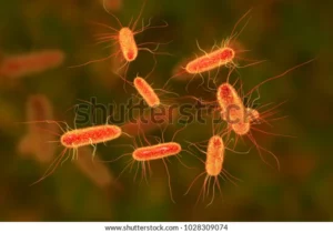
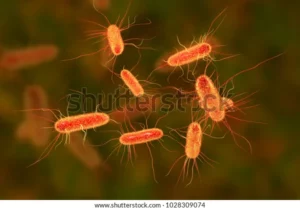
escherichia coli bacterium ecoli gramnegative 600w 1028309074 300x210 1

In this article, we will be discussing lactose fermenting bacteria examples

The bacteria that ferment lactose and produce hydrogen sulfide are called lactose fermenting bacteria.

Escherichia coli

Escherichia coli are a rod-shaped gram-negative bacterium that is usually found in the lower intestine of human beings and the gut of certain animals like deer, pig, dog, cattle, sheep, and poultry. Most the E.coli are harmless and help in keeping the digestive tract healthy but some strains cause food poisoning, urinary tract infections, and diarrhea. Certain strains of E.coli produce a toxin called Shiga which damages the wall of the intestine and causes vomiting, abdominal cramps, and kidney failure. The bacteria can be transmitted through contaminated water, uncooked meat, untreated milk, and vegetables. The bacteria is anaerobic, it ferments lactose and produces hydrogen sulfide.

lactose fermenting bacteria examples
Escherichia coli Bacteria from Shutterstock

Escherichia adecarboxylata

Escherichia adecarboxylata also called Leclercia adecarboxylata is a disease-causing bacteria that resembles E.coli. It is a motile gram-negative bacterium found in the gut of animals. They usually cause diseases like Pneumonia, cellulitis, Pharyngeal abscess, and cholecystitis in immunocompromised patients.

Citrobacter koseri

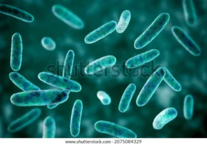
Citrobacter koseri is an anaerobic, motile, gram-negative, rod-shaped non-spore-forming bacteria normally found in the gut of animals and humans. The bacteria can cause urinary tract infections in adults and meningitis in infants. Citrobacter koseri is transmitted from mother to child during birth through vaginal infections.

scientific image bacteria citrobacter gramnegative 600w 2075084329
Citrobacter koseri from Shutterstock

Citrobacter freundii

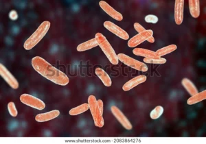
Citrobacter freundii is a gram-negative rod-shaped coliform bacterium that is transmitted through contaminated food. It usually causes diarrhea, urinary tract infection, pneumonia, and at times intracranial abscesses and meningitis.

scientific image bacteria citrobacter gramnegative 600w 2083864276
Citrobacter freundii from Shutterstock

Citrobacter farmeri

Citrobacter farmer is a gram-negative bacterium that is transmitted from infected mother to child during birth and through contaminated food and water. The bacteria usually cause sepsis, pulmonary infections, and meningitis in neonates and young children and urinary tract infections in adults.

Klebsiella pneumoniae

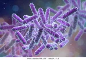
Klebsiella pneumoniae is a non-motile, encapsulated, gram-negative bacterium that usually resides in the gut but causes infection once it moves outside the gut region. They are transmitted through direct contact with an infected person and through contaminated medical equipment like catheters and ventilators. If the bacteria enter the respiratory tract it causes pneumonia and also causes other infections like meningitis and cellulitis.

enterobacteriaceae gramnegative rodshaped bacteria part 600w 1042543318
Klebsiella pneumoniae from Shutterstock

Klebsiella oxytoca

Klebsiella oxytoca is gram-negative rod-shaped that naturally occurs in the nose, mouth, and intestinal tract. If they are present inside the gut they are considered healthy bacteria but once it comes out it causes serious infections. The bacterium causes soft tissue infection, pneumonia, urinary tract infection, and septicemia that leads to septic shock.

biofilm containing bacteria klebsiella 3d 600w 585336695
Klebsiella oxytoca from Shutterstock

Klebsiella granulomatis

Klebsiella granulomatis is a rod-shaped, aerobic, gram-negative bacterium that causes a sexually transmitted disease called Granuloma inguinale. It causes chronic inflammation in the genital areas, initially, it develops as a painless lump and later it breaks down to form a sore.

stock photo bacteria klebsiella granulomatis the causative agent of sexually transmitted disease granuloma 1202581522
Klebsiella granulomatis from Shutterstock

Klebsiella variicola

Klebsiella variicola is a non-motile rod-shaped gram-negative bacterium that forms circular, convex, and mucoid colonies. They cause urinary tract infections and respiratory tract infections. These are closely related to Klebsiella pneumonia and are usually found in plants.

Klebsiella ozaenae

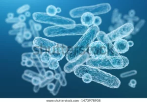
Klebsiella ozaenae is a rod-shaped gram-negative bacterium that causes a chronic disease called Ozena that destroys the mucosa of the nasal cavity. The other infections caused by the bacteria are pneumonia, otitis media, rhinitis, and urinary tract infection. The bacteria spread through person-person contact. This mostly affects immunocompromised patients in the nasopharynx region.

enterobacteriaceae gramnegative rodshaped bacteria part 600w 1473978908
Klebsiella ozaenae from Shutterstock

Serratia plymuthica

Serratia plymuthica is an anaerobic, gram-negative rod-shaped bacterium that is usually found in the roots of coconut and fig trees. They are usually transmitted through contaminated respiratory equipment and catheters. The infection caused by the bacteria is chronic osteomyelitis and sepsis.

Serratia liquefaciens

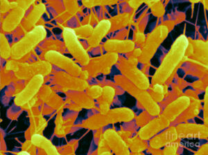
Serratia liquefaciens is a motile straight rod-shaped gram-negative bacterium that is usually found in soil, water, plants, and the gut of insects, fish, rodents, and human beings. The bacteria cause urinary tract infection, meningoencephalitis, pneumonia, bloodstream infections, and sepsis. The bacteria are transmitted through contaminated water, uncooked meat, and untreated milk.

3 sem of serratia liquefaciens scimat
Serratia liquefaciens from Pixels

Also Read:

19+ Kingdom Bacteria Examples: Detailed Explanations And Images

3d rendering ecoli contamination food 600w 1202781193 300x224 1

In this article, we will be discussing examples of different types of bacteria

Bacteria kingdom is one of the major kingdoms under the kingdom Animalia. Previously the kingdom was called monera; it contains two major domains; Eubacteria and Archaea.

Examples of bacteria based on their shape

Streptococcus pyogenes:

It is a gram-positive bacterium that grows in chains and causes many infections like cellulitis, pharyngitis, scarlet fever, tonsillitis, pneumonia, and necrotizing fasciitis. The transmission of bacteria takes place through nasal discharge, air droplets, contaminated surfaces, and contaminated food.

grampositive bacteria streptococcus s pyogenes 600w 1840873864
Streptococcus pyogenes from Shutterstock

Staphylococcus epidermidis

Staphylococcus epidermidis causes skin infections like inflammations, boils, sinus infections, and endocarditis. It is a highly contagious infection that is usually associated with implanted devices like catheters and prosthetic devices.

Enterococcus faecalis 

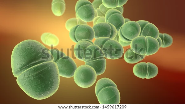
Enterococcus faecalis is a gram-positive bacteria that causes gastrointestinal tract infections in humans. The other infections caused by the bacteria are urinary tract infections, endocarditis, prostatitis, cellulitis, and wound infections. It is transmitted mainly through contaminated food.

3d illustration showing enterococcus faecalis 600w 1459617209
Enterococcus faecalis from Shutterstock

Chlamydia trachomatis 

Chlamydia is a sexually transmitted disease caused by Chlamydia trachomatis a gram-negative bacterium. The bacteria infect the epithelial wall of the cervix, rectum, and urethra. If the infection is not treated properly it will lead to infertility, ectopic pregnancy, and pelvic inflammatory disease. In infants, the bacteria causes conjunctivitis and pneumonia.

Neisseria meningtitidis

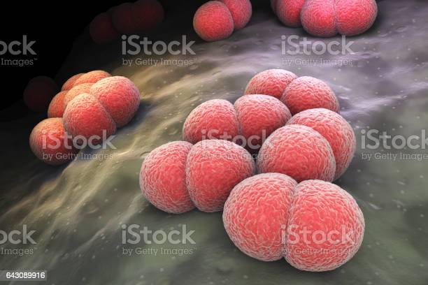
Neisseria meningitides is a gram-negative bacterium that causes meningococcal disease. The most common site of colonization of the bacteria is the nasopharynx region. The bacterium causes sepsis, pericarditis, conjunctivitis, pneumonia, sinusitis, and urethritis.

istockphoto 643089916 612x612 3
Neisseria meningtitidis from istockphoto

Staphylococcus aureus

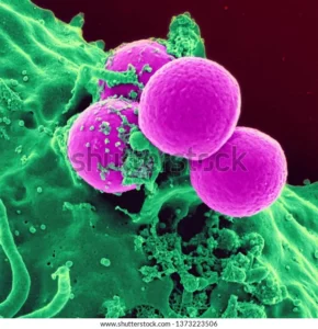
Staphylococcus aureus is a round-shaped gram-positive bacterium that is mostly found in the upper respiratory tract and skin. The bacterium is transmitted by direct contact and contaminated objects.  It travels through the bloodstream and causes endocarditis, bacteremia, pneumonia, and osteomyelitis.

staphylococcus aureus colony 600w 1373223506
Staphylococcus aureus from Shutterstock

Streptococcus pneumoniae

Streptococcus pneumonia is a gram-positive lancet-shaped anaerobic bacteria that cause pneumonia, middle ear infections, sepsis, and meningitis. The infection spreads through air droplets. The symptoms of the pneumococcal infections area shortness of breath, joint pain, fever, cough, ear pain, irritability, and sleeplessness.

streptococcus pneumoniae pneumococcus bacterias 3d 600w 572028538
Streptococcus pnemoniae from Shutterstock

Aeromonas hydrophila

Aeromonas hydrophila is a rod-shaped gram-negative bacterium that is mainly found in fresh water and belongs to the Vibrionaceae family.  It causes infections like kidney disease, cellulitis, meningitis, septicemia, pneumonia, and respiratory tract infections.

Bacillus anthracis

Bacillus anthracis is a gram-negative rod-shaped bacterium that causes a highly infectious disease called anthrax. The bacteria naturally occur in animals like sheep, goats, camels, and antelope. The infection causes multiple organ damage, and inflammation of the membrane of the brain and spinal cord leading to hemorrhagic meningitis.

bacillus anthracis grampositive spore forming 600w 477383266
Bacillus anthracis from Shutterstock

Clostridium botulinum

Clostridium botulinum is a rod-shaped gram-positive bacterium that produces a neurotoxin called botulinum. The toxin blocks nerve functions and causes muscular and respiratory paralysis. The infection is not contagious but is transmitted through contaminated food.

Escherichia coli

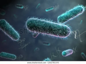
Escherichia coli is a rod-shaped gram-negative bacteria commonly found in the lower intestine. In most cases the bacteria are harmless but certain strains cause stomach pain, diarrhea, cramps, and fever. The infection is usually caused by ingesting contaminated food like ground meat.

3d rendering ecoli contamination food 600w 1202781193
Escherichia coli from Shutterstock

Legionella pneumophila

Legionella pneumophila is flagellated, rod-shaped, aerobic gram-negative bacterium that causes legionellosis disease. Legionellosis is a type of acute pneumonia that is contracted while handling potting soil or compost.

legionella pneumophila bacterium causative agent 600w 1459171178
Legionella pneumophila from Shutterstock

Mycobacterium leprae

Mycobacterium leprae is a rod-shaped, aerobic, gram-positive bacillus that causes a chronic infectious disease called Leprosy.  The bacterium affects the peripheral nerves of the nose, skin, muscles, and eyes. The disease is highly contagious and transmits through airborne droplets from the infected individual’s sneeze or cough.

mycobacterium leprae bacteria inside human 600w 453181294
Mycobacterium leprae from Shutterstock

Vibrio cholera

Vibrio cholera is gram-negative anaerobic comma-shaped bacterium that causes an intestinal infection called cholera by secreting the cholera toxin. The symptoms of cholera are extreme diarrhea, leg cramps, thirst, vomiting, and restlessness.

vibrio cholerae bacteria 3d illustration 600w 1181713009
Vibrio cholerae from Shutterstock

Salmonella typhimurium

Salmonella typhimuriums is a flagellated, rod-shaped gram-negative bacteria causing salmonella infection. The infection is caused due to the intake of raw meat, eggs, and poultry.  This is the most common intestinal tract infection that causes gastroenteritis, diarrhea, abdominal cramps, and fever.

Rickettsia typhi 

Rickettsia typhi is a small rod-shaped gram-negative bacterium that causes murine typhus infection. The bacterium is usually transmitted by fleas. The symptoms of the infection are rash, fever, headache, and chills.

Borrelia afzelii

Borrelia afzelii is a spiral-shaped gram-negative aerobic bacterium that is transmitted through ticks and causes Lyme’s disease. The common symptoms of the disease are swollen lymph nodes, fever, chills, stiff neck, fatigue, and body aches.

Campylobacter coli

Campylobacter coli is an S-shaped gram-negative bacterium that causes campylobacteriosis – a diarrheal disease. The bacteria are usually transmitted by eating undercooked or raw poultry meat, seafood, and water.

campylobacter bacteria c jejuni fetus 600w 1046052091
Campylobacter coli from Shutterstock

Helicobacter hepaticus

Helicobacter hepaticus is a spiral-shaped bacterium with bipolar single sheathed flagellum that causes liver cancer. The infection caused by the bacterium causes chronic gastrointestinal inflammation that leads to neoplasia. The morphology of the breast glands also changes that leading to adenocarcinoma.

Helicobacter pylori

Helicobacter pylori are a helical-shaped gram-negative bacterium that causes infection in the stomach and duodenum. The bacterium irritates the inner lining of the stomach causing inflammation and peptic ulcers. Stomach burning, nausea, stomach bloating, weight loss, loss of appetite, indigestion and burping are some of the symptoms of the infection.

Leptospira interrogans 

Leptospira interrogans is a gram-negative spiral-shaped bacteria with internal flagella that causes leptospirosis disease.  The infection is usually mild but if left untreated can cause liver and kidney failure. The bacterium affects both animals and humans and it usually enters the body through skin abrasions and by the bite of the infected rat. The symptoms of the infection are high fever, headache, chills, vomiting, abdominal pain, jaundice, and muscle aches.

Also Read:

19+Transplanting Plants Examples: Detailed Explanations And Images

istockphoto 1021818122 612x612 2 300x200 1

In this article we will going to discuss about the transplanting plants examples.

Transplanting is a process in horticulture where the seedlings are grown in one place and then replanted in another place during the growing season.

The plants that are grown by transplanting techniques are:

  1. Paddy
  2. Tomato
  3. Cauliflower
  4. Broccoli
  5. Cabbage
  6. Eggplant
  7. Onion
  8. Watermelon
  9. Zucchini
  10. Squash
  11. Turnips
  12. Peppers
  13. Beans
  14. Lettuce
  15. Peas
  16. Potatoes
  17. Cucumbers
  18. Beetroot
  19. Carrots
  20. Celery
  21. Brussels sprouts

Paddy

Paddy is a Kharif crop grown by transplantation process; it is first grown in nurseries to a height of 15-20 cm for 15 to 30 days and then transplanted into puddle fields. The seedlings are transplanted manually or with the help of tractor-drawn discs in straight rows. Clayey soil is required for rice cultivation and this soil helps to hold water for a longer time and helps in the growth of the crop.

transplanting plants examples
Paddy Transplanted in the field from istockphoto

Tomato

Tomatoes are summer crops which is rich in vitamin A, Vitamin C, and antioxidants it is usually grown by transplanting technique. The seeds of the tomato are grown in nurseries until they get two sets of leaves and are about 2-4 inches tall. The land in which the tomatoes are to be transplanted should be irrigated for 3 to 4 days and the seedlings should be planted deep with proper spacing.  The tomato fruit can be harvested after 60 to 100 days. The best temperature for the crop is between 21℃ to 24℃.

hand planting tomato seedling ground 600w 257996501
Tomato Transplanted in fields from Shutterstock

Cauliflower

Cauliflower is a cruciferous vegetable rich in Vitamin-b, antioxidants, and many other nutrients. The seedlings are transplanted after they are grown for 5 to 6 weeks in nurseries.  They are planted in well-drained soil about 2 to 3 feet deep. The seedlings should be well spaced. The optimum temperatures for the plants are about 16 to 18℃.

gardener planting cauliflower seedlings freshly 600w 1369733357
Cauliflower Seedlings planted in fields from Shutterstock

Broccoli

Broccoli belongs to the cabbage family and its seedlings are grown in nurseries for 3 to 4 weeks and then transplanted to well-drained soil in rows with 18 to 24 inches of space between each plant. The plants grow well in temperatures between 16℃ to 26℃.

gardener preparing broccoli seedlings planting 600w 426267529
Broccoli Transplanted in fields from Shutterstock

Cabbage

Cabbage seedlings are grown to a height of 10 to 15 cm in nurseries and then transplanted to sandy soils with pH 6-6.6 and the optimum temperature for the growth of the plant is between 15℃ to 21℃. The plant can be harvested after 90-120 days of planting.

woman working garden planting cabbage 600w 145479772
Cabbage Transplanted to fields from Shutterstock

Eggplant

Eggplant belongs to the family Solanaceae and is a perennial plant. It is also known as an aborigine. The plant is grown indoors for two weeks and then transplanted to sandy loamy soils. The best temperature for the plants to grow is between 20℃ to 30℃.

row freshly transplanted eggplant seedlings 600w 1958721133
Eggplant Transplanted from Shutterstock

Onion

Onion is a root vegetable that belongs to the genus allium. It is rich in sulfur-containing compounds and antioxidants. The onions are grown in nurseries until they reach a height of 15-20cm and then transplanted to loose loamy soil with a space of about 6 inches between the plants. The optimum temperature for the plant is 16 to 25.

woman spends early spring planting 600w 1989450293
Onion Transplanted from Shutterstock

Watermelon

Watermelon is a fruit that belongs to the gourd family. The seeds are grown in nurseries for 2 weeks and then transplanted to loamy well-drained soil. The plants should be planted 2-3 feet apart. The optimum temperature for the plants to grow is 26-28℃.

watermelon planting vegetable garden when 600w 2149647441
Watermelon Transplanted from Shutterstock

Zucchini

Zucchini belongs to the squash family; it is grown indoors until they get three to four true leaves and then transplanted outside in fertile fast-draining soil. The optimum temperature for the plant to grow is 16 to 21℃.

fresh organic zucchini seedling on 600w 1407559232
Zucchini Transplant from Shutterstock

Squash

Squash belongs to the Cucurbita genus; it has a high content of fiber, magnesium, and vitamin C that helps in repairing cells and tissue. The seeds of the squash are grown in nurseries till they have three to four true leaves and then transplanted to fertile, well-drained soil. The space between each plant should be three feet.

young squash cultivation seed trays 600w 1399213802
Squash Seedlings in trays from Shutterstock

Turnips

Turnips are root vegetables grown in temperate climates. They belong to the mustard family. The seedlings are transplanted to the felids from the nursery till true leaves appear. The soil should be well-drained and the pH should be maintained between 6 to 7.5. The space between each plant should be 6 to 8 inches apart.

Peppers

Peppers are also called capsicum that belongs to the Solanaceae family. The seedlings are grown for 7-8 weeks in nurseries and then transplanted into fields. The seedlings should be planted 1.5 apart and 3-4 inches deep. The optimum temperature for peppers to grow in between 18 to 26℃.

Beans

Beans belong to the Fabaceae family that is rich in protein, fiber, iron, vitamin A, vitamin K, and folic acid. The seedlings should be grown in nurseries till three to four true leaves appear and then transplanted to fields. The optimum temperature for beans to grow is 21- 26℃.

woman planting out broad bean 600w 1867934026 1
Beans Transplant from Shutterstock

Lettuce

Lettuce is a leafy vegetable that belongs to the family Asteraceae; it is an annual plant that is rich in vitamin C vitamin K and folic acid. The plants are grown for 2-3 inches tall indoors and then transplanted into fields.

Peas

The pea plant belongs to the family Fabaceae; it contains a seed pod that contains several peas. The seedlings should grow for 7.3cm long before being transplanted into fields. The plants should be spaced 18inches apart. The optimum temperature for peas to grow is 15 to 30℃.

man planting sugar snaps peas 600w 2120138621
Peas Transplant from Shutterstock

Potato

Potato is a starchy root vegetable that belongs to the Solanaceae family. The plants are grown for 10 to 14 days in nurseries before transplanting into fields. The best temperature for potatoes to grow is 24℃.

bushes seedlings potatoes garden irrigation 600w 1409787749 1
Potato Transplant from Shutterstock

Cucumber

The cucumber is a climber that belongs to the family Cucurbitaceae; it is rich in phosphorous and potassium.  The plants are grown in nurseries for three to four weeks and are transplanted into fields 4 to 5 feet apart.

Beetroot

Beetroot is a root plant that belongs to the family Amaranthaceae; the vegetable is rich in fiber, manganese, iron, potassium, and vitamin C. the seedlings are grown in nurseries till the first set of true leaves appears and then transplanted into fields with 10cm between them.

Carrots

Carrot is a root vegetable that is rich in carotenoids, vitamin A, iron, vitamin C, and biotin. The seedlings are grown for two weeks indoors and then transplanted into well-drained clay soil. The best temperature for carrots to grow is 21℃.

carrot seedlings planted row beds 600w 2054718911
Carrot Transplanted in fields from Shutterstock

Celery

Celery belongs to the family Apiaceae; it is a long fibrous marshland plant. The seedlings are transplanted into the fields after getting 5 to 6 true leaves. The soil should be loose, well-drained, and fertile; the plants are planted 18inches deep into the soil with 12 inches of space between the plants.

closeup growing celery plantation leaf 600w 1550085875
Celery Transplant from Shutterstock

Brussels Sprouts

Brussels sprouts are leafy vegetables that belong to the cabbage family. The vegetable is rich in Vitamin K, omega-3-fatty acids, fiber, and Vitamin C. the seedlings should grow 10-15cm tall before being transplanted into fields. They are usually transplanted in late spring or early summer.

Also Read: