Are Algae Autotrophs? A Comprehensive Guide

are algae autotrophs

Algae are a diverse group of photosynthetic organisms that play a crucial role in the Earth’s ecosystems. One of the defining characteristics of algae is their ability to produce their own food through the process of photosynthesis, making them autotrophs. In this comprehensive guide, we will delve into the details of how algae are autotrophs, the mechanisms behind their photosynthetic capabilities, and the impact they have on their environment.

Understanding Algae as Autotrophs

Autotrophs are organisms that can synthesize their own organic compounds from inorganic substances, such as carbon dioxide and water, using energy from sunlight or chemical reactions. Algae are considered autotrophs because they possess the ability to perform photosynthesis, a process in which they convert light energy from the sun into chemical energy in the form of organic compounds, primarily carbohydrates.

The Photosynthetic Process in Algae

The photosynthetic process in algae occurs within specialized organelles called chloroplasts, which contain the pigment chlorophyll. Chlorophyll is responsible for absorbing the necessary wavelengths of light, primarily in the red and blue regions of the visible spectrum, to drive the photosynthetic reactions.

During photosynthesis, algae use carbon dioxide (CO2) and water (H2O) as raw materials, and with the energy from sunlight, they produce glucose (C6H12O6) and oxygen (O2) as the primary products. The overall reaction can be represented as:

6CO2 + 6H2O + Sunlight energy → C6H12O6 + 6O2

This process not only provides the algae with the necessary organic compounds for growth and development but also releases oxygen, which is essential for the survival of other organisms in the ecosystem.

Quantifying Algal Photosynthetic Activity

The autotrophic nature of algae can be quantified by measuring their photosynthetic activity, which can be done in several ways:

  1. Oxygen Production: By measuring the rate of oxygen production during photosynthesis, researchers can determine the photosynthetic activity of algae. A study found that the oxygen production rate of the green alga Chlorella vulgaris increased by up to 50% when exposed to higher light intensities, demonstrating their ability to utilize light energy for photosynthesis.

  2. Carbon Dioxide Fixation: Another method to quantify algal photosynthetic activity is by measuring the rate of carbon dioxide (CO2) fixation. A study on the marine diatom Phaeodactylum tricornutum showed that its CO2 fixation rate increased by 30% when grown under higher light conditions, indicating its efficient use of light energy for photosynthesis.

  3. Chlorophyll Content: The amount of chlorophyll-a, a key pigment involved in photosynthesis, can be used as an indicator of algal photosynthetic activity. A study on the freshwater green alga Scenedesmus obliquus found that its chlorophyll-a content varied significantly with changes in environmental factors, such as nutrient availability and light intensity, highlighting the dynamic nature of algal photosynthesis.

Algae and Nutrient Availability

In addition to their photosynthetic capabilities, algae can also influence the availability of nutrients in their environment. Periphytic algae, which grow on surfaces such as rocks and sediments, can reduce the availability of nutrients in the overlying water, affecting the growth and development of other aquatic organisms.

A study on the impact of periphytic algae on nutrient availability in a stream ecosystem found that the presence of these algae significantly decreased the concentrations of dissolved inorganic nitrogen and phosphorus in the water column. This reduction in nutrient availability can have cascading effects on the entire aquatic community, highlighting the important role of algae in shaping the nutrient dynamics of their environment.

The Ecological Significance of Algae as Autotrophs

are algae autotrophs

Algae, as autotrophs, play a crucial role in the Earth’s ecosystems, particularly in aquatic environments. Their ability to convert inorganic substances into organic compounds through photosynthesis makes them the primary producers in many aquatic food webs, serving as the foundation for the entire ecosystem.

Primary Production and Nutrient Cycling

Algae are responsible for a significant portion of the primary production in aquatic ecosystems, converting solar energy into organic matter that can be utilized by other organisms. This primary production supports the growth and development of a diverse array of heterotrophic organisms, including zooplankton, fish, and even terrestrial animals that rely on aquatic food sources.

Moreover, algae play a vital role in the cycling of essential nutrients, such as nitrogen and phosphorus, within aquatic ecosystems. As they consume these nutrients during photosynthesis, they incorporate them into their biomass, which can then be consumed by higher trophic levels or released back into the environment through decomposition.

Oxygen Production and Carbon Sequestration

In addition to their role in primary production and nutrient cycling, algae are also crucial for maintaining the balance of oxygen and carbon dioxide in aquatic environments. Through their photosynthetic activities, algae release oxygen into the water, which is essential for the respiration of other aquatic organisms.

Furthermore, algae can act as carbon sinks, sequestering atmospheric carbon dioxide and incorporating it into their biomass. This process helps to mitigate the effects of climate change by removing greenhouse gases from the atmosphere and storing them in the form of organic matter.

Bioindicators of Environmental Conditions

Due to their sensitivity to changes in environmental conditions, such as nutrient levels, pH, and pollution, algae can serve as valuable bioindicators. By monitoring the composition and abundance of algal communities, researchers can assess the overall health and quality of aquatic ecosystems.

For example, the EPA’s “Using Algae to Assess Environmental Conditions in Wetlands” report highlights the use of algae as indicators of wetland health, providing insights into the nutrient status, pH, and other environmental factors that influence the overall ecosystem.

Conclusion

In conclusion, algae are indeed autotrophs, possessing the remarkable ability to produce their own food through the process of photosynthesis. Their photosynthetic capabilities, quantified through measures of oxygen production, carbon dioxide fixation, and chlorophyll content, demonstrate their efficient utilization of light energy to synthesize organic compounds.

Moreover, algae play a crucial role in aquatic ecosystems, serving as the primary producers that support the entire food web, cycling essential nutrients, and regulating the balance of oxygen and carbon dioxide. Their sensitivity to environmental conditions also makes them valuable bioindicators, providing insights into the overall health and quality of aquatic habitats.

As we continue to explore and understand the complex world of algae, their autotrophic nature and the myriad of ecological functions they perform will undoubtedly remain a topic of great interest and importance in the field of biology and environmental science.

References:

  1. Using Algae To Assess Environmental Conditions in Wetlands – EPA: https://www.epa.gov/sites/default/files/documents/wetlands_11algae.pdf
  2. Photoautotroph – an overview | ScienceDirect Topics: https://www.sciencedirect.com/topics/biochemistry-genetics-and-molecular-biology/photoautotroph
  3. Quantifying the impact of periphytic algae on nutrient availability for … – Quizlet: https://quizlet.com/484091562/biology-1108-lab-practical-1-flash-cards/

Do Chloroplasts Have DNA?

do chloroplasts have dna

Chloroplasts, the organelles responsible for photosynthesis in plant cells, are known to possess their own genetic material, distinct from the nuclear DNA. This chloroplast DNA (ctDNA) plays a crucial role in the functioning and maintenance of these essential cellular structures. In this comprehensive blog post, we will delve into the intricacies of chloroplast DNA, exploring its characteristics, significance, and the implications of its presence in plant cells.

The Presence and Structure of Chloroplast DNA

Chloroplasts do indeed possess their own DNA, which is present in multiple copies within each chloroplast. This chloroplast DNA is a small, circular, double-stranded molecule that is typically found in the stroma, the fluid-filled space within the chloroplast.

The size of the chloroplast genome varies among different plant species, ranging from around 120,000 to 160,000 base pairs. This genetic material is compact, with a high gene density, and it encodes a diverse array of genes essential for the chloroplast’s function.

The Copy Number of Chloroplast DNA

do chloroplasts have dna

The number of copies of chloroplast DNA (ctDNA) within each chloroplast can vary significantly, depending on the plant species and the developmental stage of the cell. Studies have shown that the copy number of ctDNA can range from as low as 3 copies per plastid to as high as 275 copies per plastid in leaf cells of different developmental stages in some species. In certain plant species, the copy number can even reach upwards of 10,000 or more copies per leaf.

This high copy number of chloroplast DNA is a crucial factor in the field of chloroplast genetic engineering. The abundance of ctDNA provides a compelling basis for the introduction and expression of foreign genes within the chloroplast, as the high copy number can theoretically lead to high expression levels of the introduced genes.

The Genetic Content of Chloroplast DNA

The chloroplast genome encodes a wide range of genes that are essential for the chloroplast’s function and the overall photosynthetic process. These genes can be broadly categorized into the following groups:

  1. Photosynthesis-related genes: The chloroplast genome contains genes that encode for proteins involved in the light-dependent and light-independent reactions of photosynthesis, such as the components of the photosynthetic electron transport chain and the enzymes involved in carbon fixation.

  2. Gene expression-related genes: The chloroplast genome also encodes genes for the machinery involved in the transcription and translation of chloroplast-encoded proteins, including genes for ribosomal RNAs, transfer RNAs, and various RNA polymerase subunits.

  3. Chloroplast division and maintenance genes: The chloroplast genome contains genes that regulate the division and replication of chloroplasts, ensuring the proper segregation and inheritance of these organelles during cell division.

  4. Other essential genes: In addition to the above-mentioned categories, the chloroplast genome also encodes genes involved in various other cellular processes, such as lipid biosynthesis, amino acid metabolism, and the synthesis of essential cofactors.

The Significance of Chloroplast DNA

The presence of chloroplast DNA has several important implications for plant biology and biotechnology:

  1. Photosynthesis and chloroplast function: The genes encoded by the chloroplast genome are crucial for the proper functioning of the chloroplast and the overall photosynthetic process. Any disruptions or mutations in these genes can have significant impacts on the plant’s ability to perform photosynthesis and maintain cellular homeostasis.

  2. Chloroplast biogenesis and division: The chloroplast genome contains the genetic information necessary for the division and replication of chloroplasts, ensuring the proper inheritance and distribution of these organelles during cell division.

  3. Chloroplast genetic engineering: The high copy number of chloroplast DNA and the presence of a distinct genetic system within the chloroplast make it an attractive target for genetic engineering. Chloroplast transformation has been explored as a means to introduce and express foreign genes, potentially leading to improved crop traits or the production of valuable biomolecules.

  4. Evolutionary significance: The presence of chloroplast DNA, along with its distinct genetic features, provides insights into the evolutionary origins of chloroplasts. Chloroplasts are believed to have originated from cyanobacteria-like organisms that were engulfed by ancient eukaryotic cells, a process known as endosymbiosis. The chloroplast genome retains remnants of this evolutionary history, offering clues about the evolutionary relationships between chloroplasts and their cyanobacterial ancestors.

Conclusion

In summary, chloroplasts do indeed possess their own DNA, which is present in multiple copies within each chloroplast. The number of copies of chloroplast DNA can vary significantly among different plant species and developmental stages, with some species having as many as 10,000 or more copies per leaf. This chloroplast DNA encodes a diverse array of genes essential for the chloroplast’s function, including those involved in photosynthesis, gene expression, and chloroplast division. The presence of chloroplast DNA has important implications for plant biology, chloroplast function, and biotechnological applications, such as chloroplast genetic engineering. Understanding the characteristics and significance of chloroplast DNA is crucial for advancing our knowledge of plant cellular biology and exploring its potential applications in various fields of study.

References:

  1. Bock, R. (2007). Structure, function, and inheritance of plastid genomes. In Cell and Molecular Biology of Plastids (pp. 29-63). Springer, Berlin, Heidelberg.
  2. Maliga, P. (2004). Plastid transformation in higher plants. Annual review of plant biology, 55, 289-313.
  3. Sugiura, M. (1992). The chloroplast genome. Plant molecular biology, 19(1), 149-168.
  4. Timmis, J. N., Ayliffe, M. A., Huang, C. Y., & Martin, W. (2004). Endosymbiotic gene transfer: organelle genomes forge eukaryotic chromosomes. Nature Reviews Genetics, 5(2), 123-135.
  5. Wicke, S., Schneeweiss, G. M., dePamphilis, C. W., Müller, K. F., & Quandt, D. (2011). The evolution of the plastid chromosome in land plants: gene content, gene order, gene function. Plant molecular biology, 76(3-5), 273-297.

Are Bacteria Negatively Charged? A Comprehensive Exploration

are bacteria negatively charged

Bacteria are indeed negatively charged, a characteristic that plays a crucial role in their surface properties, adhesion, and interactions with various surfaces and other cells. This negative charge is attributed to the presence of specific molecules on the bacterial cell surface, such as teichoic acids in Gram-positive bacteria and lipopolysaccharides (LPS) in Gram-negative bacteria.

The Molecular Basis of Bacterial Negative Charge

The negative charge on the bacterial cell surface is primarily due to the presence of negatively charged molecules, such as:

  1. Teichoic Acids: These are polymers found in the cell wall of Gram-positive bacteria, consisting of ribitol or glycerol phosphate units. The phosphate groups on these molecules contribute to the overall negative charge of the bacterial surface.

  2. For example, the Gram-positive bacterium Staphylococcus aureus has teichoic acids with a charge density of approximately -0.2 C/m^2 on its cell surface.

  3. Lipopolysaccharides (LPS): These are large molecules found in the outer membrane of Gram-negative bacteria, composed of a lipid component (lipid A) and a polysaccharide component. The phosphate and carboxyl groups present in the LPS molecules confer a negative charge to the bacterial surface.

  4. Studies have shown that the negative charge density of the LPS-coated outer surface of the Gram-negative bacterium Escherichia coli can reach up to -6.6 ± 1.3 mC/m^2.

These negatively charged molecules on the bacterial cell surface electrostatically repel other negatively charged molecules, leading to physical separation between the LPS molecules and contributing to the structural integrity of the bacterial cell.

Measuring Bacterial Surface Charge Density

are bacteria negatively charged

Researchers have employed various techniques to determine the surface charge density of bacteria, including:

  1. Second-Harmonic Light Scattering (SHS): This technique can detect the number of molecular ions adsorbed as counter-charges on the exterior bacterial surface. For example, a study by Wilhelm et al. used SHS to measure the negative charge density of the LPS-coated outer surface of E. coli, as mentioned earlier.

  2. Atomic Force Microscopy (AFM): AFM can be used to study the electrostatic interactions between the bacterial cell surface and the AFM tip, providing insights into the surface charge distribution. For instance, a study by Raiteri et al. investigated the electrostatic interactions in AFM imaging of bacteria.

  3. Zeta Potential Measurements: The zeta potential, which is the potential difference between the dispersion medium and the stationary layer of fluid attached to the dispersed particle, can be used to estimate the surface charge of bacteria. This method has been widely used to characterize the surface charge of various bacterial species.

These techniques have revealed the significant negative charge on the bacterial cell surface, which is a crucial factor in their surface properties and interactions.

Importance of Bacterial Negative Charge

The negative charge on the bacterial cell surface plays a vital role in various aspects of bacterial biology and behavior, including:

  1. Adhesion and Interaction with Surfaces: The negative charge on the bacterial surface influences their adhesion to different surfaces, such as host cells, medical devices, or environmental surfaces. Electrostatic interactions between the negatively charged bacterial surface and the surface of interest can either promote or hinder bacterial adhesion.

  2. For example, the negative charge on the surface of Pseudomonas aeruginosa cells has been shown to contribute to their ability to adhere to various surfaces, including medical implants and host tissues.

  3. Cell-Cell Interactions: The negative charge on the bacterial surface can also affect their interactions with other cells, such as host immune cells or other bacterial species. Electrostatic repulsion between negatively charged bacterial surfaces can influence cell-cell recognition, aggregation, and communication.

  4. Studies have demonstrated that the negative charge on the surface of Streptococcus mutans cells plays a role in their ability to form biofilms and interact with other oral bacteria.

  5. Antimicrobial Susceptibility: The negative charge on the bacterial surface can influence their susceptibility to certain antimicrobial agents, as the electrostatic interactions between the charged bacterial surface and the antimicrobial compound can affect the compound’s ability to penetrate the cell membrane.

  6. Research has shown that the negative charge on the surface of Staphylococcus aureus cells can contribute to their resistance to cationic antimicrobial peptides, which are attracted to the negatively charged bacterial surface.

  7. Environmental Interactions: The negative charge on the bacterial surface can also impact their interactions with various environmental factors, such as the presence of charged ions, minerals, or organic matter in their surroundings.

  8. For instance, the negative charge on the surface of Bacillus cereus cells has been found to influence their ability to adsorb and interact with charged soil particles, which can affect their transport and fate in the environment.

Understanding the negative charge on the bacterial cell surface and its implications is crucial for various fields, including microbiology, biotechnology, and environmental science, as it can provide insights into bacterial behavior, interactions, and potential applications.

Conclusion

Bacteria are indeed negatively charged, a characteristic that is primarily attributed to the presence of negatively charged molecules, such as teichoic acids and lipopolysaccharides, on their cell surface. This negative charge plays a crucial role in various aspects of bacterial biology, including adhesion, cell-cell interactions, antimicrobial susceptibility, and environmental interactions. Researchers have employed various techniques, such as second-harmonic light scattering, atomic force microscopy, and zeta potential measurements, to study and quantify the negative charge on the bacterial cell surface. Understanding the negative charge on bacteria is essential for advancing our knowledge in fields ranging from microbiology to biotechnology and environmental science.

Reference:

  1. Wilhelm, M. J., Sharifian, G. M., Wu, T., Li, Y., Chang, C. M., Ma, J., & Dai, H. L. (2021). Determination of bacterial surface charge density via saturation of adsorbed ions. Colloids and Surfaces B: Biointerfaces, 194, 111352.
  2. Auer, G. K., & Weibel, D. B. (2017). Bacterial cell mechanics. Biophysical Journal, 113(1), 15-25.
  3. Raiteri, R., Borghesi, A., Ferrari, M., & Pantarotto, D. (2015). Electrostatic interactions in atomic force microscopy of bacteria. Langmuir, 31(3), 928-935.
  4. Auer, G. K., & Weibel, D. B. (2017). Bacterial cell mechanics. Biophysical Journal, 113(1), 15-25.

21+ Spore Forming Bacteria Examples:Detailed Facts Around It

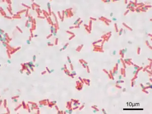
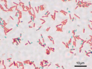
Bacillus subtilis Spore 300x225 1

This article illustrates information on “spore forming bacteria examples” with all different types of bacterial species information in detail.

The word spore or endospore means it is one kind of “non-reproductive structure” that is formed during the condition of nutrient deprivation and some unfavorable or poor condition,that helps bacterial survival and growth.

Now take a look at spore forming bacteria example in details

Clostridium spp.

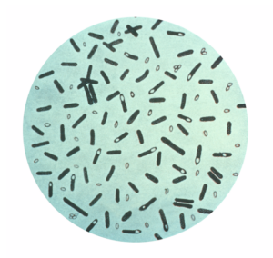
These bacteria belong to the species of the Clostridiaceae family. It is a most common spore forming bacteria examples.It is obligate anaerobes, rod-shaped and gram-negative bacteria which able to form endospores.The endospores are mostly in a bottle shape. The most common species of this bacteria are also able to form this endospores-like structure. The species are Clostridium botulinum,C.tetani and C.perfringes.

Clostridium botulinum

It is obligate anaerobes, rod-shaped and gram-negative bacteria which able to form endospores. These bacteria are mostly found in freshwater, soil, or naturally found in the environment. The endospores of this bacterial species are oval and very resistant. The vegetative cell is produced and also multiplies during the germination of endospores under favorable conditions and environments. It can grow and produce some toxin named botulinum neurotoxin that acts as a harmful agent.

Clostridium tetani

These bacteria are anaerobic, gram-positive bacilli, and endospore-forming bacteria. The species of this bacteria usually a rod-shaped but after the formation of the endospore, the shape is a change to drumstick and wider than the other species like bacillus. They are mostly found in dust, soil, and everywhere in the environment.

Clostridium perfringens

These bacteria are anaerobic, gram-positive, and endospore-forming bacteria. It is a type A strain of clostridium. They can able to survive in harsh and extreme adverse conditions for a longer period because they are more resistant to cold and heat. The germination time of endospore is very fast and short compared to other organisms and it’s around 6.3minuts.

Bacillus spp.

These bacteria belong to the species of the Bacillaceae family. It is a most common spore forming bacteria examples.It is a gram-positive, rod-shaped, and protective endospore-forming bacteria. Different species of this bacillus form endospores are Bacillus thuringiensis, Bacillus cereus and Bacillus anthracis.

Bacillus subtilis Spore
spore forming bacteria examples
Image from Wikipedia

Bacillus thuringiensis

The bacteria is a gram-positive, rod-shaped, and protective endospore-forming bacteria. The vegetative cell contains endospore with released some toxin crystals which act as a delta-endotoxin or also known as crystals of insecticidal protein toxin which have a bipyramidal shape.

Bacillus cereus

The bacteria is a motile, gram-positive, usually aerobic or facultatively anaerobic, and resistant endospore-forming bacteria. most of them form spores in the presence of oxygen. The endospores of this bacteria produce different kinds of enterotoxins like heat-stable emetic cereulide toxin and some tissue destructive enzymes.

Bacillus anthracis

The bacteria is a rod-shaped, gram-positive, aerobic spore-forming bacteria that form spores that are oval-shaped. During the non-reproductive cell have a deficiency of certain nutrients, it starts to form the spore.  The spore of this bacteria can able to survive in very harsh conditions as well. The spores of this bacteria act as infectious particles of toxin named anthrax.

Sporolactobacillus spp.

These bacteria belong to the species of the Sprolactobacillaceae family. The bacteria are gram-positive, rod-shaped, anaerobic, and form resistant and dormant forms of endospores. The most common species of this bacteria include, Sporolactobacillus dextrus, Sporolactobacillus terrae, Sporolactobacillus laevis, Sporolactobacillus vineae , Sporolactobacillus inulinus

Sporosarcina spp.

These bacteria belong to the species of the Caryophanaceae family. The bacteria are gram-positive, heterotrophic, and rod-shaped and some of the species are aerobic or facultative aerobic. They are able to form endospores. The most common species of this bacteria are Sporosarcina aquamarine and Sporosarcina halophila, Sporosarcina ureae. These all species of bacteria belong to the species of the Planococcaceae family. It is a gram-positive, motile, aerobic, and spore-forming bacteria.

Thermolithobacteria

These bacteria belong to the Thermolithobacteraceae family. It is a rod-shaped gram program. The most common species of this bacteria are Thermolithobacter ferrireducens and Thermolithobacter carboxydivorans.

Thermoactinomyces vulgaris

These bacteria belong to the Thermoactinomycetaceae family.It is a most common spore forming bacteria examples.It is a gram-positive bacteria. It can also able  to form endospores where In a natural habitat with extreme longevity, it can form viable endospores.

Do Spore-Forming Bacteria Have Different Dietary Requirements Than Other Bacteria?

Spore-forming bacteria’s nutritional habits differ from other bacteria. While some bacteria can adapt to various diets, spore-forming bacteria have specific dietary requirements. These bacteria have a unique ability to form resistant spores that allow them to survive harsh conditions. Understanding their nutritional needs is essential in controlling their growth and preventing harmful effects.

Does Glycolysis Occur in Spore-Forming Bacteria?

Spore-forming bacteria and glycolysis facts to know: Glycolysis is a universal metabolic pathway that occurs in most organisms, including bacteria. However, not all bacteria follow the same energy-generating pathway. While glycolysis is a common process, some spore-forming bacteria may have modified or alternative metabolic strategies. Understanding these differences provides valuable insights into the diverse biochemical adaptations of bacteria.

Sporohalobacter

These bacteria belong to the Haloanaerobiaceae family. It is an anaerobic and spore-forming bacteria. It can able to live and grow in hypersaline environments. The most common species of this bacteria that can form endospores are Sporohalobacter salinus  and Sporohalobacter morettii.

Oxobacter

These bacteria belong to the Clostridiaceae family. It is a rod-shaped, obligatory anaerobic, and endospore-forming microorganism. They can able to do catabolize activity to convert private into acetic acid and carbon dioxide. The most common species of this bacteria is Oxobacter pfennig.

Also Read:

19+ Soil Bacteria Examples:Detailed Facts Around It

300px ZBDYF00Z 150x150 1

This article illustrate all around information about “soil bacteria examples” in detail with some facts.

Some bacteria can able to make a symbiotic relationship with plants for the transfer and breakdown of nutrients from the soil to the plants, for the protection of plants among other pathogenic attacks or harmful microbes refer to as a soil bacteria.

Now take look on soil bacteria example in a brief

Pseudomonas fluorescens

These bacteria belong to the family Pseudomonadaceae.It is a most common soil bacteria examples. It is a rod-shaped, obligate anaerobe and gram-negative soil bacteria usually found in water and soil. It has an antimicrobial mechanism. The most important role of this bacteria is to act as plant growth-promoting bacteria and plant health and also as a biocontrol that helps to suppress infection on the seed and root of the plant by fungus and pathogenic microorganisms.

Rhizobium spp.

These bacteria belong to the family Rhizobiaceae. It is a most common soil bacteria examples.It is a gram-negative soil bacteria. They help in promoting the growth and development of plants and also improve the fertility of the soil.

The most important species of this Rhizobium bacteria include,

The main role of this bacteria is to fix the atmospheric nitrogen in association with the plant host. The bacteria make symbiotic relationships with the root nodules of leguminous plants. The bacteria help to fix the nitrogen,do nitrification and Dinitrification process and convert into ammonia and then convert it into a useful organic compound that is helpful for the fertility of the soil and the growth of the plants.

300px ZBDYF00Z
soil bacteria examples
Image from Microbewiki

Flavobacterium

These bacteria belong to the family Flavobacteriaceae. It is a motile and nonmotile, rod-shaped and free-living, gram-negative and most common soil bacteria examples,mostly found in water and soil. The main role of this bacteria is it can act as a decomposer. It can convert complex compounds like pectin and other polysaccharides into simple forms.

Azotobacter spp.

These bacteria belong to the family Pseudomonadaceae. It is an aerobic, free-living, spherical, or oval-shaped, and gram-negative soil bacteria. Most of these soil bacteria act as an enhancer for the plant yield stability and crop nutrition It has a very important role in nitrogen fixation, and fertility of the soil by increasing the amount of nitrogen.it can also synthesize some active biological substances such as phytohormone and act as an Azotobacterium nitrogen biofertilizer.

Azotobacter cells
soil bacteria examples
Image from Wikipedia

Bacillus subtilis

These bacteria belong to the family bacilliaceae. It is a rod-shaped, obligate aerobe and gram-positive soil bacteria. The most important role of this bacteria is to activate some mechanisms for the plant defense to reduce harmful pathogenic bacteria which infect the plants and make the plant resistant to infection and enhance crop immunity by secreting active substances.

Bacillus megaterium

These bacteria belong to the family Bacilliaceae.It is a most common soil bacteria examples.It is a spore-forming, rod-shaped, and gram-positive soil bacteria. The most important role of these bacteria is to act as plant growth-promoting bacteria and as a bio-organic fertilizer for plant improvement and pathogen inhibitor. They produce some potassium fixing and phosphate fixing fertilizer for solubilization of potassium and phosphate.

800px Bacillus megaterium DSM 90 cells
soil bacteria examples
Image from Wikipedia

Agrobacterium spp.

These bacteria belong to the family Rhizobiaceae. It is a gram-negative soil bacteria. It can be found in water, soil, and most of the surface of the root. The most common species of this bacteria is Agrobacterium tumefaciens. They are mostly used in modern biotechnology applications for the horizontal gene transfer for the production of transgenic plants.

Klebsiella spp.

These bacteria belong to the family Enterobacteriaceae. It is a rod-shaped, facultatively anaerobic, gram-negative bacteria mostly found in water, soil, or on plants. The most common species of this bacteria klebsiella varicella can able to create a rhizosphere microenvironment in alkali-saline conditions for the improvement of the soil environment and to promote plant growth. It can also act as a biofertilizer and nitrogen fixation.

Arthrobacter

These bacteria belong to the Micrococcaceae family.it is an obligate, aerobic gram-positive soil bacteria. The most common role of this bacteria is to reduce the number of harmful chemicals and pesticides that are present in soil and improve the fertility of the soil and promote the ggrowthof the plgrowth ofost common species of this bacteria named Arthrobacter agilis, A.albus, A.cumminsii, A.globiformis, and many more. one of the species name Arthrobacter agilis acts as bioremediation in the agricultural industry.

Frequently asked questions:

What are bacteria in the soil?

Some bacteria can act as a shield and also help to plant with different processes like nutrients and recycling of nitrogen, sulfur, carbon, phosphorus, mineral supply, Nitrogen fixation, etc. these bacteria can able to grow and adapt themselves in the different microenvironments and can able survive in changing soil conditions.

Such types of soil bacteria can binding soil particles together by their secretions and can form some micro-aggregates.For the improvement of soil structure and to keep the soil productive and healthy, for water-holding capacity, and water infiltration, these micro-aggregates act as a building block that only forms through the help of these different types and species of soil bacteria.

What are the most common bacteria in the soil?

The most common region of soil is known as a Rhizosphere where we can isolate the different bacteria. There are most common bacteria in the soil are Agrobacterium, Micrococcus, Flavobacterium, Pseudomonas, Bacillus, Clostridium, Mycobacterium, Arthrobacter, Corynebacterium, and many more.

Based on energy consumption and carbon utilization, some Phototrophs, Chemoorganotrophs, Chemolithotrophs, Autotrophs and Heterotrophs, and also some other metabolic active microorganisms are found.

How to make soil bacteria?

To make good bacteria within the soil, it is important to keep soil healthy with a good amount of extra composite and useful nutrients with the availability of water so that the bacteria can able to do their work for the growth of plants and make the soil more fertile.

The soil microbes are naturally present in the soil for the different activities of growth of the plant, nutrient recycling, and fixation of carbon, and nitrogen.

How do measure bacteria in soil?

Most of the soil contains around 100 million to 1 Billion bacteria. There are different methods are available to measure bacteria in the soil. The most common methods which act as an indicator are Fumigation-extraction, substrate-induced respiration, and fumigation-incubation.

For the testing of microbial activity in the soil, the fluorescein diacetate test is the modern method by which it can measure the bacteria via the presence of some secretions like enzymes bacteria and some other activity of microorganisms present in the soil. For the identification of soil microbes, there are other simple techniques are used by their ecological characteristics, physiology, molecular characteristics, genetic characteristics, and morphology.

How to get bacteria from the soil?

There are different microbial techniques are used to easily get bacteria from the soil. To isolate bacteria, it is required to dilute the sample of soil and need to do some streaking and spread plate, and pour plate method to isolate the different colonies of bacteria separately.

The spread plate and serial dilution are the best starting points to get bacteria from the soil. and Another most common method generally use to get bacteria is the simple centrifugation-blending procedure.


Also Read:

19+ Commensal Bacteria Examples: Detailed Explanations And Images

1024px Streptococci 300x185 1

These articles illustrate all-around information on the “commensal bacteria example”in detail

Some bacteria can produce some signals to inhibit the growth of pathogens or they can able directly act on the host’s immune system to prevent colonization and invasion by pathogens by inducing protective responses termed commensal bacteria.

Now take a look at this all commensal bacteria example in detail.

Staphylococcus salivarius

These bacteria belong to the species of the Streptococcaceae family.It is the most common commensal bacteria examples. It is facultatively anaerobic, gram-positive, and spherical-shaped bacteria. It acts as a regulator of host inflammatory response and may contribute to the establishment of immune homeostasis.

These bacteria also act as one of the most common and first colonizers of the gut and oral cavity of humans after birth. It is usually considered harmless. A certain strain of this bacteria has various benefits which act as probiotics, one living organism give health benefit to the host.

Streptococcus spp.

These bacteria belong to the species Streptococcaceae family. It is a gram-positive bacteria. It can induce protection against several respiratory pathogens. It acts as an oral commensal such as streptococcus oralis, can induce protection against some host inflammatory responses, and also act as a colonizer of the oral cavity and a part of the normal flora for the human health.

1024px Streptococci
commensal bacteria examples
Image from Wikipedia

Corynebacterium spp.

These bacteria belong to the species of the Corynebacteriaceae family. It is the most common commensal bacteria examples.The bacteria are mostly aerobic, bacilli mean having rod-shaped, and some are also club-shaped and gram-positive bacteria. It can provide an advantage for the most beneficial because it can directly attack the infectious pathogen and enhance some immune responses It has a probiotic potential. it aslo have an antimicrobial and antibiofilm potential.

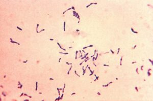
800px Corynebacterium diphtheriae Gram stain
commensal bacteria examples
Image from Wikipedia

Staphylococcus epidermidis

These bacteria belong to the species of the Streptococcaceae family and are permanent members of the human microbiota. It is a gram-positive bacteria. The bacteria can able to produce some proteins and provide direct protection against infections and pathogens via working with endogenous host antimicrobial peptides. It can also diminish Inflammation and also give benefit to the skin because they act as a skin microbiome to promote host defense.

Bifidobacterium spp.

These bacteria belong to the species of the Bifidobacteriaceae family.It is the most common commensal bacteria examples It is a gram-positive bacteria. it is also nonmotile and acts as a branched anaerobic. The bacteria act as the most common gastrointestinal microbiota and give benefits as probiotics.

There is a strain of this bacteria including B.bifidum, B.animalis, B.commune, B.breve, and many more that have several functions as a human microbiota. It can act as inhibition of papathogensegulate the intestinal microbial homeostasis and also modulate the systematic and local immune responses.

Faecalibacterium spp.

These bacteria belong to the species of the Oscillospiraceae family. It is a rod-shaped, mesophilic, gram-positive, and anaerobic bacteria. It is one of the most important and abundant commensal bacteria of human gut microbiota. some strain of this bacteria produces some protein which gives some anti-inflammatory effects. The most common species of this bacteria is Facecalibacterium prausnitzii.

Lactobacillus spp.

These bacteria belong to the species of the Lactobacillaceae family. It is the most common commensal bacteria examples. The bacteria have rod-shaped, aerotolerant anaerobes, microaerophilic, non-spore-forming, and gram-positive bacteria.

These bacteria have a very significant role in animal and human microbiota. It can protect against potential invasion by pathogens because these bacteria exhibit a mutualistic relationship with the human body. It can also provide a source of nutrients. The species include L.brevis, L.acidophilus, L.plantarum, L.casei, L.sanfranciscensis, and many more which produce lactic acid as a primary byproduct.

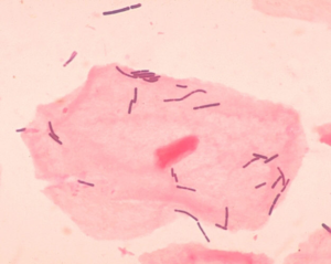
Lactobacillus sp 01
commensal bacteria examples
Image from Wikipedia

Actinobacteria

These bacteria are members of Actinomycetota. It acts as an important member of the normal microbiota and most common commensal bacteria examples It is a gram-positive bacteria that is distributed in the terrestrial ecosystems as well as aquatic. It can also act as a decomposer of organic substances. It can also produce a variety of secondary metabolites and can produce different antibiotics for human health. The actinobacteria also act as immunomodifires which enhance the immune responses.

Clostridium spp.

These bacteria belong to the species of the clostridiaceae family.it is a gram-positive, rod-shaped, and spore-forming anaerobes bacteria. They can act as probiotics to directly interact with the gut or are strongly involved in the maintenance of overall gut function, intestine, and the immune system. It can modulate metabolic and immune processes and also interact with other resident microbial populations to provide essential and specific functions.

Clostridium botulinum 01
commensal bacteria examples
Image from Wikipedia

Ruminococcus spp.

These bacteria belong to the species of the Oscillospiraceae family. These bacteria are gram-positive gut microbes and usually anaerobic and important commensal bacteria examples It can provide a beneficial effect on human health, especially to gut microbiota. It acts a provide strong immune responses toward infection and pathogenicity.it can help and play a major role in digesting complex. high fiber and resistant starch.

Ruminococcus photo
commensal bacteria examples
Image from Microbewiki

Bacteroids fragilis

These bacteria belong to the species of the Bacteroidaceae family.It is the most common commensal bacteria examples. It is an anaerobic bacteria. The bacteria are rod-shaped, it is a gram-negative bacteria. The most important role of these bacteria as a part of normal microbiota and to prevent inflammation in the part of the intestine and also act as an antibiotic resistance.

Streptococcus pyrogens

These bacteria belong to the species of the streptococcaceae family. They are aerotolerant, non-motile, and gram-positive bacteria. It is a part of the skin microbiota in humans. but they are pathogenic as well. They are also known as a “microbiome” because they are a part of normal microflora as well.

Streptococcus pyogenes
commensal bacteria examples
Image from Wikipedia

Lactobacillus rhamosus

These bacteria belong to the species of the Lactobacillaceae family. It is the most common commensal bacteria examples.The bacteria are aerotolerant anaerobes,non-spore-forming, gram-positive bacteria, and microaerophilic. It has a probiotic potential which is generally found in the part of the intestine that acts as a common microflora. They can survive in both conditions either alkaline or acidic. It also has antimicrobial properties and the ability to reduce the harmful and unwanted growth of bacteria.

Neisseria meningitides

These bacteria belong to the species of the Neisseriaceae family. the bacteria have round-shaped,  nonmotile, and gram-negative. without any infection, they can act as a colonizer and act like normal flora. It is the most common commensal bacteria examples.The species of this bacteria is harmless to humans.It can act as a phagocytotic inhibitor and antibody via resistance via the part of their capsule.

Also Read:

Are Bacteria Microorganisms? A Comprehensive Guide

are bacteria microorganisms

Bacteria are microscopic, single-celled organisms that are ubiquitous in nature and play a crucial role in various ecological systems. They are quantifiable and measurable using various techniques, which can provide both qualitative and quantitative information about their structure, function, and behavior.

Understanding Bacteria as Microorganisms

Bacteria are classified as microorganisms due to their microscopic size, typically ranging from 0.2 to 10 micrometers (μm) in diameter. They are prokaryotic, meaning they lack a true nucleus and membrane-bound organelles like eukaryotic cells. Bacteria are found in almost every habitat on Earth, from the deepest ocean trenches to the highest mountain peaks, and they are essential for the functioning of many ecosystems.

Characteristics of Bacterial Cells

Bacterial cells have a unique structure that sets them apart from other microorganisms. They possess a cell wall, which provides structural support and protection, and a cell membrane, which regulates the flow of nutrients and waste in and out of the cell. Bacteria also have a circular DNA molecule, called a chromosome, which contains their genetic information.

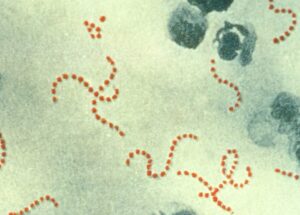
Bacteria can be classified based on their shape, which can be spherical (cocci), rod-shaped (bacilli), or spiral (spirilla). They can also be arranged in different patterns, such as single cells, pairs, chains, or clusters. These characteristics are important in the identification and categorization of different bacterial species.

Quantifying and Measuring Bacteria

are bacteria microorganisms

Bacteria can be quantified and measured using various techniques, each with its own advantages and limitations. Here are some of the most common methods:

Standard Plate Count Method

The standard plate count method is one of the most widely used techniques for quantifying bacteria. It involves diluting a sample with sterile saline or phosphate buffer diluent until the bacteria are dilute enough to count accurately. The final plates in the series should have between 30 and 300 colonies, and the number of colonies should give the number of bacteria that can grow under the incubation conditions employed. This method is an indirect measurement of cell density and reveals information related only to live bacteria.

Spectrophotometric (Turbidimetric) Analysis

Another method for quantifying bacteria is spectrophotometric (turbidimetric) analysis, which is based on turbidity and indirectly measures all bacteria (cell biomass), dead and alive. This method is faster than the standard plate count but is limited because sensitivity is restricted to bacterial suspensions of 10^7^ cells or greater.

Microscopic Observation

Microscopic observation of bacteria can reveal a wealth of qualitative information, such as their shape, size, and arrangement. Bacteria can be applied to a slide as a so-called smear, which is then allowed to dry on the slide. The dried bacteria can be stained to reveal whether they retain the primary stain in the Gram stain protocol (Gram positive) or whether that stain is washed out of the bacteria and a secondary stain retained (Gram negative). Examination of such smears will also reveal the shape, size, and arrangement (singly, in pairs, in chains, in clusters) of the bacteria, which are important in categorizing bacteria.

Quantitative PCR (qPCR)

Quantitative PCR (qPCR) is another technique that can be used to determine the bacterial load and estimate the absolute taxon abundance from NGS data. This method is a simple and cost-effective alternative to flow-cytometry, which is another technique that can be used to determine the total bacterial load of stool samples.

Other Techniques

In addition to these methods, there are many other quantitative and qualitative techniques that can be used to analyze bacteria, such as immunoelectrophoresis, immunoelectron microscopy, biochemical dissection of metabolic pathways, the molecular construction of cell walls and other components of microorganisms, and mutational analysis.

Importance of Quantifying and Measuring Bacteria

Quantifying and measuring bacteria is crucial for understanding their role in various ecological systems and for developing strategies to control their growth and behavior. Bacteria are involved in a wide range of processes, from nutrient cycling and decomposition to the production of biofuels and the development of new antibiotics.

By understanding the abundance, distribution, and activity of bacteria in different environments, researchers can gain insights into the complex interactions between microorganisms and their surrounding ecosystems. This information can be used to develop more effective strategies for managing and manipulating bacterial populations, such as in the treatment of infectious diseases, the remediation of contaminated environments, and the optimization of industrial processes.

Conclusion

Bacteria are microorganisms that are ubiquitous in nature and play a crucial role in various ecological systems. They can be quantified and measured using a variety of techniques, each with its own advantages and limitations. Understanding the structure, function, and behavior of bacteria is essential for developing effective strategies to control their growth and behavior, and for harnessing their potential in various applications.

References:
– Qualitative and Quantitative Analysis in Microbiology. (n.d.). Retrieved from https://www.encyclopedia.com/science/encyclopedias-almanacs-transcripts-and-maps/qualitative-and-quantitative-analysis-microbiology
– How to Count Our Microbes? The Effect of Different Quantitative Microbiome Profiling Approaches. (2020, August 7). Retrieved from https://www.ncbi.nlm.nih.gov/pmc/articles/PMC7426659/
– Bacterial Numbers – Biology LibreTexts. (2021, August 1). Retrieved from https://bio.libretexts.org/Learning_Objects/Laboratory_Experiments/Microbiology_Labs/Microbiology_Labs_I/11:_Bacterial_Numbers
– Madigan, M. T., Martinko, J. M., Bender, K. S., Buckley, D. H., & Stahl, D. A. (2015). Brock Biology of Microorganisms (14th ed.). Pearson.
– Tortora, G. J., Funke, B. R., & Case, C. L. (2018). Microbiology: An Introduction (13th ed.). Pearson.

Do Bacteria Have a Nucleus? A Comprehensive Guide

do bacteria have a nucleus

Bacteria, the microscopic single-celled organisms, are known for their unique cellular structure and organization. One of the fundamental differences between bacteria and eukaryotic cells is the presence or absence of a nucleus. In this comprehensive guide, we will delve into the intricacies of bacterial cell structure, explore the reasons behind the absence of a nucleus, and understand the implications of this crucial difference.

The Absence of a Nucleus in Bacteria

Bacteria are prokaryotic organisms, meaning they lack a membrane-bound nucleus. Instead, their genetic material, in the form of a circular DNA molecule, is located in the cytoplasm of the cell. This arrangement is known as the “nucleoid” and serves as the central control center for the bacterial cell.

The Nucleoid: Bacteria’s Genetic Hub

The nucleoid in bacteria is a highly organized structure that serves as the repository for the cell’s genetic information. Unlike the membrane-bound nucleus in eukaryotic cells, the bacterial nucleoid is directly exposed to the cytoplasm, allowing for efficient access to the genetic material.

  • The nucleoid is typically a single, circular DNA molecule that can range in size from 0.58 million base pairs in Mycoplasma genitalium to 12.2 million base pairs in Sorangium cellusum.
  • The DNA in the nucleoid is organized in a compact, supercoiled structure, which is maintained by various DNA-binding proteins and enzymes.
  • The physical location of the nucleoid within the bacterial cell is often connected to the sites of RNA and protein synthesis, facilitating the efficient flow of genetic information.

The Advantages of a Nucleoid-Based Genome

The absence of a membrane-bound nucleus in bacteria offers several advantages that contribute to their success as diverse and adaptable organisms:

  1. Rapid Replication and Division: Without the need to traverse a nuclear membrane, the bacterial genome can be replicated and segregated more efficiently during cell division, allowing for faster growth and proliferation.

  2. Direct Access to Genetic Information: The proximity of the nucleoid to the sites of RNA and protein synthesis enables a more direct and immediate response to environmental cues and changes, enhancing the bacteria’s ability to adapt and thrive.

  3. Energy Conservation: The lack of a nuclear membrane and associated organelles reduces the energy requirements for bacterial cells, allowing them to allocate more resources towards other essential cellular processes.

  4. Simplified Cellular Architecture: The absence of a nucleus and other membrane-bound organelles simplifies the overall cellular structure of bacteria, contributing to their smaller size and reduced complexity compared to eukaryotic cells.

The Cytoskeleton and Spatial Organization in Bacteria

do bacteria have a nucleus

While bacteria lack a dynamic cytoskeleton like that found in eukaryotic cells, they do possess a variety of cytoskeletal structures that play crucial roles in their cellular organization and function.

Bacterial Cytoskeletal Polymers

Bacteria have several types of cytoskeletal polymers that contribute to their structural integrity and organization:

  1. Actin-like Proteins: Bacteria possess actin-like proteins, such as MreB and FtsA, which form filamentous structures involved in cell shape maintenance, chromosome segregation, and cell division.

  2. Tubulin-like Proteins: Bacterial cells contain tubulin-like proteins, known as FtsZ, which assemble into a ring-like structure at the site of cell division, guiding the process of cytokinesis.

  3. Intermediate Filament-like Proteins: Some bacteria, such as Caulobacter crescentus, have intermediate filament-like proteins that provide structural support and help maintain the cell’s shape.

Spatial Organization in Bacterial Cells

While the absence of a nucleus in bacteria limits their capacity for spatial organization compared to eukaryotic cells, they have developed various mechanisms to maintain the proper positioning and segregation of their genetic material and other cellular components:

  1. Chromosome Positioning: The bacterial nucleoid is often positioned in a specific orientation within the cell, with the origin of replication located near the cell center and the termination region near the cell poles.

  2. Chromosome Segregation: During cell division, the replicated bacterial chromosomes are actively segregated to the daughter cells, ensuring the equal distribution of genetic material.

  3. Subcellular Localization of Proteins: Bacteria can localize specific proteins to particular regions of the cell, such as the cell poles or the division site, through the use of cytoskeletal structures and other spatial cues.

  4. Membrane-Associated Complexes: Bacteria can form membrane-associated complexes, such as the divisome and the flagellar motor, which help organize and coordinate various cellular processes.

The Evolutionary Significance of the Absence of a Nucleus in Bacteria

The absence of a nucleus in bacteria is not merely a structural difference but has profound evolutionary implications. This fundamental distinction between prokaryotic and eukaryotic cells has shaped the divergent paths of these two domains of life.

The Emergence of Eukaryotes

The evolution of eukaryotic cells, with their membrane-bound nucleus and complex cytoskeletal structures, is believed to have arisen from the endosymbiotic integration of an alpha-proteobacterium (the precursor of mitochondria) and an archaeal host cell. This event, known as the “endosymbiotic theory,” marked a significant turning point in the diversification of life on Earth.

The Advantages of a Membrane-Bound Nucleus

The presence of a membrane-bound nucleus in eukaryotic cells has conferred several advantages that have contributed to their increased complexity and adaptability:

  1. Spatial Organization: The sequestration of the genetic material within a nucleus allows eukaryotic cells to better organize their cellular components and processes, leading to the development of specialized organelles and more intricate cellular architectures.

  2. Regulation of Gene Expression: The nuclear membrane provides an additional layer of control over gene expression, enabling eukaryotic cells to fine-tune the transcription and translation of their genetic information.

  3. Genome Complexity: The larger and more complex genomes of eukaryotes, which can span millions to billions of base pairs, are better accommodated within the membrane-bound nucleus, facilitating the evolution of multicellular organisms.

  4. Cellular Compartmentalization: The presence of a nucleus and other membrane-bound organelles in eukaryotic cells allows for the spatial separation of various cellular processes, leading to increased efficiency and specialization.

Conclusion

The absence of a nucleus in bacteria is a fundamental difference that sets them apart from eukaryotic cells. This structural distinction has profound implications for the organization, function, and evolution of these two domains of life. By understanding the reasons behind the nucleoid-based genome of bacteria and the advantages conferred by the membrane-bound nucleus in eukaryotes, we gain valuable insights into the diverse strategies that have shaped the remarkable diversity of life on our planet.

References:

  1. Cao, Q.; Huang, W.; Zhang, Z.; Chu, P.; Wei, T.; Zheng, H.; Liu, C. The Quantification of Bacterial Cell Size: Discrepancies Arise from Varied Quantification Methods. Life 2023, 13, 1246.
  2. Potvin-Trottier, L.; Luro, S.; Paulsson, J. Why are bacteria different from eukaryotes? BMC Biology 2013, 11, 119.
  3. AAT Bioquest. Do bacteria cells have a nucleus? https://www.aatbio.com/resources/faq-frequently-asked-questions/Do-bacteria-cells-have-a-nucleus.
  4. Sender, R.; Fuchs, S.; Milo, R. Revised Estimates for the Number of Human and Bacteria Cells in the Body. PLoS Biol. 2016, 14, e1002533.

Do Bacteria Have Genetic Material?

do bacteria have genetic material

Bacteria, the microscopic single-celled organisms, are known to possess genetic material in the form of DNA (deoxyribonucleic acid). This genetic material is essential for their growth, development, and reproduction, as it contains the instructions necessary for these vital processes.

Bacterial Chromosomes: The Genetic Powerhouse

Bacteria typically have a single circular chromosome, although some species may contain two chromosomes or even have linear chromosomes. The amount of DNA in bacterial chromosomes varies significantly, ranging from as little as 580,000 base pairs in some bacteria to as much as 13 million base pairs in others. For instance, the chromosome of the well-known bacterium Escherichia coli (E. coli) contains approximately 4.7 million base pairs, while the chromosome of Myxococcus xanthus contains roughly 9.45 million base pairs. If the E. coli chromosome were to be stretched out to its fullest extent, it would measure approximately 1.2 millimeters in length.

Genetic Information Storage: The DNA Sequence

do bacteria have genetic material

The genetic information of bacteria is stored in the sequence of nitrogenous bases within the DNA molecule. These bases include adenine (A), cytosine (C), guanine (G), and thymine (T). The rules of base pairing in double-stranded DNA molecules dictate that the number of adenine and thymine bases must be equal, and the number of cytosine and guanine bases must also be equal.

The relationship between the number of G-C base pairs and A-T base pairs is an important indicator of evolutionary and adaptive changes within an organism. This ratio, known as the G+C content or molar ratio, can be calculated as the percentage of G+C bases out of the total number of bases (A+T+G+C). The G+C content in prokaryotes (including bacteria) can vary significantly, ranging from around 25% in most Mycoplasma species to approximately 50% in E. coli, and up to nearly 75% in Micrococcus, actinomycetes, and fruiting myxobacteria.

Plasmids: Auxiliary Genetic Elements

In addition to the main chromosome, bacteria can also carry smaller, circular DNA molecules called plasmids. These plasmids can carry auxiliary genetic information and can be replicated and passed on to the bacterial offspring during cell division. Plasmids often encode fitness-enhancing features, such as antibiotic resistance or the ability to degrade specific compounds. However, some bacteria also harbor “cryptic” plasmids that do not confer any obvious advantages.

Genetic Material Transfer: Transformation and Beyond

Bacteria can acquire new genetic material through a process called transformation, where they take up DNA from their environment, often from other bacteria that have shed their genetic material. If the DNA is in the form of a circular plasmid, it can be replicated and passed on to the receiving bacterial cell and its descendants. This process of horizontal gene transfer allows bacteria to acquire new traits and adapt to changing environmental conditions.

In addition to transformation, bacteria can also exchange genetic material through other mechanisms, such as conjugation (direct cell-to-cell transfer of genetic material) and transduction (transfer of genetic material via bacteriophages, or viruses that infect bacteria).

Implications and Significance

The presence of genetic material in bacteria has profound implications for their biology, evolution, and interactions with their environment. The diversity of bacterial genomes, as reflected in the wide range of G+C content, provides insights into the evolutionary adaptations and ecological niches occupied by different bacterial species. Additionally, the ability of bacteria to acquire new genetic material through processes like transformation can contribute to the spread of antibiotic resistance and the emergence of novel pathogenic strains.

Understanding the genetic makeup of bacteria is crucial for various fields, including microbiology, biotechnology, and medicine. It enables researchers to study the genetic basis of bacterial functions, develop targeted antimicrobial strategies, and harness the potential of bacteria for industrial and environmental applications.

In conclusion, bacteria do possess genetic material in the form of DNA, which is stored in a single circular chromosome and sometimes in smaller, circular plasmids. This genetic information is essential for the growth, development, and reproduction of bacteria, and can be transferred between bacterial cells, allowing them to adapt to changing environments and acquire new traits.

References:

  1. Impact of recombination on bacterial evolution – PMC – NCBI
  2. Bacterial Gene Transfer byNatural Genetic Transformation in … – NCBI
  3. Bacteria – Genetic Content, DNA, Prokaryotes – Britannica
  4. Conjugation, transformation & transduction | Bacteria (article)
  5. A highly conserved and globally prevalent cryptic plasmid is … – NCBI

Comprehensive Guide to Measuring and Quantifying Bacterial Pathogens

are bacteria pathogens

Bacterial pathogens are microorganisms that can cause infectious diseases in humans, animals, and plants. Accurately measuring and quantifying these pathogens is crucial for understanding their prevalence, virulence, and potential impact on public health and the environment. This comprehensive guide delves into the various methods used to assess bacterial pathogens, their virulence, and the strategies for monitoring and detecting them in the environment.

Measuring and Quantifying Bacterial Pathogens

Culturability

One of the most common methods for measuring bacterial pathogens is culturability, which involves determining the ability of bacteria to produce a colony on solid media. This technique provides a direct count of viable, culturable bacteria and is widely used in microbiology laboratories. However, it is important to note that some bacteria may enter a viable but nonculturable (VBNC) state, where they are still alive but unable to grow on traditional culture media.

Metabolic Activity

Measuring the metabolic activity of bacterial pathogens can provide insights into their viability and potential for causing infection. Techniques such as ATP bioluminescence, which measures the amount of adenosine triphosphate (ATP) present in bacteria, can be used to assess the metabolic activity of pathogens. ATP is a crucial energy currency in living cells, and its presence indicates the bacteria are metabolically active and potentially infectious.

Membrane Integrity

Assessing the integrity of the bacterial cell membrane is another approach to quantifying pathogens. Techniques like propidium monoazide (PMA) treatment selectively enter and stain bacteria with damaged membranes, allowing for the quantification of viable but nonculturable (VBNC) bacteria. This method is particularly useful for detecting pathogens that may have entered a dormant or stress-induced state, which can be missed by traditional culturing techniques.

Virulence Quantification

The virulence of bacterial pathogens can be quantified using the infectious dose (ID) or lethal dose (LD) on a given population. The ID is the number of bacteria required to infect a certain percentage of a population, while the LD is the number of bacteria required to kill a certain percentage of a population. These values can be used to compare the virulence of different bacterial strains or species and assess their potential impact on human or animal health.

Monitoring and Detecting Bacterial Pathogens in the Environment

are bacteria pathogens

Monitoring and detecting bacterial pathogens in the environment is crucial for public health and environmental protection. The Environmental Protection Agency (EPA) recommends monitoring for microbial pathogens and indicators in watershed projects, especially when combined with microbial source tracking (MST) for pathogen source assessment.

Grab Sampling

Grab sampling is the most common approach for collecting samples to test for fecal indicator bacteria (FIB) or pathogens. The timing of sample collection for FIB may be tied to known or suspected seasonal patterns or to compliance monitoring requirements for dry vs. wet-weather sampling.

Microbial Source Tracking (MST)

Microbial source tracking (MST) is a powerful tool for identifying the sources of bacterial pathogens in the environment. MST techniques, such as genetic fingerprinting or host-specific marker analysis, can help distinguish between human, animal, or environmental sources of pathogens, allowing for more targeted mitigation efforts.

Emerging Technologies

Advances in technology have led to the development of new tools for the detection and quantification of bacterial pathogens. Nano-biosensors, for example, can be used to rapidly and accurately detect the presence of pathogenic bacteria in environmental samples. These innovative approaches can complement traditional culturing and molecular methods, providing a more comprehensive understanding of the prevalence and distribution of bacterial pathogens.

Conclusion

Measuring and quantifying bacterial pathogens is a critical aspect of understanding their impact on public health and the environment. By employing a range of techniques, including culturability, metabolic activity, and membrane integrity assessments, researchers and public health professionals can gain valuable insights into the viability and virulence of these microorganisms. Additionally, the monitoring and detection of bacterial pathogens in the environment, through methods like grab sampling and microbial source tracking, are essential for identifying and mitigating the risks they pose. As technology continues to evolve, new tools and approaches will undoubtedly emerge, further enhancing our ability to measure, quantify, and manage bacterial pathogens effectively.

References:

  1. Trinh, K. T. L., & Lee, N. Y. (2022). Recent Methods for the Viability Assessment of Bacterial Pathogens: Advances, Challenges, and Future Perspectives. Journal of Microbiological Methods, 182, 116554.
  2. Shen, Y., Xu, J., & Zhang, Y. (2021). Nano-biosensors for the detection of pathogenic bacteria. Nano Today, 36, 101082.
  3. Microbiology (OpenStax). (2024). How Pathogens Cause Disease. Retrieved from https://bio.libretexts.org/Bookshelves/Microbiology/Microbiology_%28OpenStax%29/15:_Microbial_Mechanisms_of_Pathogenicity/15.02:_How_Pathogens_Cause_Disease
  4. Monitoring for Microbial Pathogens and Indicators. (2013). EPA. Retrieved from https://www.epa.gov/sites/default/files/2016-05/documents/tech_notes_9_dec2013_pathogens.pdf