Antiparallel Dna Strands: What, Why, Structure, Significance And FAQs

400px AT GC 300x164 1

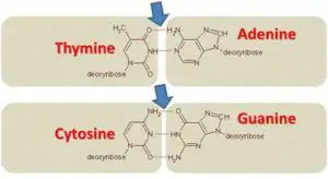
Antiparallel DNA strands refer to the arrangement of the two strands in a DNA molecule. In this arrangement, the two strands run in opposite directions, with one strand running in the 5′ to 3′ direction and the other running in the 3′ to 5′ direction. This antiparallel orientation is crucial for DNA replication and transcription processes. The antiparallel arrangement allows for complementary base pairing between the strands, where adenine (A) pairs with thymine (T) and guanine (G) pairs with cytosine (C). This complementary base pairing is essential for maintaining the genetic code and ensuring accurate DNA replication and protein synthesis.

Key Takeaways

Fact Description
Antiparallel DNA strands Two DNA strands running in opposite directions
Direction of the strands One strand runs in the 5′ to 3′ direction, while the other runs in the 3′ to 5′ direction
Complementary base pairing Adenine (A) pairs with thymine (T), and guanine (G) pairs with cytosine (C)
Importance in DNA processes Crucial for DNA replication and transcription
Maintenance of genetic code Ensures accurate DNA replication and protein synthesis

Understanding the Structure of DNA

DNA, short for deoxyribonucleic acid, is a molecule that carries the genetic instructions for the development, functioning, and reproduction of all living organisms. It is often referred to as the “blueprint of life.” Understanding the structure of DNA is crucial in the field of molecular biology as it provides insights into how genetic information is stored and transmitted.

The Antiparallel Nature of DNA Strands

One of the key features of DNA is its antiparallel nature. This means that the two strands of DNA run in opposite directions. One strand runs in the 5′ to 3′ direction, while the other runs in the 3′ to 5′ direction. This antiparallel arrangement is essential for DNA replication and the accurate transmission of genetic information.

Structural Features of DNA

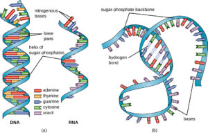
DNA has a double helix structure, resembling a twisted ladder. This structure is formed by two complementary strands of nucleotides held together by hydrogen bonds. The nucleotides consist of a sugar molecule (deoxyribose), a phosphate group, and one of four nitrogenous bases: adenine (A), thymine (T), cytosine (C), and guanine (G).

The base pairing between the nitrogenous bases is specific and follows the rules of complementary base pairing. Adenine always pairs with thymine, forming two hydrogen bonds, while cytosine always pairs with guanine, forming three hydrogen bonds. This base pairing ensures the stability and integrity of the DNA molecule.

The Watson-Crick model, proposed by James Watson and Francis Crick in 1953, provided the first accurate description of the DNA structure. According to this model, the two DNA strands are twisted around each other in a helical fashion, with the nitrogenous bases facing inward. This arrangement allows for efficient base pairing and easy access to the genetic information.

DNA replication is a fundamental process in which the DNA molecule is duplicated. It occurs during cell division and is essential for the transmission of genetic information to daughter cells. The replication process involves the action of various enzymes, including DNA polymerase, DNA helicase, and DNA ligase.

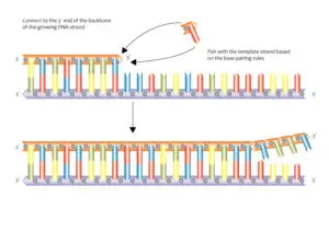
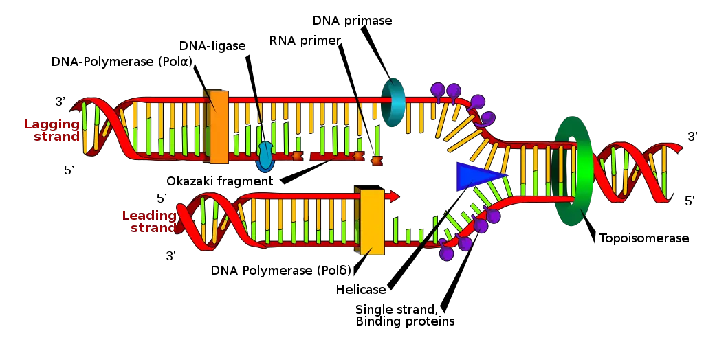
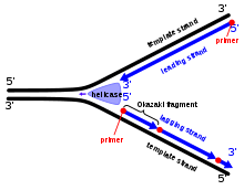
During replication, the DNA strands separate at specific sites called replication forks. DNA helicase unwinds the double helix, creating two single strands. DNA polymerase then adds complementary nucleotides to each single strand, following the 5′ to 3′ direction. The leading strand is synthesized continuously, while the lagging strand is synthesized in short fragments called Okazaki fragments. These fragments are later joined by DNA ligase to form a continuous strand.

DNA topology refers to the three-dimensional arrangement of DNA in space. DNA supercoiling is a common phenomenon in which the DNA molecule becomes twisted upon itself. DNA gyrase is an enzyme that helps relieve the tension caused by supercoiling, ensuring the proper functioning of DNA.

Understanding the structure of DNA and its various features is crucial for unraveling the mysteries of the genetic code. It provides insights into how genetic information is stored, replicated, and transmitted, paving the way for advancements in fields such as medicine, agriculture, and biotechnology.

The Antiparallel Arrangement of DNA Strands

The antiparallel arrangement of DNA strands is a fundamental characteristic of the double helix structure, which is the iconic shape of DNA. This arrangement refers to the orientation of the two strands running in opposite directions. In other words, while one strand runs from the 5′ to 3′ direction, the other strand runs from the 3′ to 5′ direction. This antiparallel arrangement plays a crucial role in DNA replication and other essential cellular processes.

How are DNA Strands Antiparallel?

The antiparallel arrangement of DNA strands is a consequence of the base pairing between nucleotides. Each nucleotide consists of a sugar molecule, a phosphate group, and a nitrogenous base. The two strands of DNA are held together by hydrogen bonds formed between complementary base pairs. Adenine (A) pairs with thymine (T), and guanine (G) pairs with cytosine (C).

In the Watson-Crick model of DNA, the two strands are oriented in opposite directions. One strand has its 5′ end (containing the phosphate group) at the top, while the other strand has its 3′ end at the top. This arrangement allows for the complementary base pairing to occur, ensuring the stability and integrity of the DNA molecule.

Why are DNA Strands Antiparallel?
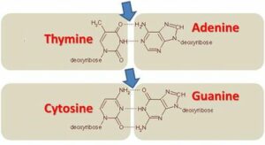
400px AT GC
Wikipedia

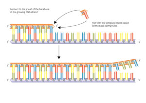
The antiparallel arrangement of DNA strands is essential for DNA replication. During replication, the DNA molecule unwinds at specific sites called replication forks. DNA helicase enzymes separate the two strands by breaking the hydrogen bonds between the base pairs. As the strands separate, DNA polymerase enzymes synthesize new strands by adding complementary nucleotides.

The antiparallel arrangement allows DNA polymerase to synthesize new strands in the 5′ to 3′ direction. The leading strand is synthesized continuously in the 5′ to 3′ direction, following the replication fork. However, the lagging strand is synthesized discontinuously in small fragments called Okazaki fragments. These fragments are later joined together by DNA ligase.

The antiparallel arrangement also plays a role in DNA topology and supercoiling. DNA gyrase enzymes help relieve the tension caused by the unwinding of the DNA molecule during replication. They introduce negative supercoils by temporarily breaking and rejoining the DNA strands.

In summary, the antiparallel arrangement of DNA strands is a crucial aspect of the double helix structure. It facilitates base pairing, DNA replication, and other essential processes in molecular biology. Understanding this arrangement helps us unravel the mysteries of the genetic code and the intricate workings of life itself.

The Significance of Antiparallel DNA Strands

Antiparallel DNA strands play a crucial role in various biological processes, particularly in DNA replication and genetic information transfer. Understanding the significance of antiparallel DNA strands is essential for comprehending the intricate mechanisms that govern molecular biology.

Role in DNA Replication

During DNA replication, the double helix structure of DNA unwinds to expose the two complementary strands. The antiparallel nature of these strands is vital for the accurate replication of genetic material.

DNA replication occurs in a 5′ to 3′ direction, meaning that the new DNA strand is synthesized in the opposite direction to the parental template strand. The antiparallel arrangement of the DNA strands allows for the continuous synthesis of one strand, known as the leading strand, while the other strand, called the lagging strand, is synthesized in short fragments known as Okazaki fragments.

The process of DNA replication involves several key enzymes and proteins. DNA helicase unwinds the double helix, creating a replication fork where the two strands separate. DNA polymerase then adds nucleotides to the growing DNA strand, following the complementary base pairing rules. The antiparallel arrangement ensures that the DNA polymerase can synthesize the new strand in the correct direction.

To join the Okazaki fragments on the lagging strand, DNA ligase plays a crucial role. It seals the gaps between the fragments, resulting in a continuous DNA strand. The antiparallel nature of the DNA strands is essential for the proper functioning of DNA ligase and the seamless completion of DNA replication.

Importance in Genetic Information Transfer

Antiparallel DNA strands also play a vital role in the transfer of genetic information. The complementary base pairing between the two strands allows for the accurate transmission of the genetic code during processes such as transcription and translation.

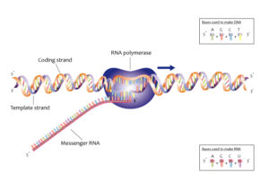
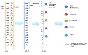
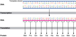
During transcription, the DNA sequence is transcribed into RNA by RNA polymerase. The antiparallel arrangement ensures that the RNA molecule is synthesized in a complementary manner to the DNA template strand. This process allows for the faithful transfer of genetic information from DNA to RNA.

In translation, the mRNA molecule is used as a template to synthesize proteins. The antiparallel nature of DNA strands ensures that the mRNA sequence is complementary to the DNA template strand, allowing for the correct translation of the genetic code into amino acids.

Furthermore, the antiparallel arrangement of DNA strands also plays a role in DNA topology and supercoiling. DNA gyrase, an enzyme involved in DNA topology, helps relieve the torsional strain caused by the winding of DNA strands. The antiparallel arrangement allows DNA gyrase to efficiently manage the supercoiling of DNA, ensuring the proper functioning of the genetic material.

In conclusion, the significance of antiparallel DNA strands cannot be overstated in molecular biology. From DNA replication to genetic information transfer, the antiparallel arrangement ensures the accurate transmission and faithful replication of the genetic code. Understanding the role of antiparallel DNA strands provides valuable insights into the fundamental processes that govern life itself.

Exploring the Antiparallel DNA Structure

The double helix structure of DNA is a fundamental concept in molecular biology. It consists of two complementary strands that run in opposite directions, known as antiparallel strands. In this article, we will delve into the intricacies of the antiparallel DNA structure, including the labeling of antiparallel DNA strands and the potential disadvantages associated with them.

Labeling Antiparallel DNA Strands

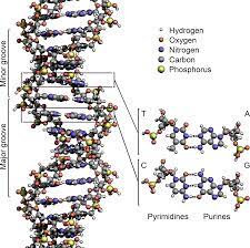
DNA structure and bases
Wikipedia

Labeling antiparallel DNA strands is crucial for understanding the structure and function of DNA. The labeling process involves identifying the orientation of the strands and determining the 5′ to 3′ directionality. The 5′ end of a DNA strand has a phosphate group attached to the 5th carbon of the sugar molecule, while the 3′ end has a hydroxyl group attached to the 3rd carbon. By labeling the strands, scientists can analyze the nucleotide sequence and study the interactions between the strands.

To label antiparallel DNA strands, researchers often use fluorescent dyes or radioactive isotopes. These labels allow for visualization and tracking of the DNA strands during experiments. Additionally, specific techniques such as DNA sequencing rely on accurate labeling to determine the order of nucleotides in a DNA molecule.

Potential Disadvantages of Antiparallel DNA Strands

While the antiparallel DNA structure is essential for DNA replication and other cellular processes, it does present some potential disadvantages. One such disadvantage is the formation of DNA knots and tangles due to the topological properties of the antiparallel strands. DNA topology refers to the different ways in which DNA can be twisted, coiled, or knotted.

During DNA replication, the unwinding of the double helix by DNA helicase creates a replication fork. As the replication fork moves along the DNA molecule, the antiparallel strands can become tangled, leading to the formation of knots. These knots can impede the progress of DNA replication and cause errors in the genetic code.

To overcome these knots and tangles, cells employ enzymes like DNA gyrase, which can introduce temporary breaks in the DNA strands and relieve the tension. Another mechanism involves the synthesis of short DNA fragments, known as Okazaki fragments, on the lagging strand during DNA replication. These fragments are later joined together by DNA ligase.

In conclusion, the antiparallel DNA structure plays a crucial role in maintaining the integrity and stability of the genetic material. By labeling the antiparallel strands and understanding their potential disadvantages, scientists can gain valuable insights into the intricate workings of DNA and its role in molecular biology.

Frequently Asked Questions

Does DNA have Antiparallel Strands?

Yes, DNA does have antiparallel strands. This means that the two strands of DNA run in opposite directions. One strand runs in the 5′ to 3′ direction, while the other runs in the 3′ to 5′ direction. The antiparallel nature of DNA is an essential feature of its structure.

Why do DNA Strands need to be Antiparallel?

The antiparallel arrangement of DNA strands is crucial for several reasons. Firstly, it allows for the formation of the double helix structure. The complementary strands of DNA are held together by hydrogen bonds between the nitrogenous bases. The antiparallel arrangement ensures that the bases can pair up correctly, with adenine (A) always pairing with thymine (T) and guanine (G) always pairing with cytosine (C).

Secondly, the antiparallel nature of DNA is essential for DNA replication. During replication, the DNA strands separate, and each strand serves as a template for the synthesis of a new complementary strand. The 5′ to 3′ directionality of one strand allows for continuous replication, while the 3′ to 5′ directionality of the other strand leads to the formation of short Okazaki fragments on the lagging strand.

What does it mean that DNA is Antiparallel?

When we say that DNA is antiparallel, we mean that the two strands of the DNA molecule run in opposite directions. The 5′ end of one strand is aligned with the 3′ end of the other strand. This arrangement is known as the Watson-Crick model, named after the scientists who proposed the structure of DNA.

The antiparallel nature of DNA is crucial for the functioning of enzymes involved in DNA replication, such as DNA polymerase. These enzymes can only add nucleotides to the 3′ end of a growing DNA strand. Therefore, the antiparallel arrangement ensures that DNA replication can occur smoothly and accurately.

In addition to DNA replication, the antiparallel strands of DNA also play a role in DNA topology and supercoiling. The winding and twisting of DNA strands can result in the formation of knots and tangles. Enzymes like DNA gyrase help relieve these topological stresses by introducing temporary breaks in the DNA strands and allowing them to rotate.

In summary, the antiparallel nature of DNA strands is a fundamental aspect of DNA’s structure and function. It enables base pairing, DNA replication, and proper DNA topology. Understanding the antiparallel arrangement is essential for studying molecular biology and deciphering the genetic code.

Conclusion

In conclusion, antiparallel DNA strands play a crucial role in the structure and function of DNA. The antiparallel arrangement of the two strands allows for efficient replication and transcription processes. The complementary base pairing between the strands ensures the accurate transmission of genetic information during DNA replication and protein synthesis. Additionally, the antiparallel orientation of the strands contributes to the stability and integrity of the DNA molecule. Understanding the concept of antiparallel DNA strands is essential in comprehending the fundamental mechanisms of genetics and molecular biology.

References

In the field of molecular biology, understanding the structure and function of DNA is crucial. The discovery of the double helix structure by Watson and Crick in 1953 revolutionized our understanding of genetics and paved the way for further research in the field. This groundbreaking model explained how DNA’s base pairing and complementary strands allow for accurate DNA replication and the transmission of genetic information.

To comprehend the intricate process of DNA replication, it is essential to grasp the concept of the 5′ to 3′ direction. DNA replication occurs in this specific direction, where new nucleotides are added to the growing DNA strand. The nucleotide sequence is precisely maintained through base pairing, where adenine (A) pairs with thymine (T) and cytosine (C) pairs with guanine (G) through hydrogen bonds.

The Watson-Crick model of DNA replication elucidates the role of various enzymes in this process. DNA polymerase is responsible for synthesizing new DNA strands by adding nucleotides to the existing template strands. This enzyme ensures the accuracy of DNA replication by proofreading and correcting any errors that may occur.

During DNA replication, the double helix unwinds at specific sites called replication forks. DNA helicase plays a crucial role in this unwinding process by breaking the hydrogen bonds between the complementary strands. As the replication fork progresses, the leading strand is synthesized continuously, while the lagging strand is synthesized in short fragments known as Okazaki fragments.

To connect these Okazaki fragments and complete the synthesis of the lagging strand, an enzyme called DNA ligase is required. This enzyme seals the gaps between the fragments, resulting in a continuous DNA strand. Additionally, primer DNA is necessary to initiate DNA replication, providing a starting point for DNA polymerase to begin synthesizing new DNA strands.

DNA topology and supercoiling also play significant roles in DNA replication. DNA gyrase is an enzyme that helps relieve the tension and strain caused by the unwinding of the DNA double helix during replication. It achieves this by introducing temporary breaks in the DNA strands, allowing them to unwind and prevent the formation of knots or tangles.

In conclusion, the process of DNA replication is a complex and highly regulated mechanism that ensures the accurate transmission of genetic information. Understanding the key players, such as DNA polymerase, DNA helicase, DNA ligase, and the role of DNA topology, is essential in unraveling the mysteries of the genetic code and advancing our knowledge in the field of molecular biology.

What are the Detailed Explanations of the 5 Coliform Bacteria Examples?

Coliform bacteria explained in detail refers to the thorough explanations of the five examples of these bacteria. These include Escherichia coli, Enterobacter aerogenes, Klebsiella pneumoniae, Citrobacter freundii, and Enterobacter cloacae. Each bacterium has its own distinct characteristics, habitats, and potential health implications. Understanding these details helps in identifying and managing their presence in water, food, and other environments.

[]

Are antiparallel DNA strands and the pyrimidine nature of adenine connected?

Adenine, one of the four nucleobases found in DNA molecules, has long been recognized as a purine base. However, recent research has shed light on its pyrimidine nature as well. Adenine’s classification as both a purine and a pyrimidine base stems from its ability to form two different tautomeric forms. This discovery has interesting implications, particularly when considering the arrangement of DNA strands. The antiparallel orientation of DNA strands, where one strand runs in the opposite direction to its complementary strand, may interact with adenine’s pyrimidine nature in intriguing ways. To gain a deeper understanding of how these concepts intersect, delve into the article Adenine: Understanding Its Pyrimidine Nature.

Frequently Asked Questions

Q1: Does DNA have antiparallel strands?

Yes, DNA does have antiparallel strands. In the double helix structure of DNA, the two strands run in opposite directions, one from 5′ to 3′ and the other from 3′ to 5′. This is what is referred to as antiparallel.

Q2: What is the significance of antiparallel DNA strands?

The antiparallel nature of DNA strands is crucial for DNA replication. It allows the DNA polymerase to add nucleotides to the 3′ end of the new strand, ensuring accurate and efficient replication of the genetic code.

Q3: What is meant by the term ‘antiparallel structure of DNA strands’?

The term ‘antiparallel structure of DNA strands’ refers to the orientation of the two strands in a DNA molecule. One strand runs in the 5′ to 3′ direction, while the other runs in the 3′ to 5′ direction. This is a key feature of the double helix structure of DNA.

Q4: Why are the two DNA strands antiparallel?

The two DNA strands are antiparallel to facilitate the process of DNA replication. This orientation allows enzymes like DNA polymerase and DNA helicase to function properly, ensuring the accurate copying of the genetic code.

Q5: What does it mean when we say ‘DNA is antiparallel’?

When we say ‘DNA is antiparallel’, we are referring to the orientation of the two strands in a DNA molecule. In the double helix structure, one strand runs from 5′ to 3′ and the other runs from 3′ to 5′. This antiparallel arrangement is crucial for processes like DNA replication and transcription.

Q6: What are the ‘antiparallel strands of DNA’?

The ‘antiparallel strands of DNA’ refer to the two complementary strands in a DNA molecule that run in opposite directions. This means one strand runs from 5′ to 3′ and the other from 3′ to 5′. These strands are held together by hydrogen bonds between complementary base pairs.

Q7: Why do DNA strands need to be antiparallel?

DNA strands need to be antiparallel for the process of DNA replication to occur. The enzymes involved in DNA replication, such as DNA polymerase and DNA helicase, require this antiparallel structure to function correctly.

Q8: How does the antiparallel nature affect DNA replication?

The antiparallel nature of DNA strands affects DNA replication by dictating the direction in which the new strands are synthesized. DNA polymerase can only add nucleotides to the 3′ end of the new strand, resulting in one strand (the leading strand) being synthesized continuously, while the other (the lagging strand) is synthesized in fragments, known as Okazaki fragments.

Q9: What is the role of antiparallel DNA strands in the Watson-Crick model?

In the Watson-Crick model of DNA, the antiparallel strands form the double helix structure. The strands are held together by hydrogen bonds between complementary base pairs, with one strand running from 5′ to 3′ and the other from 3′ to 5′. This model explains how DNA replicates and how the genetic code is preserved.

Q10: Why are DNA strands antiparallel to each other?

DNA strands are antiparallel to each other to ensure accurate DNA replication. This orientation allows the enzymes involved in replication to function properly, ensuring the genetic code is accurately copied and passed on to the next generation.

Also Read:
DNA Transcription Diagram: Detailed Explanations

mm 1 300x169 1

DNA transcription is the method of getting the segment of DNA copied into RNA with having the process segmented.

The very method of getting the DNA codes coped into RNA and also for further carrying to the new model is transcription. The inclusion of DNA transcription diagram consist of the steps involved in the process being initiation, elongation and the termination.

The segments of the DNA that are to be transcribed in the molecules of RNA are able to have proteins encoded within them and are said to make messenger RNA. The rest of the DNA pieces are made to copy RNA and are called the non-coding ones. It is mostly the work of RNA needed here.

The general number of the messenger RNA is more than usually 10 times the actual tissue of the ncRNA and is valid for all cells despite the genomic percentage to be less than two and can also be converted to messenger RNA. The genome of mammals can have themselves converted actively along with the major part being 80.

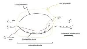
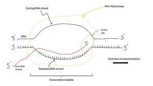
It is not just in DNA but also the RNA where it used up base pairs for the nucleotides as the complementary code. At the time for transcription, a sequence of DNA is read by any of the RNA polymerase that gets to make a complementary, antiparallel RNA strand called the primary transcript. This method does allow involvement of quite a lot of steps.

Transcription is actually one of the basic methods that happen in the genome. It is the method of converting DNA to RNA. It refers to the first phase of central dogma with transcribing bits of RNA to specific areas of RNA. The most places for its act are the one that codes for proteins. There are places where an entire host is seen.

dna transcription diagram
Image Credit-
DNA TranscriptionWikipedia

Steps shown in the DNA transcription Diagram

There are quite a few steps in the process of DNA transcription involved along with involvement of few enzymes as well.

There is many function of the DNA that gets involved Initiation considering it as the first step. It is followed by elongation termination, 5’ Capping, polyadenylation and splicing. There are six of the steps shown in the DNA transcription diagram.

DNA transcription is the method by which any genetic data inside the DNA which is re written for the messenger RNA. There is a messenger RNA that stays inside the nucleus where it acts as a base for this process of DNA. By having the control over the making of messenger RNA inside the nucleus, the cell gets to regulate the rate of genes being expressed.

There is a split in the process of transcription and is done in three vital phases. These are the base steps seen in DNA termination. They are-

Initiation

It is the first step at where the very first nucleotides in the chain of RNA are made to synthesize. Transcription results in an RNA complement.

It is a step of multiple processes that begins when the RNAP holoenzyme links with the template of DNA and ends while the core polymerase escapes at the promoter after the first approximately synthesis on the nine types of nucleotides.

The entire method of DNA transcription is made to get catalysed by an enzyme called the RNA polymerase that links and makes itself mobile with the molecule of DNA till it gets seen by a sequence of promoter. This part of the DNA shows the beginning area of the method and there is possibility of much sequence of promoters here. As a result, transcription has a lower copying fidelity than DNA replication.

The factors of this method are the proteins that actually get to control the rate of this process and then link with the promoter sequence along with the RNA polymerase. After it linking with the promoter chain, the enzyme of RNA polymerase unwinds a segment of DNA that helps in getting the DNA strand exposed. Transcription has some proofreading mechanisms, but they are fewer and less effective than the controls for copying DNA.

DNA transcription is a method in which all genetic data within DNA is transcribed into messenger RNA. There is a messenger RNA remaining inside the nucleus that serves as the basis for this DNA process. By controlling messenger RNA production within the nucleus, cells can regulate the rate of gene expression.

ik 1
Image credit-
InitiationWikipedia

Elongation

There are the presences of two strands of DNA that are seen and used for the process of DNA transcription.

One of the two strands of DNA or the template strand is actually read in the way of 3’ to 5’ path and thus it provides the strand of template of the new made molecule of messenger RNA. The other strand available is called the coding strand.

The reference of both of the strands of DNA is this as for the purpose being the base sequence is said to be identical that is referred to synthesize the messenger RNA except for getting the thiamine base replaced with the uracil. The bases can be added by the three prime way and in 5’ to 3’ path.

The enzyme of RNA polymerase uses the inside ribonulcleotide to make a new messenger RNA strand. It is done by getting the making of the bonds called phosphodiester bond catalyzed in between the places ribonucleotides that are adjacent. It follows the rule of complementary base paring being adenine with uracil, thymine to adenine, cytosine to guanine and vice versa.

Termination

The method of elongation takes place till the enzyme for RNA polymerase encounters a message for it to stop.

During this phase, the process of transcription stops the sequencing and here the entire method comes to a halt and the RNA polymerase enzyme gets to release the RNA template.

The very way of getting the process of RNA transcription terminated is called the termination. It happens only one time and at the very point when the polymerase gets to transcribe the sequence of the DNA called the terminator. The same for terminating is seen at the end of any gene that is used and can work in much number of paths.

If the stretch of DNA is transcribed into an RNA molecule that encodes a protein, the RNA is termed messenger RNA (mRNA); the mRNA, in turn, serves as a template for the protein’s synthesis through translation. Other stretches of DNA may be transcribed into small non-coding RNAs such as microRNA, transfer RNA (tRNA), small nucleolar RNA (snoRNA), small nuclear RNA (snRNA), or enzymatic RNA molecules called ribozymes as well as larger non-coding RNAs such as ribosomal RNA (rRNA), and long non-coding RNA (lncRNA).

During transcription, the DNA sequence is read by any RNA polymerase that receives a complementary antiparallel RNA strand called the primary transcript. This method allows you to use a few steps. Here the ribosome is made to reach the stop codon to get the process to an end. There are three codons to stop- UAA, UGA and UAG. Overall, RNA helps synthesize, regulate, and process proteins; it therefore plays a fundamental role in performing functions within a cell.

mm 1
Image credit-
Transcription bubbleWikipedia

mRNA processing before translation

The messenger RNA is made to transcribe to a point where there is a reference to the pre mRNA.

There need to be a method of processing that shall be valid up to help converting the made up messenger RNA to the mature type mRNA. There are steps involved during the method of processing.

5’ Capping- Capping means getting in adding of a methylated cap of guanine to the end of 5’ messenger RNA. Its vital presence if for getting recognized to the molecule of ribosomes and then protect the immature molecule from getting degraded.

Polyadenylation- It is described as the addition of poly A tail to the end of 3’ of the messenger RNA. This poly A tail has many molecules of adenosine triphosphate. It helps in getting the RNA stabilized which is needed for the RNA and is also unstable than DNA. This mRNA then exits the nucleus, where it acts as the basis for the translation of DNA. By controlling the production of mRNA within the nucleus, the cell regulates the rate of gene expression.

Splicing- It allows the genetic sequence of the only pre mRNA to code many other proteins that serve as a genetic material. The method is based on inside the transcript. It involves getting the introns removed and then joining of the exons by the way of ligation. By the end of transcription, mature mRNA has been made.

This acts as the messaging system to allow translation and protein synthesis to occur. Within the mature mRNA, is the open reading frame (ORF). This region will be translated into protein. It is translated in blocks of three nucleotides, called codons. At the 5’ and 3’ ends, there are also untranslated regions (UTRs). These are not translated during protein synthesis.

Also Read:

Uracil In DNA Replication: Why And Detailed facts

DNA Replication from 3 to 5 prime 13061371753 300x212 1

Uracil is one among the five nitrogenous bases in the nucleic acids- DNA, RNA. (Adenine, Guanine, cytosine, thymine and uracil).

Uracil in DNA replication- Uracil does not have a predominant role in DNA Replication but yes, recent studies have shown that the presence of uracil is noted in DNA replication. Uracil arises sometimes due to incorporation of Deoxyuridine Monophosphate during the process of DNA replication.

Is uracil present in a DNA Strand or Uracil in DNA Replication?

DNA has four nitrogenous bases namely Adenine, Guanine, Cytosine, Thymine.

No, Uracil is not present in a DNA strand, instead Thymine is present in DNA on behalf of uracil.

uracil in dna replication
DNA Synthesis
Image Credits- Wikimedia

Uracil is present only in RNA (Adenine, cytosine, guanine, uracil) strands.

Uracil acid
Uracil Acid
Image Credits- Wikimedia

Read More on Nucleotide Excision Repair and Single Nucleotide Polymorphism | An Important discussion

Does DNA use uracil?

DNA being the genetic material for most of the organisms, do not have uracil as one of nitrogenous bases in them.

Though they are not present as one of the nitrogenous bases (Adenine, Guanine, Cytosine, thymine) in rare cases uracil is formed due to the deamination  of cytosine by hydrolytic deamination process.

This leads to U:G mispairing which leads to mild impact in mutations.

This formation is very rare and will have an impact in evolutionary change.

Read More on Biosynthesis of Purines and Pyrimidines | An important part of cellular metabolism

Why is uracil not present in a DNA strand ?

Uracil is present in RNA and not in DNA.

The reason behind the Uracil being present in RNA and not in DNA is that uracil is not that well resistant towards any photochemical mutations which is the base of stability in the nucleic acids.

Thymine is more resistant to photochemical mutation and thus is very stable and is present in DNA.

The stability is the base for the protection of the genetic message.

Retaining the genetic messages and protecting them without any changes or mutations is the ideal job role of a DNA, this is why DNA has thymine.

Whereas the RNA like mRNA is short lived and also any mistakes or errors due to the instability will not result in any long lasting damages, this is also the reason why uracil is present in RNA and not in DNA.

And the other reason is thymine can undergo oxidation process easily when compared to uracil. Thymine which is usually present only inside the nucleus is protected by the oxygen molecule inside the nucleus.

While, RNA is present outside the nucleus and they are resistant to oxidation process.

Read More on Sequence Of Nitrogenous Bases In DNA: What, Why, Purpose, Detailed Facts

Uracil in DNA polymerase:

DNA Polymerase is a family of enzymes that synthesis the nucleotides of DNA during DNA Replication.

Uracil arises sometimes due to incorporation of Deoxyuridine Monophosphate during the process of DNA replication and due to the deamination of cytosine forming U:G mismatches, which is rare.

Read More on Channel Proteins Passive Transport: How, Why, Types, Detailed Facts

Properties/ Characteristics of Uracil:

  • Uracil is a colorless organic compound that is present in RNA– Ribonucleic Acid.
  • Uracil falls under the pyrimidine class of compounds.
  • Uracil is also involved in the transport of hereditary property.
  • RNA molecule have a five carbon sugar which is called as the ribose sugar, a highly negatively charged phosphate molecule which is the backbone of the entire molecule and the nitrogenous base which can be Adenine, uracil cytosine and guanine.
  • The complementary strand for uracil is Adenine, which is a purine.
  • Example: One strand of RNA is

A U U G C A U A G G G G C C U U U A A C C U G G C A U A G G

The complementary strand will be

U A A C G U A U C C C C G G A A A U U G G A C C G U A U C C

Also Read:

DNA Replication vs Polymerase: Comparative Analysis

jj

The polymerase is used up for getting the process of replication work being an enzyme and also fasting up the process.

The very basic difference for DNA replication vs polymerase is its definition being that replication is the process where the DNA gets itself replicated at cell division and while polymerase is an enzyme helping in getting the chains of the nucleic acids synthesized.

The structure of DNA is of a double helix with two stands coming up coiled together to make a character of double helix. There are nucleotides present on each stands. Nucleotides are referred to have a group of phosphate, a deoxyribose sugar and a nucleobase.

The pairing of the base is done following the complimentary base pairing system. Adenines pairs up with guanine and are the purine bases. The rest of the two being cytosine pairs with thymine in DNA and are the pyrimidine base. They help in forming the backbone for the DNA.

dna replication vs polymerase
Image Credit-
DNAWikipedia

DNA Replication vs Polymerase

DNA replication is the process while polymerase is an enzyme having its use in this process fasting up the reaction.

Just like the rest of the polymerization process in biology, DNA replication begins in to catalyze the one of three enzymes and have its steps coordinated. Polymerase helps in adding of the nucleotide to the DNA stand.

The polymerases are of the family enzymes that are used for all the forms of replication in DNA. It is in general approach the one that can’t have the initiation of the new stands to be synthesized but also can help in extending the existing RNA or DNA strands to be paired inside the template strand.

The ultimate known difference for DNA replication vs polymerase between them stands for what they are replication being a process and the polymerase being an enzyme with having its presence in both RNA and DNA. Each of the DNA strand have nucleotide that are four in its types. The four types of them are adenine, guanine, cytosine and thymine or uracil.

For the process of the cell to get it divided is it needs to first replicate itself. It needs to be ended after its strands with no stop in middle proceeding with absolute completion. One the process of replication is done; there is no repetition of this process within the similar cell cycle.

A DNA primase, which is a specialized DNA-dependent RNA polymerase that is capable of synthesizing a short RNA strand starting from a single-stranded DNA as a template. This RNA oligonucleotide is then transferred to the active site of the DNA polymerase, functioning as a primer for subsequent incorporation of the deoxyribonucleotide triphosphate also termed to be dNTPs.

DNA Replication

The process of allowing the genetic material called to be DNA to have itself copied during cell division is called DNA replication.

The copying of DNA is done to make two molecules of DNA that can be identical. Replication is the process that is vital for while there is a division of cell the new daughter cells being two in number shall have the equal genetic data from parent.

The very motif of the process is to get the exact information of genes passed on to the next generation. This process is based on the knowledge that each of the DNA stand shall help in acting as a template for getting itself duplicated. This process of DNA replication showcases definite points called origins. Because for polymerase can add a nucleotide only onto a preexisting 3′-OH group, it needs a primer to which it can add the first nucleotide.

Origins are the specific areas that help in getting the double helical stricture of DNA unwound. There is little section of the RNA also called as ribonucleic acid which is called a primer. Primer is the area that points to the start for synthesizing DNA of any new stands. It is needed for replication as then it shall help polymerase in adding up the parts of DNA.

After the use of primer is done an enzyme called polymerase starts its work by helping the DNA to replicate itself by paring of the bases to the parent stands. After the process of synthesis is done, the RNA primers are then replaced along with the DNA. DNA replication is a short process by needs quote vitals to work.

If there is a presence of any gap seen between the new fragments of DNA that is synthesized then they shall be sealed up together with the enzymes. The process is quite vital and thus need no mistakes or any sort of mutation. To get it checked, the cell reads the new DNA stands. After it, the cell divides and identical copy is made.

The result of DNA replication is two DNA molecules consisting of one new and one old chain of nucleotides. This is why DNA replication is described as semi-conservative, half of the chain is part of the original DNA molecule, half is brand new. DNA replication requires other enzymes in addition to DNA polymerase, including DNA primase, DNA helicase, DNA ligase, and topoisomerase.

rr
Image credit-
DNA ReplicationWikipedia

The process of replication includes-

  • Unzipping of the helical structure of the DNA molecule
  • This is done by enzyme called helicase that helps in getting the hydrogen bonds broke down by holding the base together.
  • The separating of the stand ceased a shape of Y called replication fork and acts as a template.
  • One of the strands is direction to 3’ to 5’ called the leading stands and the other is away called the lagging stands. Thus both the stands are differently replicated.
  • Then a short part called primer comes to plat helping the lagging part bind acting as the staring part.
  • DNA polymerase the binds the leading and works with it adding the complementary stands.
  • The process of DNA replication is continuous.

Polymerase

The researches take the help of the power that this enzyme holds to copy the molecules in tubes with polymerase chain reaction.

The basic role of this enzyme is to efficiently and by all accuracy help in replicating the genome so that it shall ensure the originality of the genetic data and it’s faithful to transfer via generation. It is useful during the process of DNA replication.

It is useful in gathering the molecules of both DNA and RNA by tallying the template stands and using the method of base paring interaction or also by using the half ladder replication process of RNA. It also can take part in polymerase chain reaction. Taq DNA polymerase is the most common enzyme used for PCR amplification. This enzyme is extremely heat resistant with a half-life of 40 minutes at 95°C. 

This is an enzyme that can be either is independent or dependent of template. The classification of it can be based on its structure or functions. As mentioned in the model of base paring by Watson and Crick, they helps in catalyzing the synthesis of both RNA and DNA after paring via complement base to the parent template. On April 16, 1956, about 60 years ago, Arthur Kornberg and his team of biochemists were the first to isolate and later characterize the enzyme which is now known as DNA polymerase I.

pp
Image credit- PolymeraseWikipedia

It is not just useful in getting the replication of DNA done but also helps in repair and in some times also helps in getting the cell differentiated. It helps in getting the synthesis of polydeoxyribonucleotides from the mono-deoxyribonucleoside triphosphates in DNA.

DNA polymerase is an essential component for PCR as to its key role in synthesizing new DNA strands. Consequently, understanding the characteristics of this enzyme and the subsequent development of advanced DNA polymerases is critical for adapting the power of PCR for a wide range of biological applications

Also Read:

Sequence Of Nitrogenous Bases In DNA:What, Why, Purpose, Detailed Facts

image

DNA is a chemical molecule that has all the genetic information of specific organisms in which they are present. Here arises a question, Explanation on the sequence of nitrogenous bases in DNA. Let’s see in this article.

The sequence of nitrogenous bases in DNA do not follow a specific or particular order but yes, they are COMPLEMENTARY to each other to which the amino acids get coded, that is the nucleotides pair up specifically making the strand complementary to each other.

DNA strands have four nitrogenous bases which are guanine, adenine, thymine and cytosine.

Before heading into the concept of Sequence Of Nitrogenous Bases In DNA, We need to understand the structure of DNA.

The DNA has three major components

  • The Nitrogenous Base– The subunits of DNA which are discussed in detail in this article.
  • The Sugar Molecule- The deoxyribose sugar
  • The Phosphate Group– The highly negative portion and the backbone of the complete structure.
sequence of nitrogenous bases in dna
DNA Strand Structure
Image Credits: Wikimedia

What is the Pairing Sequence of Nitrogenous Bases in DNA?

There are 2 classes of compounds- Purine and Pyrimidine. In DNA, there are about  four nitrogenous bases.

The chemical molecule- Purine in DNA are Guanine – G and Adenine – A. The chemical molecule- Pyrimidine in DNA are Thymine – T and Cytosine – C. The pairing up of these nitrogenous bases in DNA is the purine pairs up with the pyrimidine molecule and Pyrimidine molecule with the Purine molecule.

So Adenine (Purine) pairs up with Thymine (Pyrimidine) and Cytosine (Pyrimidine) pairs up with Guanine (Purine).

This explains the pairing sequence of DNA’s nitrogenous bases.

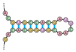
OSC Microbio 10 03 RNAStruct2
Pairing if nitrogenous base in DNA
Image Credits: Wikimedia

Read More on DNA Structure | A detailed insight with all crucial aspects

What is the Main Purpose of the Sequences of Nitrogenous Bases in DNA?

The nitrogenous bases are like the foundation of DNA and RNA which is collectively known as Nucleic acids.

The main purpose of sequences of nitrogenous bases in DNA is to store the genetic information of the organisms in them.

What does the Order of Nitrogenous Bases in DNA Determine?

The order of nitrogenous bases in DNA is complementary to each other.

The order of nitrogenous bases in DNA is arranged in such a way that they code for the protein molecule (that is Amino acids). 

Protein TranscriptionTranslation
Coding Protein
Image Credits: Wikimedia

Example:

One strand of DNA is

A T T G C A T A G G G G C C T T T A A C C T G G  C A T A G G

The complementary strand will be

T A A C G T A T C C C C G G A A A T T G G A C C G T A T C C

229 Nucleotides 01
DNA Structure
Image Credits: Wikimedia

Read More on DNA Replication Steps and Critical FAQs

Why is the sequence of nitrogenous bases in dna important?

Nitrogenous bases are simply known as nitrogen bases, that is the molecules are made up of nitrogen atoms and are a replica of base. 

Proteins are the fundamental unit or the primary molecule of every cell in the living organisms. The significance of the DNA’s nitrogenous bases is only when the right or relevant nitrogenous bases pairs up with the relevant one or the right one, the protein synthesis takes place and the right amino acid is coded.

The nitrogen in these nitrogen bases are the constructive material of the nucleic acids.

When the right nitrogenous bases are paired up and the coding of protein is done that’s when the organism’s protein- cellular mechanism is fulfilled.

Read More on Do Bacteria Have DNA :Why,How And Detailed Insights

How genetic information can be stored in a sequence of nitrogenous bases in DNA?

It looks so fascinating that how come DNA stores the genetic information of specific organisms.

The nitrogenous bases in the DNA stores that genetic information. The minute component in the cell stores so much information. The nitrogenous bases code for specific amino acid molecules which collectively make up protein and the complete living mechanisms take place.

The central dogma: DNA is converted into mRNA (Transcription process) and then transformed to Protein molecule- Amino acids subunits (Translation process).

0324 DNA Translation and Codons
Central Dogma
Image credits: Wikimedia

Here are a few bases and their corresponding amino acids.

  • T T T and T T C – Codes for Phenylalanine Amino Acid Molecule.
  • T T A and T T G- Codes for Leucine Amino Acid Molecules.
  • T C T , T C C , T C A , T C G- Codes for Serine Amino Acid Molecules.
  • T A T and T A C- Codes for Tyrosine amino acid molecule.
  • T G T and T G C- Codes for Cysteine Amino Acid Molecule
  • G G T , G G C , G G A , G G G- Codes for Glycine Amino Acid Molecule.

Also Read:

Is Prokaryotic DNA a double Helix: Why, How and Detailed Insights

dna

The prokaryotes are referred to the organism that has no specific nucleus and no rest number of organelles with inside membranes.

With concern to the response for is prokaryotic DNA a double helix, the reply to it is yes. The DNA of all the prokaryotes that includes the bacteria and the archaea is circular in their structure and is double stranded.

Just not similar to that of the eukaryotes, these are the structures having no specific region or are boundary less and have a double stranded DNA being circular and are smaller than that of the DNA of the eukaryotes in their cell. There is a formation of a loop in the prokaryotes which is the plasmids and is of no specific use in the cell growth.

The molecule of the DNA can be long and get stretched where the DNA in the cell of a human may be of length of 2m. Thus, the cell DNA need to be packed in such a way that it can be fitted in the cell and function inside its boundary and shall not be seen with naked eyes.

There is also a presence of a second type of nucleic acid called the ribonucleic acid. Just like the DNA, RNA is also a polymer of the nucleotides. Each of these is made up of the nitrogenous base with a phosphate group and also a five carbon one called the ribose.

Cell arrangement to prove is prokaryotic DNA a double helix

DNA is referred to as the deoxyribose nucleic acid and is the concerned molecule that works and gets replicated.

The model for the DNA suggests that prokaryotic DNA is a double helix which is double helix was given by Watson and Crick where the DNA is a polymer of the nucleotides. Each of the nucleotides includes the nitrogen bases and is made of two strands.

There are two of the purines which are adenine and guanine, the two of the pyrimidine which are the thymine and cytosine. Along with the composition of nitrogenous bases they also have a phosphate group and a five carbon sugar called the deoxyribose.

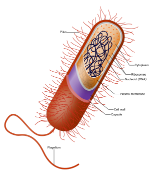
Each of the DNA stand is made up of nucleotide bonds attached together which are covalent in between the phosphate group of the one and the deoxyribose of the next sugar seen. This is the actual backbone for the DNA and here is the base extension point. The prokaryotes have cytoplasm, ribosomes, cell wall, flagella and cell membrane.

The strand having one of the bases does get to bond with the base of the strand that comes second along with the hydrogen bonds. Adenine always gets to bond with thymine then with cytosine and then gets to bond with guanine. Most prokaryotes have circular, single chromosome placed beside nucleoid.

It gets into the part of duplication when the cell is ready to get divided and shall read in order to generate the molecules like that of proteins to let the functions of the cell be carried out smooth. This is the absolute reason for the DNA getting to be packed and let protected in many ways. The genome is made of double stranded DNA ns is single in form of a circle.

is prokaryotic dna a double helix
Image credit- DNAWikipedia

Why is prokaryotic DNA a double helix?

The prokaryotes are seen with a single chromosome which is circular and is double stranded. The DNA of the prokaryotes are free floating and not in the nucleus.

With response to is prokaryotic DNA a double helix, The adenine always gets to bond with thymine and cytosine gets to pair with guanine. This is the cause that makes two of the strands to become spiral around each of them in a shape that forms the double helix.

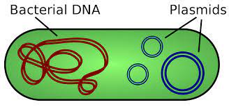
With the reply to is prokaryotic DNA a double helix they have a chromosome that covers the area of the cytoplasm known as the nucleoid and is single being circular. They are also seen to be made up of tiny rings that look like the double stranded chromosomal DNA that are extra and are called plasmids. 

The eukaryotes have the one with being linear packed up in the chromosomes. The helix of DNA is enclosed around the proteins to make the nucleosomes. The coils of proteins and made to coil up and during division the chromosomes are seen to be coiled up more to help in movement.

The chromosomes of the prokaryotes are likely to have two specific areas that shall separate them by reflecting certain degrees of packing, staining, and then getting to determine if the DNA in any of the area is being euchromatin (expressed) or heterochromatin (not expressed). Some of the prokaryotes make loops called plasmids which are not useful for any normal growth of cell.

The DNA of the prokaryotes seems to be circular for the circular DNA did evolve at the first even before the linear eukaryotic DNA. This is also for the prokaryotes were one among the common ancestors that descended with circle polymerase and getting to replicate DNA in circle. Thus, the part of answer for is prokaryotic DNA a double helix or not, its an yes.

Also Read:

Do Prokaryotes Have DNA Replication: Detailed Insights and Facts

dna1

Prokaryotes are the one that lack a nucleus and also the rest of the organelles. They are mostly single celled and are small.

With the question for do prokaryotes have DNA replication, the answer is yes. Inside the prokaryotes there is just a single origination point where replication takes places in two paths and is always at same time. It takes place inside the cytoplasm.

The replication in DNA for the prokaryotes is the process where the organism do duplicate the DNA in a on other copy and is passed on to the daughter cells. It is usually generalized for E.Coli but is also shown by bacteria.

Replication of DNA is however a bi-directional one and generates at the one point for the purpose of replication. It also has there steps that includes the initiation, the elongation and the termination.

What is DNA replication in prokaryotes called and why?

The time when the prokaryotes do DNA replication, there is a theta like structure seen at the site of replication.

The process for DNA replication in the prokaryotes is called as Theta replication. The reason for this is that during the process it makes a resemblance of a Greek letter called theta (θ).

A theta structure is an intermediate structure that is formed during the process for replication of DNA that is circular. There is an unwinding if the helix that is double stranded and makes a loop which is called the replication bubble.

In the theta replication, the DNA which is double stranded unwinds itself at the site of replication that helps makes the stands for nucleotides helping them to serve as the templates which shall be the base for DNA synthesizing.

While the unwinding takes places there is a formation of loop and s called the replication bubble. The process for unwinding may take place in both the ends or in one side of bubble that makes it quite large. The process for DNA replication on both the strands goes equal with unwinding.

The point where there is unwinding, the pace where the strands for nucleotides gets separated from the circular DNA helix that is double stranded is termed as the replication fork.

do prokaryotes have dna replication
Image credit- DNA ReplicationWikipedia

Where does DNA replication occur in prokaryote?

Inside the prokaryotes there is only one point for replication where the process takes place in two separate paths in the similar time. DNA replication takes place before binary fission.

The answer for do prokaryotes have DNA replication is yes and the process of DNA replication for these organism takes place inside the cytoplasm of the cell. The DNA replication is seen inside the nucleus of the cell. 

The process for DNA replication occurs prior to that of cell division which helps in ensuring that both the cells do receive the same copy of the genetic substance from the parent. The division in animals have different time period of completion.

The division in prokaryotes and eukaryotes do have similarities and differences for the size and the molecules being complex. The cells of the prokaryotes are quite in the structure with only little DNA and no organelles or nucleus.

The replication for DNA can also take place in a test tube and thus this process can be called to be self-replication. The mechanism for the process of DNA replication inside a test tube is the same as the way in the eukaryotes and the prokaryotes and mitosis or meiosis in the eukaryotes.

pro1
Image credit- ProkaryoteWikipedia

How do prokaryotes replicate?

The replication in DNA is so that there is a duplicate formation of the genetic material from the parent to the other getting passed to the daughter cells.

But on the other hand the reproduction process of the prokaryotes takes place via the process of cell division which is known as binary fission. It is just like the process of mitosis in the eukaryotes.

This procedure includes copying of the chromosomes an also separating of a single cell into two others. The process for binary fission is asexual mode of reproduction which involves no egg or sperm making.

There is no generation of sperm or egg or any type if mixing of the genetic material from the two organisms. There is an exception for a rare type of mutation or DNA sequence change, where the binary fission makes the daughter cells that are same to the mother cell.

The reproduction in prokaryotes is much faster than compared to the eukaryotes. The speed of division can be mentioned in regards to generation time or the time length from the making of one generation to the birth of the next one.

Well, with regards to do prokaryotes have DNA replication, not can all bacteria be quick in reproduction with some of that are pathogenic as well. Mycobacterium tuberculosis has a generation time for about 12 hours. They are still the fast multipliers in natural conditions and test tub in lab.

How do prokaryotes make DNA?

The reproduction in the prokaryotes is asexual and takes place generally by the binary fission, they have circular chromosome and do not go for mitosis.

The chromosome or the DNA of the prokaryotes is replicated and there are two copies as outcome which is different from each other for the reason being the mobility of the cell membrane to which they are adhered.

Prokaryotes are the organisms that often do lack any of the organelles and also the nucleus. But, they do have DNA which is circular and is stored in the central part of the call which is called the nucleoid that s in turn surrounded by the nuclear membrane.

The prokaryotes have a singular chromosome which is actually single and usually occupies a region of the cytoplasm and is called the nucleoid. They also include of many rigs that are small in size and are usually double stranded with having extra chromosome and are the plasmids.

A phenomenon in the prokaryotes is referred to as supercoiling that helps the organism in reducing the DNA and allows them for more space and so that the DNA can be kept packed easily. This is quite plectonemic as the chromosomes are actually small.

pal
Image credit- PlasmidsWikipedia

What are the characteristics of prokaryotic DNA replication?

The deoxyribonucleic acid and the chromosome in the prokaryotes is circular in shape with being located inside the cytoplasm of the cell.

The replication of the DNA in the prokaryotes is bi directional and generated from the single place of origin for replication. It takes place in the cytoplasm and occurs in the direction of 5’to 3’ direction.

 The stands of the DNA are considered individually and are made in several directions that are to be followed. This gives them a lead and while generating of the lead it also makes a lagging strand.

The replication of DNA is actually a semiconservative process. It means that the each of the DNA stand is a double helix one and works as a template for getting a new one synthesized. This method takes it from one molecule to start with and then ending up for two daughter molecules.

The two new daughter molecules that are formed as a result are also double helix and have one old and one new strand carrying the copy of the genetic material from the parent. There is also formation of replication fork.

Why do prokaryotes only have one origin of replication?

In the prokaryotes, there are there vital types of polymerases that are called DNA pol I, DNA pol II, DNA pol III. These have only one site of replication origin.

The cell of the prokaryotes is much smaller and thus can get away with any process having only one site of origin. Mostly all the prokaryotes do have a single place of replication however the rate for the replication is also very high.

In the prokaryotes, the single place of replication does have many of the adenine and thiamine bases that have the hydrogen bonds on the weaker sides than compared to the guanine and cytosil pairs. It helps in making the DNA strands easier to separate.

There is an enzyme that is called the helicase that helps in unwinding of the DNA by getting the hydrogen bonds to break amongst all the nitrogenous base makings. With concern to this, eukaryotes and prokaryotes that different replication site.

What is unique about prokaryotic DNA replication?

There is only one point of replication of DNA for the prokaryotes which takes place in two directions and is concerned within the cytoplasm.

The uniqueness given by the prokaryotes are concerned with the replication site. The site of replication in length is approx. 245 bases in pairs in length and is quite in rich within Adenine and Thiamine.

The sequence of AT together is seen by some of the proteins that bind to the sites. There is an enzyme known as the helicase that helps in getting the DNA unwind itself with breaking of the hydrogen deals in the nitrogen pairs. The strands help in preventing the rewinding of the double helix.

After the DNA keeps on opening, the Y-shaped structures are called to be the replication forks. They are located in the site of replication and are usually bi directional as they proceed. There is also a single strand that binds the proteins which covers the single strands of DNA.

Another enzyme linked with it is the DNA polymerase II that adds up to the nucleotides taking each at a time on the DNA chain that is growing. The nucleotides that are added need energy, this energy is taken from the nucleotides that have phosphates linked with them and are three in number.

Is prokaryotic DNA replication unidirectional?

There is only one site for the part of replication for the concern in prokaryotes still the replication is rapid.

In regards with the prokaryotes the DNA replication process is nit unidirectional. They have only single site of origin for the purpose of replication and is bidirectional.

Bidirectional replication of the DNA is any mechanism that is ensured to all the eukaryotes and mostly all the prokaryotes cells. The replication that is unidirectional is rare and do is seen to take place only in very limited number of the cells of prokaryotes.

The reason for the DNA replication to be bidirectional is that the enzymes related to unwinding of the DNA called the DNA helicases cause the two of the parent DNA strands to get itself unwinded and then separates from one to the other in both ways at the same site that forms the Y-Shaped replication forks.

How many origins of replication do prokaryotes typically have?

The eukaryotes cells have DNA replication that is needed during the formation of replication forks with the prokaryotes being rapid.

There are three polymerases that has the single site for replication on the specific chromosome which is similar to many prokaryotes. It is approx. of length of 245 base pairs in length and is quite rich in AT pairs.

The cells for the prokaryotes uses up the single variety for the rapid replication propose. There are actually approx. about 350 origins of replication in the entire the genomes.  On the contrary there is an estimation of 40,000 to 80,000 origins that get distributed via the human genome.

The replication in bacteria is regulated at the time of initiation stage. DNA is hydrolyzed into inactive by RIDA which is the regulator inactivation of the DnaA that gets converted to DnaA-ATP by the DARS. The complementary strand for 3’ to 5’ gets synthesized all time as the polymerase can add to nucleotides.

There are the three models that can be suggested for DNA replication. These are dispersive, conservative and semi-conservative. The method that is conservative suggest that the parental DNA and remains together and gets to form the parental DNA along with it together.

Is prokaryotic DNA replication conservative or semiconservative?

The genetic material needs to get itself replicated to get assure the policy for hereditary.

It has already been seen that DNA is replicated by the process which is semi conservative in the WT-4 cells that grow in 34 degrees Celsius or at the 38.5 degree covering the entire logarithmic phase and gets into the stationary phase.

The base system applied for the process of DNA replication is same from the microbes to the eukaryotes, there are a lot of variations that to make up the final product mode. The microbe DNA replication for some of the microbes do have ailments that are single stranded.

For this way, the replication of the genetic product shall be kept in consideration through the complementary strand inside the intermediate step for double stranded in the form of replication. All the form of DNA does need a region for replication to get the strands separated.

A very general and common character for the origins responsible for the separation of strands in the semiconservative replication process is that they are much rich in the A=T. Not so common about it is the prevalence of the G over the C and the T over the A in the prokaryotes which is also the leading strand.

com
Image credit- Complementary strandWikipedia

Why is DNA replication faster in prokaryotes than eukaryotes?

The process of replication is much fast in prokaryotes than the other. Some of the bacteria take 40minies while animals cell take 400 hours.

The DNA polymerase is much rapid in regards to the base that is replicated per second; however it does have only one origin of replication. Eukaryotes have distinct way for replication.

On the other hand there are many site for replication in the eukaryotes, there are many parallel running DNA polymerases that make up the process of DNA replication easy and much faster in the eukaryotes.

The reason for the eukaryotes having many sites for working out the way from DNA replication is that they are much larger in size than the prokaryotes and also there are multiple functions being performed by them which mostly cover mobility.

Is prokaryotic DNA replication semi discontinuous?

The experiments show that DNA replication is semi discontinuous on the lagging strand while leading has to be continuous.

The process for DNA replication is semi discontinuous as for one among the strands that is synthesized on regular basis and on the other hand another reason is for the formation of the Okazaki fragments which is formed discontinuously.

Semi discontinuous process is the double helix of the DNA that unwinds taking the replication on the 3’ to 5’ stand which gets to process itself easy in the 5’ to 3’. Here the leading strand is worked on continuous and the lagging one is taken for being discontinuous.

The process of replicating DNA in the prokaryotes is semi discontinuous. In some of the microbe, both of the stands can be made to copy on the 5’ to 3’ way together with no need for discontinuous process of replication.

Okazaki fragment EN.svg
Image credit- Okazaki fragmentsWikepedia

How do you tell if a DNA sequence is prokaryotic or eukaryotic?

The prokaryotic cell do have only one strands of DNA that is circular while eukaryotes have maay linear type DNA.

The gene expression for the prokaryotes do take place in the cytoplasm for there is no definite nucleus and thus the DNA is free in the cytoplasm while the one in eukaryotes take places in the cytoplasm and nucleus.

The DNA that the prokaryotes have is in size small and is circular which is located in the cytoplasm. They are chromosomally arranged and are situated in the cell’s nucleus. The eukaryotic cell is more dense and complex and has organelle having membranes like the nucleus.

Unlike the DNA that is circular in the prokaryotic cells, it in general has only one site for getting the DNA replicated and in the eukaryotes there is a liners DNA in the eukaryotes cell that has many site for the replacing.

FAQs-

What is Replication fork?

The replication fork is formed inside the DNA within its long helical during the process of DNA replication. It is made up of helicases.

This helps in breaking of the hydrogen bonds that holds the stands of the DNA together inside the helix itself. The structure formed as a outcome has two of the prongs branched with each made up of the DNA that is single strand.

Do eukaryotes also have single replication site?

The chromosomes for the eukaryotes have multiple origin sites while just prokaryotes have only one.

This is so as the eukaryotic chromosome are bigger and having multiple origins saves them time. The eukaryotes stores the DNA in chromosomes of nucleus.

Without having many replication sites, the process for replication would take long; it would slow down the cell growth part and also cause degradation of quite few cells.

What is complementary strand?

They are either of the any two chains that do catch up with the double helix of the DNA with regards to the two chains that have been made from the complementary base pairs.

Also Read:

Chromatin Organization: The Intricate Dance of DNA Packaging

chromatin organization impact on packaging of dna

Chromatin organization plays a pivotal role in the intricate process of DNA packaging within the nucleus of eukaryotic cells. The precise arrangement and compaction of chromatin fibers directly impact the accessibility, transcription, and overall genomic function. This comprehensive blog post delves into the measurable and quantifiable data that showcases the profound influence of chromatin organization on the packaging of DNA.

Chromatin Packing Density and Fiber Width

One of the key aspects of chromatin organization is the packing density of the chromatin fibers. A study by Bajpai and Padinhateeri (2020) found that the packing density of chromatin is influenced by the length of the interacting region and intrachromatin electrostatic interactions. These factors determine the clustering of nucleosomes and the overall width of the chromatin fiber.

Using computational simulations, the researchers examined how the interplay between DNA-bending nonhistone proteins, histone tails, intrachromatin electrostatic, and other interactions shape the packaging of chromatin. They discovered that the packing density of chromatin can vary significantly, with the fiber width ranging from 10 nm to 30 nm, depending on the specific conditions and interactions within the chromatin structure.

Equation 1: Chromatin Packing Density = f(Interacting Region Length, Intrachromatin Electrostatic Interactions)

This finding highlights the dynamic and complex nature of chromatin organization, where the packing density and fiber width are not fixed but rather influenced by a delicate balance of various molecular interactions.

Fractal Packaging Domains in Chromatin

chromatin organization impact on packaging of dna

Another important aspect of chromatin organization is the presence of fractal packaging domains (PDs) within the chromatin structure. A study by Wang and Wang (2021) measured the radius of these PDs and found that the median value is 96.0 nm, which aligns with the upper bound of the fractal regime calculated from the average mass scaling curve.

Furthermore, the researchers estimated the average genomic size of the PDs to be 352.6 kilo-base pair (kbp) based on the median PD radius. Interestingly, they observed that each PD had a unique packing efficiency factor, indicating that there is no universal constant to describe the functional relationship between PD packing properties.

Equation 2: PD Radius = 96.0 nm (Median Value)
Average Genomic Size of PDs = 352.6 kbp

This finding suggests that chromatin organization is not a one-size-fits-all phenomenon but rather a highly complex and heterogeneous process, with each packaging domain exhibiting its own unique packing characteristics.

Chromatin Packing Domains and Transcription

To further understand the impact of chromatin organization on DNA packaging, a study by Wang and Wang (2021) developed a nanoscale chromatin imaging and analysis platform. This platform allowed for the quantification of chromatin organization at broad spatial and temporal scales and the exploration of its relationship with transcription.

The study revealed that chromatin is localized into spatially separable packing domains, with an average diameter of around 200 nanometers, sub-megabase genomic size, and an internal fractal structure. Interestingly, the chromatin packing behavior of these domains exhibited a complex bidirectional relationship with active gene transcription.

Equation 3: Chromatin Packing Domain Diameter ≈ 200 nm
Chromatin Packing Domain Genomic Size ≈ Sub-megabase

This finding underscores the intricate interplay between chromatin organization and gene expression, where the packing of chromatin not only influences but is also influenced by the transcriptional activity within the genome.

Chromatin Structure and DNA Damage

The organization of chromatin also plays a crucial role in the distribution and extent of DNA double-strand breaks (DSBs) induced by ionizing radiation. A study by Venkatesh et al. (2016) compared the chromatin structure and DSB distribution in human embryonic stem cells (hESCs) and differentiated cell lines.

The researchers found that the chromatin structure in hESCs was more open and less compacted compared to differentiated cells. This difference in chromatin organization led to a more uniform distribution of DSBs in hESCs, while the more compact chromatin in differentiated cells resulted in a non-random distribution of DSBs.

Figure 1: Chromatin Structure and DNA Double-Strand Break Distribution
[A schematic diagram illustrating the relationship between chromatin structure and the distribution of DNA double-strand breaks induced by ionizing radiation in hESCs and differentiated cells.]

This study highlights the critical role of chromatin organization in determining the susceptibility and response of cells to DNA damage, which has important implications in the fields of radiation biology and cancer research.

Conclusion

In conclusion, the studies presented in this blog post provide a wealth of measurable and quantifiable data on the profound impact of chromatin organization on the packaging of DNA. From the packing density and fiber width of chromatin to the fractal packaging domains and their relationship with transcription, the intricate dance of chromatin organization is a crucial factor in understanding genome structure and function.

These findings underscore the importance of continued research and exploration in this field, as a deeper understanding of chromatin organization can shed light on the complex mechanisms underlying cellular processes, DNA damage response, and potential therapeutic interventions.

References:

  1. Bajpai, G., & Padinhateeri, R. (2020). Irregular Chromatin: Packing Density, Fiber Width, and Occurrence of Heterogeneous Clusters. Journal of Molecular Biology, 432(4), 812-829. https://doi.org/10.1016/j.jmb.2019.12.023
  2. Venkatesh, P., Panyutin, I. V., Remeeva, E., Neumann, R. D., & Panyutin, I. G. (2016). Effect of Chromatin Structure on the Extent and Distribution of DNA Double Strand Breaks Produced by Ionizing Radiation; Comparative Study of hESC and Differentiated Cells Lines. International Journal of Molecular Sciences, 17(1), 58. https://doi.org/10.3390/ijms17010058
  3. Wang, Y., & Wang, X. (2021). Nanoscale chromatin imaging and analysis platform bridges 4D chromatin organization and transcription. Nature Communications, 12(1), 1-14. https://doi.org/10.1038/s41467-021-21246-2
  4. Elia, M. C., & Bradley, M. O. (1992). Influence of chromatin structure on the induction of DNA double strand breaks by ionizing radiation. Cancer Research, 52(7), 1580-1586. https://cancerres.aacrjournals.org/content/52/7/1580
  5. Radulescu, I., Elmroth, K., & Stenerlow, B. (2004). Chromatin organization contributes to non-randomly distributed double-strand breaks after exposure to high-let radiation. Radiation Research, 161(1), 1-8. https://doi.org/10.1667/RR3102