7 Hypertonic Solution Example: Detailed Insights

Osmosis is the method by which the molecules of the solvent tend to pass form the solution of less concentration to the solution that has more concentration.

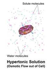
A hypertonic solution is the one that have more of the concentration of solute out of the cell than in it. Some of the Hypertonic solution example consists of-

Osmosis is termed to be a passive method and takes place without any use of energy. It contains the involvement of the molecules from the area of high concentration to low area till there is no different seen in both the concentration and is equal on either sides for membrane. Any of the solvent are able to go via this method being gas or liquid.
Hypertonic solution example
Image credit-Hypertonic solutionWikipedia

In more simple terms the hypertonic solution is a specific type of solution that has more solute concentration on the outer part while being compared to the within of the cell. This leads to leaving of the water of the cell and then flowing in the way of the solution around it. It is easy to not find it elsewhere rather presuming it to be found in the lab.

Sea water

Hypertonic solutions help in getting to keep the food safe. Also, the method of reverse osmosis is used to get the solutions purified.

Sea water has more amount is salt particles when compared to the freshwater that even makes it a good hypertonic solution example. The fish there in the clear water are not able to love in the salt water.

It is so as the water shall rush from the fish cells onto the nearby salt water and thus cause the freshwater fish to die from dehydration. The fish in saltwater on the other part, have adapted themselves to this hypertonic solution example and rely on this to have their cells regulated.

image 32
Image credit-Sea waterWikipedia

The same thing gets to work with the plants present in the salt water. The usually plants tend to need water to get into the cells, while the plants in seawater like the seaweed or the mangroves tend to already have salt from the nearby area and with adaptability tend to best survive in the same level.

Sugary drinks

In very simple terms, sugary drinks are said to be beverages that are made with either adding sweeteners or sugar.

Sometimes, we taste a lot of sugar and this is because there is more sugar in the drink that there is water that makes it a hypertonic solution and a good hypertonic solution example. 

The lips tend to pucker as the water from the mouth tend to rush in the drink and then gets the mouth dehydrated. The sugar drinks can be drawn for water from the cells of the intestine that helps prevent the nutrients from getting absorbed. This is also a reason behind the sports drink tend to have less sugary drinks.

image 33
Image credit-IV drip-Wikipedia

The sugar drinks also have electrolytes that come in the body fluid and then diminish as one sweats or exercises. Hypertonic ones have less salt and also sugar than the human body. In general, it is taken after workout and can be a taken at large events with being good at energy while losing a lot of fluid that gets replaced.

IV Drips and the injections

IV fluids are mostly the liquids that have been formulated and are injected in the vein to help get rid of dehydration.

These IV Drips and the injections as hypertonic solution example are actually administered in the hospital and the injection IV drips. They are provided to the patient with edema to draw water.

These draw water away from the patients having body issues from the tissue that are bloated and then back to the blood. They can be sued up to replace the electrolytes in body for the people who are injured and sick and that cannot take in food in any form healing themselves.

Hypertonic saline

A hypertonic solution contains a higher concentration of solutes compared to another solution. The opposite solution with a lower concentration is known as the hypotonic solution. Scientists must describe cell contents compared to the environment. If a cell is placed in a hypertonic solution, the cell is considered hypotonic.

If the cytosol of the cell is a hypertonic solution, it means the environment is hypotonic, or more weakly concentrated. This is of great importance because solutes and water tend to flow or diffuse along their gradients. Two solutions mixed together will eventually become a single solution. Two solutions mixed together will eventually become a single solution.

Some of the example that support these to be a good example of hypertonic solution example are-

3% and 2% Hypertonic saline

The saline that is hypertonic describes any preparation of saline that has over a concentration of 0.9% of sodium chloride which is the normal saline. The doctors are seen to prescribe 3 or 2% of the hypertonic saline to the ones that need to help loosen the thick mucus. These patients can have cystic fibrosis or dangerous bronchitis.

23% and 7% Hypertonic saline

The one that is in specific of about 23 or 7% hypertonic saline is used up to help the one that have injuries with the brain being severe. It helps in getting the fluid drawn out of the cells to help with cerebral edema. Mostly, the hypertonic saline that is over 3% is used in the central base rather than any other IV drip.

Dextrose in Water

It is also referred to as the D10W as it has 10% dextrose . Water that has a large amount of dextrose in it can be much effective on the way to have the fluids replaced up and then also works with the calories in the IV drip. This sample of hypertonic solution example works will with the babies that are at risk for the blood sugar being low.

image 37
Image credit-DextroseWikimedia

Human Kidney

To regulate the amount of water in the body, the human brain has special proteins called osmoreceptors, which can measure the osmolarity of the environment surrounding the cell

If the environment becomes a highly hypertonic solution, it is because there is not enough water in the blood to dilute the solutes. The hypothalamus releases hormones while increasing the permeability of membranes in the kidney. 

The hypothalamus releases hormones while increasing the permeability of membranes in the kidney. The kidney resorbs the water that would have been excreted and adds it back to the bloodstream. The blood becomes more isotonic compared to the cells, and normal processes can continue.
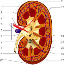
image 34
Image credit-KidneyWikipedia

Extracellular fluids

In biology the extracellular fluids are the ones that is not seen in the cells. IT so seen in the lymph.

The blood cells that are good and healthy do have the equal quantity of water around them. Yet, of we tend to sweat a lot or might lose a lot of water that sodium in certain ways the extracellular fluid shall be hypertonic and one shall be dehydrated.

Osmosis takes place at this time in between the fluids and the ted blood cells that are depleted prevents them from getting to carry the oxygen. Hypertonic dehydration can be mild and also sever like dry mouth, thirst to poor function of kidney and muscle cramp. These fluid is made of lymph, the blood plasma and interstitial fluid.

There is also the availability of two other type of solution depending on the concentration of what they serve for and they are-

  • Isotonic solution
  • Hypotonic solution

Isotonic solution

These are the solution that have the same level of osmolality or the concentration of solute as the other solution.

If there is a separation via the semipermeable membrane, then the water tends to flow in same part of either solutions and then to the other. The example for isotonic are lot different from the Hypertonic solution example.

There is zero flow of water in the solutions despite water moving in both the ways. In terms of biology, some of the cells shall be maintained in the isotonic part to have a support for the cell functions. Man of the animal cell tend to lack cell wall that help minimize the water pressure reply on the outer area to have its shape in form.
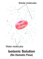
image 35
Image credit-Isotonic solutionWikipedia

Most of the animals tend to balance the osmolality and ph of the fluids by making of the isotonic solutions to have the cells bathe in. The solution tends to carry the water and the nutrients with having them in proportions to have them maintained well in the cell. Same concentration of the solute in and out of the cell with exchange of the molecules of water via the cell membrane.

This can be seen contrasting with the Hypertonic solution example and the hypotonic ones. Blood cells is not a Hypertonic solution example yet a good sample for the isotonic solution example. It is vital for the water to move in and out of the cell to have the function of transferring oxygen in place and the rest of the nutrients to be other portion of body.

If the cell tends to be hypertonic they shall be plasmolyzed. The osmoregulators and conformers are also an example for this. Nature has two type of organism one that conforms the environment osmolality and the other that tend to regulate it in its body separate from the surrounding. These animals are in isotonic state as they evolve its concentration as per the environment and its need. It is mostly in lower level.

The other form of osmoregulators do not place in the isotonic way. This means that the water wants to enter and then leave the body with using several methods for getting leaked. In of the cell they cell exist in an isotonic way and the cell needs to maintain its use. Both the conformers and regulators need to remain functional and with different use to serve.

Hypotonic solution

A solution can be either of the three being isotonic, hypertonic or hypotonic. A solution having less solutes us hypotonic.

A solution that has less quantity of the solute as compared to the concentration of solute in the rest of the solutions across is called to be hypotonic. The example of it differs from Hypertonic solution example.

It is vital to get into minds the importance of solution in science. A solution in term of scientific manner refers to the mixture system that is homogenous and is made up of two or many constituents. They have two parts in any form of mixture being solutes and solvent. The quantity that gets dissolved is the solute and the other is solvent.
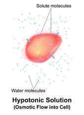
image 36
Image credit-Hypotonic solutionWikipedia

Hypotonicity is the term that is related to the property of the solution to have a well-defined and relative solution. In the biology lines, the major time the solution is compared to the cytosolic fluid is the fluid seen inside the cell. Thus, a solution can be said to be hypotonic when it has less solute amount that present fluid in the cell.

The membrane of the cell is called to be semi permeable. Thus, when a cell is made exposed to the hypotonic surrounding and shall have an influx in the water and as an outcome the swelling shall ensue the cell. There is indeed less of the osmotic pressure that the other solution to which it is actually compared. A hypotonic solution uses a semi permeable membrane and the cell seem to eventually swell up as the molecules of water tend to move at the cell by passive way.

Hypo means low. It has solutes that are non-penetrating when compared to the rest solution across the membrane. Thus, if a cell is placed in it with the solutes being non penetrating and less the concentration of water is said to be high than the one in the cell. Thus there is an osmotic gradient seen with reference to the solvent movement or water that swells the cell up.

Also Read:
9 Analogous Structures Examples: Detailed Insights

In the world of biology, it is said always evolutionary and thus any there are forms that can be termed to be analogous in its own way.

Any two or more forms in biology that is same in looks or has the same function with yet not being of the equal origin is called to be analogous. Some of the analogous structures examples are-

  • Butterfly and bird’s wings
  • Bills of duck and platypus
  • Water conservation and cactus
  • The flippers of the Dolphin and penguins
  • Eye for the mammals and the octopus
  • Potato and sweet potato
  • Bats and bee wing
  • Beaks of bird and turtle
  • Coloring of the Dolphin and shark
  • Shells of turtle and crab

The analogous structures examples are mostly the examples that have evolved separate in two of the independent life form to provide with the same use. The very word analogous comes from the basic word analogy being a device in English that refers to 2 separate things based on its similarities. The analogous structures examples are good correspondence for the evolution type called as convergent.

In this type of evolution, the analogous structures examples are where 2 of the organism are separate and are here to solve the similar problems in evolution like that of keeping itself hidden, conserving of water, swimming, or flying. These analogous structures examples also help in making of body formation that are formed independently.

analogous structures examples
Image credit-Analogous structuresWikipedia

Analogous structures examples

The wings via the ages of butterfly and birds

There are many creatures that are said to have its own wings. All of the wings have been seen to evolve in order to get the solution for only a single problem being flying.

Yet it is said that wings being an analogous structures examples have been seen to evolve via the whole history for different purposes. The 1st analogous structures examples tend to be the insects having the same form to help themselves push via the air down to get its body propelled via the air and making it easy for its tiny body to move.

The insects being also an analogous structures examples have seen to use its various body parts in order to have itself fly using parts in the protective exoskeletons to have itself propelled themselves via the air. There are many analogous structures examples that via the million year, have learned to adapt the same thing like the reptiles.

The analogous structures examples have got skin membrane that stretched between the ankle bones and the finger and is also capable of getting itself propelled via the air. After many years as well the dinosaurs being an analogous structures examples have tended to narrow down the approach of flying using feathers they helped them keep itself warm and push into the sky.

Bills of Duck and Platypus

The very 1st specimen of the platypus was seen in the British museum by the Australian with being simple stuck being a duck’s bill having a beaver like animal.

The truth was much more interesting that getting evolved. The analogous structures examples of platypus had evolved with almost having the same form like that of the ducks to get the problem of accumulating food solved such like that of the aquatic plants and fish in water. These two analogous structures examples are not mostly related to each other.

The duck billed platypus are mostly an animal as well that have analogous structures examples and are mostly small with being much shy animals. They do have a head that is flat and also has a body to have itself glide via the water. They have fur which is mostly dark on the top and is linked to that of the birds and reptile.

220px Schnabeltier 1
Image credit-PlatypusWikipedia

These analogous structures examples are not mostly related to each other with platypus being a mammal that have been seen to evolve long later than the birds and also the rest mammals with having different ways to have its path taken by evolution. Yet both of them have developed a good same form to move in water and also land and also both via analogous structures examples.

Water conservation and Cactus

Some of the members of the plant genres are a good analogous structures examples being the Astrophytum and the Euphorbia which look same.

Both of the analogous structures examples have same ball shape body with also being round and having itself divided into 8 of the same length wedges being pointy, hard having thorn that’s stick out in a row along with idle of every wedge. These analogous structures examples help in protecting themselves from eating.

While one seeing it via normal eye shall find it belonging to the same species being analogous structures examples. They can be distinguished by its feature of not being genetically related and also tend to live in 2 complete separate rea in world. Astrophytum is from North America and its example is cactus living in the south part of the western desserts.

On the other hand, the other analogous structures examples being Euphorbia tend to be of the genus that has poinsettias as well with some of the desserts seen in the desserts of Africa. Both of these analogous structures examples are the ones that ted to conserve water by reducing its surface area and also results in being ball round shape and having a thick waxy layer on skin being prickly.

Flippers of Dolphins and penguins

These are a good analogous structures examples for they have same function with being much different in its origin and also thus called so.

With mush research or not, some can say that penguins and dolphins are not linked yet do have same traits that do represent an evolution called as convergent and thus are analogous structures examples. They do just tend to share only one trait which are the flippers and thus are analogous structures examples.

The flippers of penguin are most feathers with mostly being of secondary of primary in its nature and are much these analogous structures examples are much critical to that of its flight with having the flippers feature to be small, short and also much denser. This helps in having a wing that is streamline form and minimizes the underwater drag and has insulation to protect itself.

The flippers of the dolphin being an analogous structures examples are one of the principal feature that controls the cetaceans due to its way in front of the mass center and also has its mobility that makes it three degrees in its freedom. Cetaceams are the porpoises, the whales and the dolphins. The flipper are analogous structures examples and o help them swim being a modified paddle.

Emperor Penguin Manchot empereur.jpg
Image credit-Penguin-Wikipedia

Eye of mammals and octopus

This analogous structures examples tend to show the same type of evolution like the rest being the convergent evolution and tend to be analogous as well.

The eye of the mammals and those of the octopus are analogous structures examples and thus also have octopus showing a good evolution for the same usage and thus also have same function. Thus they tend to be analogous structures examples and do thus same a common ancestor with being an evolution.

The octopus has an eye called the camera eye that is same to that of the human’s eye in both type of analysis being embryological and also phylogenetic and thus said that to have the camera eye on its own. It is also an example of analogous structures examples and also for convergent evolution. There is always an entire gene set needed to get this straight.

The eye of the octopus has an iris, with the vitreous cavity, the cells for pigments, the photoreceptor cell and also has a lens that is circular and helps in having the light translated from the part of the retina which is light sensitive and also has its nerve signals that travel along the optic nerve to its brain being same to that of human in its nature and thus also analogous structures examples.

Potato and sweet Potato

Both of these analogous structures examples are said to be much rich in fiber and also has many vitamins like C and B6. Both sweet potato and potatoes are tuberous plants and look more or less similar 

Sweet potato has more of the vitamin A and the potato on the other hand have more potassium with looking alike and thus analogous structures examples. The general potato is white the sweet ones are orange in appearance. Both have brown outside skin making it look the same but doffers in variety.

There are more than about 4000 of the potatoes seen and more than 1000 variety of the sweet ones that around around the globe. Both also tend to grown underground and thus analogous structures examples. Only one common feature of it has been taken to chance of it being the same in appearance. Both also have same function and stores same food.

Both of these analogous structures examples tend to store food in the manner of starch an also differ in its origin of formation and thus the sweet potato is a root tuber with the other analogous structures examples being a stem tuber. Sweet potato is said to be a root modification and also thus have same use but different formation and are thus analogous structures examples.

Bats and bee wing

A great analogous structures examples can be the bat’s wing and a bee’s wing. Bats and bees do not share common ancestry, so the structures cannot be homologous. Both bat wings and bee wings serve a common purpose.

Sometimes it is unclear whether similarities in structure in different organisms are analogous or homologous. An example of this is the wings of bats and birds. These structures are homologous in that they are in both cases modifications of the forelimb bone structure of early reptiles being an analogous structures examples.

Interestingly, though bird and bat wings are analogous as wings, as forelimbs they are homologous. Birds and bats did not inherit wings from a common ancestor with wings, but they did inherit forelimbs from a common ancestor with forelimbs. Analogous since the animals are not closely related, so the wings likely developed independent from each other rather than from a common ancestor thus forming analogous structures examples.

image 3
Image credit-Bat wingWikipedia

Bat wings are made up of flaps of skin which is stretched between the bones of the fingers and the arm while the wings of bird are made up of feathers extending all along the arm. This dissimilarity in the structure of their wings shows that the wings of the bird and bat are not inherited from a common ancestor thus making analogous structures examples. Bat’s wings are made of a supple, hairless, elastic skin stretching from the edge of the forelimb all the way to the tip of an elongated little finger.

Beaks of bird and turtle

Shells aren’t the turtle’s only analogous structure! Both turtles and tortoises eat with hard beaks that are similar to a bird’s beak, and used for the same purpose: to cut and chew food.

Analogy, in biology, similarity of function and superficial resemblance of structures that have different origins making it analogous structures examples. An analogous pair is a pair of words, numbers or objects that are connected by some rule or by similarity. The word analogy means comparison between two things for the sake of clarification.

For example, a couple names their first child A, their second child as B, their third child as C and so on. This form of evolution is referred to as convergent evolution. Convergent evolution is a kind of evolution wherein organisms evolve structures that have similar or analogous structures or functions in spite of their evolutionary ancestors being very dissimilar or unrelated.

Thus, analogous structures of unrelated species would have similar or corresponding functions although they evolved from different evolutionary origins. Reptilia while birds belong to the Class: Aves. Reptiles have scales all over the body, whereas birds have scales on the legs and the rest of the skin is covered with fluffy feathers. All the present-day reptiles are carnivore, but birds have many different types of food habits.

Coloring of the Dolphin and shark

An interesting example of analogous structures is the shared coloring of sharks and dolphins. Even though these two predators share the same oceanic habitat, they are from different animal classes making a good analogous structures examples

Another example of analogous structures are dolphins and sharks as whole species. Although we might think of dolphins and sharks as being relatively similar, their morphology says otherwise. Dolphins are mammals that have live births and have fins with a homologous structure to human arms. Sharks and dolphins are similar in construction because they need their “parts” to complete many of the same functions.

 Sharks and dolphins are both greyish in color. However, other physical similarities they have include a lighter underside, dorsal fins and located on the back, and pectoral fins is located on their sides. Also, as mentioned above, both dolphins and (most) sharks give birth to live young. Dolphins and sharks both have dorsal fins on their backs, two pectoral fins on their sides, and a tail making it a good analogous structures examples.

350px Dolphintursiops
Image credit-DolphinWikipedia

Sharks are closely related to rays, and dolphins are closely related to cows and other mammals. Streamlined bodies and fins are traits that dolphins and sharks evolved separately, both as adaptations for swimming. These fish have skeletons of cartilage and streamlined bodies and are among the scariest predators in the sea. Metaphorically, they’re often hustlers talking about the pool shark or other kinds of greedy cheaters.

Shells of turtle and crab

Crab and turtle shells are analogous structures; they evolved from different structures. Crabs and turtles have more differences than similarities. They are not closely related and thus are good analogous structures examples.

Both crabs and turtles have shells that grow from their body and that cannot be removed. The shells protect them from predators and allow them to keep their fleshy bodies safe. Crabs and turtles also both shed the outer layers of their shells during their lifetimes as they grow and thus are good analogous structures examples.

The wings of a bird and of an insect are analogous organs. Both of these species have wings that they use for flight and yet their wings came from dissimilar ancestral origins. Analogous structures are features of different species that are similar in function but not necessarily in structure and which do not derive from a common ancestral feature making it analogous structures examples.

The carapace is the shell on back of the crab that is made of a hard bone called chitin. Chitin is a polymer which is the main component of arthropod’s exoskeletons such as crabs. The exoskeletons of arthropods – crabs, shrimp, lobsters – are largely made up of chitin, a biomaterial.

Conclusion

There are many analogous structures examples with sharing same physical appearance and also yet nor related generically and also not concerned with being of the same use yet the analogous structures examples solve the same function.

Also Read:

Function of Flagella: Detailed Facts

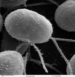
function of flagella 0

Flagella is the organelle of motility which is also a tight apparatus of 20 several kinds of proteins.

The function of flagella can depend up on the types of the cell in it concerned with like that of the algae, bacteria or the prokaryotes and also the animals’ cells which are the eukaryotes. The flagella is not only the one to be there but also cilia can be seen.

The flagella of a bacteria is much spoken about and is quite complex. The body of it transverses the cell wall while the hook shaped in curve connect the basal body to the flagella filament which is whip like and then makes several micrometer of the bacterial body.

The flagella quite is basic is considered to be just useful for nobility of the organism in any kind of cell but is recently seen to perform more than just mobility and serve other biological usage. There are different parts of the bacteria that serve as being useful. The cell type that has the presence of flagella is the sperm cell which is commonly the male sex cell.

The major unit of the flagella plays a good role in innate system and has antigen which is dominant to that of the adaptive response to immunity. They also seem to as work taking part in adhesion as adhesions. The entire flagella are seen to be vital in response to cell adhesion and getting itself invaded into the host cell.

Function of flagella in prokaryotic cell

  • There are many gram negative and positive species of bacteria which are said to have flagella. The function of flagella in the cells of prokaryotes is to help in the movement of the bacteria and also helps them enabling the process of chemotaxis. The cells of them can either have only one flagellum or more.
  • There are also any other uses of a flagella apart from movement which is quite different in bacteria and during the entire cycle of bacteria’s life. The number of bacteria differs can also be polar or peritrichous. When it comes to being a polar it means only one flagellum at a spot and the later means many of them in single spot.
  • The bacterial flagella can be useful in getting to participate in the formation of biofilm along with getting the protein exported as in adhesion. The movement of the flagella is mostly prerequisite for invasion or for adhesion. Some of the bacteria can be E.coli or one in pathogenicity.
  • Considering the size of the filament of the flagella in the prokaryotic cell to be small they help in locomotion and also helps acts as a sensory organs that is used in getting to detect the changes in temperature and ph. of the surrounding. There are quite a few researchers that are going to find out many possible uses of flagella but are yet to be found.
  • The flagella are actually said to be surrounded by the extra area of the cell membrane and thus are also sensed to detect any changes in the ph. or the temperature while being in close contact with the environment and also detects any outside cell chemical changes.
function of flagella
Image credit- FlagellaWikipedia

Function of flagella in eukaryotic cells

  • The flagella in the eukaryotes are much different from that of the prokaryotes in serving its usage. The function of flagella in the eukaryotes strives to be a conserved one and serves for the use of getting transport system for proteins, to serve motility, and works also as a sensory.  
  • The flagella of the eukaryotes like the animals and the plants helps in serving as the motility purpose that shall help in movement and also in chemotaxis. The bacteria have only one flagella and also can have many of them ad can be polar as well. The flagella show a rotationally movement which is counterclockwise that helps in the cell to propel forward and then move.
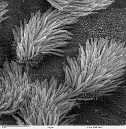
  • There are not only the flagella but also the cilia that extend on the surface of the eukaryotes. They are commonly in terms associated with locomotion and is by technique linked with the inside of the cell is covered by the extra of the cytoplasmic membrane.
  • The flagella of the eukaryotes unlike the ones that are in the prokaryotes are located in the cytoplasmic membrane. They give the organism the freedom to move back and forth with the flagella spins. There are not found in all eukaryotes but some. While there is a clockwise movement there is a tumble and this helps in getting the movement of organism changed.
  • Both the flagella present in the eukaryotes and the prokaryotes do run in movement being circular in motion of the filaments that helps in propelling the cell or the fluids inside the cell to move past the cell. The movement can also be whip like on addition to being circular as well.
cilia
Image credit- CiliaWikipedia

Function of flagella in algae

  • The flagella is an organelle that helps in the cell to move back and forward On addition to this is also serves uses in organism. In an aqueous surrounding, the flagella mechanism even shows its reaction to the chemical, the mechanical, the light and the gravitational stimulus of then cell. Flagella overall has the same function to perform in any type of cell yet differs in few respect.
  • The flagella on the algae also help in playing a vital role in the sexual fission of the oogamous, the anisogamous and the isogamous species of the algae and mostly the green algae species. They are also called the flagellates for having the whip like flagella. There can be a creation of powers stroke.
  • There is the presence of adenosine triphosphate seen in the algae, the dynein molecule are then activated and the whip like flagella bends as the dynein arms on one side of the dynein tends to cross the bridge and gets them activated and helps in moving up the tube.
  • The species of Chlamydomonas having the flagella has a character that can get converted into the sexual organelle at the time of gametogenesis. They show a specific species adhesion or a reaction of agglutination while in between the cells for the opposite type of mating. This happens due to the presence of a molecule called agglutination that is seen on its surface.

Function of flagella in cells

  • Flagella are said to be a hair like structure that is microscopic and is involved in getting to contribute in the cell movement.
  • The very basic function of flagella is to help the cells or rather any type of cell in their movement. Yet in some of them, the flagella can be seen to serve much other function like as sensory part and more. The definition or types of flagella depend on the usage of them in cells.
  • There are two type of cell seems which the eukaryotic and prokaryotic ones. Both the cells have flagella where the prokaryotes tend to have only one or more and while there are only few eukaryotes that have flagella. There are many types of flagella and are not named after structure but there roles.
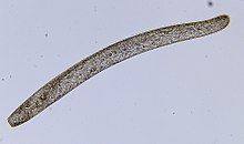
  • The flagellum is mostly classified to be a characteristic of the cells of the prokaryotes like the archaea and the bacteria. Along with the prokaryotic cells it is also linked up with the group of protozoan that is also seen in the gametes of the animals, the algae, mosses, the slime molds and the mosses.
  • The motion of the flagella is the cause for the water currents that are needed for the process of circulation and respiration of the sponges and the coelenterates. Most of the bacteria that are considered to be motile are cause of the presence of flagella that helps in the movement.
  • Yet, there is a difference between the pattern of structure for the prokaryotes and the eukaryotes and thus are different. The character of them that is likely is the movement being whip like. The flagella quite resemble that of the cilium in the structure. They have nine pairs of microtubules with each of them having a protein.
pr
Image credit- ProtozoanWikipedia

Function of flagella in animal cell

  • The flagella are stricture to be defined by their use in the different types of cells rather than their structure.
  • The function of the flagella in the cells of animal is same to that of the purpose of the flagella in the cells of eukaryotes. They helps in locomotion which is the most common function and is then also termed to be a apart of sensory organelle.
  • The animals’ cells do not only have one or more flagella for locomotion but also cilia. They are actually the appendages that are found in almost all the microbes and the animals bit not in the higher level plants. For any of the eukaryotes that have only one cell, both flagella and cilia are needed for movement of them.
  • The use of cilia is to get the water keep moving in relative speed inside the cell concerned with regular process of movement of the cilia. This process can give an end of water the cell moving via the water which is mostly concerned with the single cell species or the second one can be the contents of the moving water across the cell surface.
  • The movement of the flagella for the eukaryotic cells is actually based on the adenosine triphosphate for their energy while in the prokaryotic cells they are based on the energy that is derived from the prokaryotic proton motive force or is termed as the ion gradient across the membrane of the cell.
  • The shape of the flagella is helical in the prokaryotic bacteria and has inclusion like the protein flagella. The flagella base which is called the hook is seen near the surface of cell and is linked with the basal body within the cell envelope. It gets a motion which is circular and is the clockwise way.

Also Read:

Does Mitochondria Have Circular DNA?

does mitochondria have circular dna

Mitochondria, the powerhouses of eukaryotic cells, possess their own genetic material known as mitochondrial DNA (mtDNA). Interestingly, this mtDNA exhibits a unique circular structure, which is a crucial feature for its function and maintenance within the mitochondria. The Circular Structure of Mitochondrial DNA Mitochondrial DNA is a closed, circular, double-stranded DNA molecule that is typically … Read more

Do Fungi Have a Mitochondrion?

do fungi have a mitochondrion

Fungi, like most eukaryotic organisms, possess mitochondria – the powerhouses of the cell responsible for energy production through oxidative phosphorylation. Fungal mitochondria play a crucial role in various cellular processes, including energy metabolism, apoptosis, and signaling pathways. Understanding the structure, function, and diversity of fungal mitochondria is essential for unraveling the complex biology of these … Read more

Do Stem Cells Have a Nucleus?

do stem cells have a nucleus

Stem cells, like other eukaryotic cells, possess a nucleus that houses their genetic material, which is essential for their growth, development, and reproduction. The nucleus is a membrane-bound organelle that plays a critical role in regulating gene expression and maintaining the undifferentiated state of stem cells. The Importance of the Nucleus in Stem Cells The … Read more

Is Kinase an Enzyme? A Comprehensive Guide

is kinase an enzyme

Summary

Kinases are a class of enzymes that play a crucial role in cellular signaling and regulation. They catalyze the transfer of a phosphate group from ATP to a specific substrate, a process known as phosphorylation. This post delves into the intricate details of kinases, their enzymatic properties, and their significance in various biological processes.

What are Kinases?

is kinase an enzyme

Kinases are a family of enzymes that catalyze the transfer of a phosphate group from ATP (adenosine triphosphate) to a specific substrate, typically a protein. This process, known as phosphorylation, can modulate the activity, localization, or interaction of the target protein, thereby regulating a wide range of cellular processes.

Kinase Classification and Diversity

Kinases can be classified into several subgroups based on the specific amino acid residues they target for phosphorylation:

  1. Serine/Threonine Kinases: These kinases phosphorylate serine or threonine residues on their target proteins. Examples include protein kinase A (PKA), protein kinase C (PKC), and cyclin-dependent kinases (CDKs).

  2. Tyrosine Kinases: These kinases phosphorylate tyrosine residues on their target proteins. Examples include the epidermal growth factor receptor (EGFR) and the Src family of kinases.

  3. Dual-Specificity Kinases: These kinases can phosphorylate both serine/threonine and tyrosine residues on their target proteins. Examples include mitogen-activated protein kinases (MAPKs) and cyclin-dependent kinase-activating kinases (CAKs).

The human genome encodes over 500 different kinases, making them one of the largest enzyme families. This diversity allows kinases to regulate a vast array of cellular processes, including cell growth, cell division, metabolism, and signal transduction.

Kinase Enzymatic Activity and Regulation

Kinases are highly regulated enzymes, and their activity can be modulated by various mechanisms, such as:

  1. Allosteric Regulation: Binding of regulatory molecules, such as small molecules or other proteins, can induce conformational changes in the kinase, either activating or inhibiting its enzymatic activity.

  2. Phosphorylation/Dephosphorylation: Kinases can be phosphorylated or dephosphorylated by other kinases or phosphatases, respectively, which can alter their catalytic activity or substrate specificity.

  3. Subcellular Localization: The localization of a kinase within the cell can determine its access to specific substrates and, consequently, its overall function.

  4. Protein-Protein Interactions: Kinases can form complexes with other proteins, which can modulate their enzymatic activity or substrate specificity.

These regulatory mechanisms ensure that kinase activity is tightly controlled and coordinated to maintain cellular homeostasis and respond appropriately to various stimuli.

Kinases and Cellular Signaling

Kinases play a central role in cellular signaling pathways, acting as molecular switches that transduce extracellular signals into intracellular responses. By phosphorylating specific target proteins, kinases can activate or inactivate them, leading to the propagation of signaling cascades that ultimately influence cellular processes such as:

  1. Cell Growth and Proliferation: Kinases like receptor tyrosine kinases (RTKs) and mitogen-activated protein kinases (MAPKs) are key regulators of cell growth and division.

  2. Cell Survival and Apoptosis: Kinases like phosphoinositide 3-kinase (PI3K) and protein kinase B (PKB/Akt) are involved in promoting cell survival and inhibiting apoptosis (programmed cell death).

  3. Metabolism and Energy Homeostasis: Kinases like AMP-activated protein kinase (AMPK) and glycogen synthase kinase 3 (GSK3) play crucial roles in regulating cellular metabolism and energy balance.

  4. Immune Response and Inflammation: Kinases like Janus kinases (JAKs) and spleen tyrosine kinases (Syks) are essential for the proper functioning of the immune system and the regulation of inflammatory responses.

  5. Cell Migration and Adhesion: Kinases like focal adhesion kinase (FAK) and Rho-associated protein kinases (ROCKs) are involved in the regulation of cell motility and cell-cell/cell-matrix interactions.

The dysregulation of kinase activity has been implicated in the pathogenesis of various diseases, including cancer, neurological disorders, autoimmune diseases, and metabolic disorders. As a result, kinases have become important targets for the development of therapeutic interventions, with numerous kinase inhibitors approved for clinical use or in various stages of drug development.

Quantifying Kinase Enzymatic Activity

Measuring the enzymatic activity of kinases is crucial for understanding their role in cellular signaling and for the development of kinase-targeted therapies. Traditional methods for assessing kinase activity often relied on indirect measurements, such as the incorporation of radioactive phosphate or the use of antibodies that recognize phosphorylated substrates.

However, these methods have limitations, as they may not provide a direct and quantitative assessment of kinase activity in the presence of multiple endogenous kinases. To address this, researchers have developed more advanced techniques, such as the use of CSox-based sensors, which offer a direct and selective approach for measuring kinase enzymatic activity.

CSox-based Sensors for Kinase Activity Quantification

The CSox-based sensor system utilizes a phosphorylation-sensitive amino acid, termed CSox, to create kinase-selective biosensors. These sensors are capable of detecting phosphorylation through a process known as chelation-enhanced fluorescence (CHEF), which allows for the direct monitoring of kinase activity in unfractionated cell lysates.

The key features of the CSox-based sensor system include:

  1. Kinase Selectivity: The selectivity of the CSox-based substrate can be tuned to target a specific kinase of interest (KOI), enabling the direct measurement of that KOI’s activity in the presence of the endogenous kinome.

  2. Quantitative Readout: The CHEF-based detection mechanism provides a direct and quantitative assessment of kinase enzymatic activity, overcoming the limitations of using activity proxies to infer kinase activity.

  3. Unfractionated Cell Lysates: The CSox-based sensors can be used to measure kinase activity in complex biological samples, such as unfractionated cell lysates, without the need for extensive sample preparation or purification.

This advanced technique has significantly improved our ability to study kinase enzymatic activity in a more direct and quantitative manner, providing valuable insights into the regulation and dysregulation of kinases in various biological and disease contexts.

Conclusion

In summary, kinases are a diverse family of enzymes that play a crucial role in cellular signaling and regulation. By catalyzing the transfer of phosphate groups from ATP to specific substrates, kinases can modulate the activity, localization, and interactions of their target proteins, thereby influencing a wide range of cellular processes. The development of advanced techniques, such as CSox-based sensors, has enabled the direct and quantitative assessment of kinase enzymatic activity, further enhancing our understanding of these important enzymes and their potential as therapeutic targets.

References

  1. Beck, J. R., Peterson, L. B., Imperiali, B., & Stains, C. I. (2013). Quantification of protein kinase enzymatic activity in unfractionated cell lysates using CSox-based sensors. Nature protocols, 8(5), 1057-1070.
  2. ScienceDirect Topics. (n.d.). Kinase Assay. Retrieved from https://www.sciencedirect.com/topics/medicine-and-dentistry/kinase-assay
  3. Smyth, L. A., & Collins, I. (2012). Measuring and interpreting the selectivity of protein kinase inhibitors. Chemical Society Reviews, 41(16), 5147-5160.
  4. Promega Corporation. (n.d.). PKC zeta Kinase Enzyme System. Retrieved from https://ch.promega.com/products/cell-signaling/kinase-assays-and-kinase-biology/pkc-zeta-kinase-enzyme-system/
  5. R&D Systems. (n.d.). Methods for Detecting Protein Phosphorylation. Retrieved from https://www.rndsystems.com/resources/articles/methods-detecting-protein-phosphorylation.

Is DNA Polymerase an Enzyme?

is dna polymerase an enzyme

DNA polymerase is indeed an enzyme that plays a crucial role in the replication and synthesis of DNA. This well-established fact in the field of biochemistry and molecular biology has been extensively studied and characterized through various experimental techniques. DNA polymerase catalyzes the addition of nucleotides to the 3′ end of an existing DNA strand, using the complementary base pairing rule to ensure accurate replication of genetic information.

The Enzymatic Activity of DNA Polymerase

The enzymatic activity of DNA polymerase has been extensively studied and characterized through various experimental techniques. These studies have provided valuable insights into the mechanisms and dynamics of DNA polymerase enzymes.

Chip-based Investigations

A study published in Nature in 2015 used a chip-based method to investigate the interactions between DNA polymerases and nucleic acids. The researchers observed changes in DNA extension and polymerase conformation during enzymatic activity. This study demonstrated the high sensitivity of the method in revealing previously unidentified tight binding states for Taq and Pol I DNA polymerases, as well as the incorporation of label-free nucleotides in real-time.

Kinetics and Mechanism of High-fidelity DNA Polymerases

Another study published in the Journal of Biological Chemistry in 2010 used stopped-flow fluorescence spectroscopy to investigate the kinetics and mechanism of high-fidelity DNA polymerases. The researchers found that the substrate-induced structural change plays a key role in the discrimination between correct and incorrect base pairs. This study also highlighted the importance of conformational changes in polymerase specificity and the role of enzyme dynamics in catalysis.

Structural Insights into DNA Polymerase Enzymes

Structural studies have also provided valuable insights into the mechanisms of DNA polymerase enzymes. For example, a study published in the Proceedings of the National Academy of Sciences in 2008 used X-ray crystallography to determine the structure of the Bacillus stearothermophilus DNA polymerase I (Bst DNAP I) in complex with a DNA template and incoming nucleotide. The researchers observed that the enzyme undergoes a conformational change upon binding to the DNA template, which helps to position the incoming nucleotide for efficient catalysis.

Fidelity and Processivity of DNA Polymerase Enzymes

The high fidelity and processivity of DNA polymerase enzymes are crucial for their role in DNA replication and synthesis. These properties have been extensively studied and characterized through various experimental techniques.

Fidelity of DNA Polymerase Enzymes

The fidelity of DNA polymerase enzymes refers to their ability to accurately replicate DNA by minimizing the incorporation of incorrect nucleotides. This is achieved through a combination of mechanisms, including:

  1. Base pairing specificity: DNA polymerase enzymes have a high degree of specificity for the correct base pairing, ensuring that only complementary nucleotides are incorporated into the growing DNA strand.
  2. Proofreading activity: Many DNA polymerase enzymes possess a 3′ to 5′ exonuclease activity, which allows them to detect and correct any mismatched or incorrectly incorporated nucleotides.
  3. Mismatch repair: Cellular mechanisms, such as the mismatch repair system, can further correct any errors that may have occurred during DNA replication.

The high fidelity of DNA polymerase enzymes is crucial for maintaining the integrity of the genetic information during DNA replication and repair.

Processivity of DNA Polymerase Enzymes

The processivity of DNA polymerase enzymes refers to their ability to catalyze the addition of multiple nucleotides to the growing DNA strand without dissociating from the template. This property is essential for efficient and rapid DNA replication.

DNA polymerase enzymes achieve high processivity through various structural features, such as:

  1. Sliding clamp: Many DNA polymerase enzymes are associated with a sliding clamp protein, which encircles the DNA and tethers the polymerase to the template, preventing it from dissociating.
  2. Exonuclease domain: The presence of a 3′ to 5′ exonuclease domain in some DNA polymerase enzymes allows them to proofread and correct any errors during DNA synthesis, further enhancing their processivity.
  3. Interactions with other proteins: DNA polymerase enzymes often interact with other proteins, such as helicase and primase, which can help to maintain their association with the DNA template and increase their processivity.

The high processivity of DNA polymerase enzymes is crucial for the efficient and accurate replication of the entire genome during cell division.

Commercial Applications of DNA Polymerase Enzymes

is dna polymerase an enzyme

In addition to the extensive research on the enzymatic activity of DNA polymerase, there are also various commercial products available that utilize these enzymes for specific applications.

KAPA HiFi HotStart ReadyMix

One example is the KAPA HiFi HotStart ReadyMix from Roche Sequencing Store. This DNA polymerase-based system exhibits high fidelity and performance for PCR amplification of DNA targets. The KAPA HiFi HotStart ReadyMix is designed to:

  1. Improve performance on GC- and AT-rich templates
  2. Amplify longer targets with greater sensitivity
  3. Achieve the highest fidelity with an error rate that is 100 times lower than wild-type Taq DNA polymerase

The high fidelity and processivity of the KAPA HiFi HotStart ReadyMix make it a valuable tool for various applications in molecular biology and genetics, such as next-generation sequencing, gene expression analysis, and diagnostic assay development.

Conclusion

In summary, DNA polymerase is an enzyme that plays a crucial role in the replication and synthesis of DNA. Its enzymatic activity has been extensively studied and characterized through various experimental techniques, providing valuable insights into the mechanisms and dynamics of these enzymes. The high fidelity and processivity of DNA polymerase enzymes make them essential tools for a wide range of applications in molecular biology and genetics.

References:

  1. Pandey, M., Syed, S., Donmez, I., Patel, G., Ha, T., & Patel, S. S. (2009). Coordinating DNA replication by means of priming loop and differential synthesis rate. Nature, 462(7275), 940-943.
  2. Hohlbein, J., Gryte, K., Heilemann, M., & Kapanidis, A. N. (2010). Surfing on a new wave of single-molecule fluorescence methods. Physical biology, 7(3), 031001.
  3. Kuchta, R. D., & Stengel, G. (2010). Mechanism and evolution of DNA primases. Biochimica et Biophysica Acta (BBA)-Proteins and Proteomics, 1804(5), 1180-1189.
  4. Beese, L. S., Derbyshire, V., & Steitz, T. A. (1993). Structure of DNA polymerase I Klenow fragment bound to duplex DNA. Science, 260(5106), 352-355.
  5. Roche Sequencing Store. (n.d.). KAPA HiFi HotStart ReadyMix. Retrieved from https://rochesequencingstore.com/catalog/kapa-hifi-hotstart-readymix/

Do Eukaryotic Cells Have Enzymes? A Comprehensive Guide

do eukaryotic have enzymes

Eukaryotic cells are known for their complex and diverse cellular machinery, which includes a wide range of enzymes that play crucial roles in various cellular processes. From regulating the cell cycle to replicating DNA, eukaryotic enzymes are essential for maintaining the delicate balance of cellular function. In this comprehensive guide, we will explore the different types of enzymes found in eukaryotic cells, their specific roles, and the quantifiable data that supports their importance.

Cyclin-dependent Kinases (Cdks): The Guardians of the Cell Cycle

Cyclin-dependent kinases (Cdks) are a family of enzymes that are responsible for regulating the cell cycle in eukaryotic cells. These enzymes are essential for ensuring that the cell cycle progresses in a coordinated and controlled manner, preventing uncontrolled cell division and potential cellular dysfunction.

  • There are nine known Cdk family members in humans, each with distinct roles in cell cycle progression.
  • The activity of Cdks is regulated by the binding of cyclins, which increase or decrease in distinct phases of the cell cycle.
  • Cdk1, also known as Cdc2, is the most well-studied Cdk and is required for the transition from G2 to M phase during the cell cycle.
  • Cdk2 is involved in the transition from G1 to S phase, while Cdk4 and Cdk6 are important for the G1 phase.
  • Dysregulation of Cdk activity has been implicated in various types of cancer, making them important targets for therapeutic interventions.

DNA Polymerases: The Replication Maestros

do eukaryotic have enzymes

Eukaryotic cells have multiple DNA polymerases that are responsible for replicating the genetic material during cell division. These enzymes work in a coordinated manner to ensure the accurate and efficient replication of the genome.

  • Pol ε and Pol δ are the leading and lagging strand DNA polymerases in eukaryotes, respectively.
  • Pol ε has a higher processivity and is responsible for the majority of DNA synthesis on the leading strand.
  • Pol δ has a lower processivity and is primarily involved in the synthesis of the lagging strand.
  • The activity of these DNA polymerases is stimulated by different levels of PCNA (Proliferating Cell Nuclear Antigen), a protein that acts as a processivity factor.
  • Mutations in DNA polymerases have been linked to various genetic disorders, such as Werner syndrome and Bloom syndrome, which are characterized by premature aging and an increased risk of cancer.

Protein Kinases: The Cellular Signaling Maestros

Protein kinases are enzymes that play a crucial role in cellular signaling by adding phosphate groups to proteins, thereby altering their activity and function.

  • There are over 500 known protein kinases in the human genome, making them one of the largest enzyme families in eukaryotic cells.
  • Protein kinases are involved in a wide range of cellular processes, including signal transduction, metabolism, and cell cycle regulation.
  • Some of the most well-studied protein kinases include the mitogen-activated protein kinases (MAPKs), which are involved in the transmission of extracellular signals to the nucleus, and the cyclin-dependent kinases (Cdks), which regulate the cell cycle.
  • Dysregulation of protein kinase activity has been implicated in various diseases, including cancer, neurodegenerative disorders, and autoimmune diseases, making them important targets for therapeutic interventions.

Phosphatases: The Cellular Signaling Balancers

Phosphatases are enzymes that remove phosphate groups from proteins, reversing the action of protein kinases and playing a crucial role in regulating cellular signaling and metabolism.

  • There are over 150 known phosphatases in the human genome, making them an essential component of the cellular signaling network.
  • Phosphatases can be classified into different families based on their substrate specificity and catalytic mechanisms, including serine/threonine phosphatases, tyrosine phosphatases, and dual-specificity phosphatases.
  • Phosphatases are involved in a wide range of cellular processes, including signal transduction, cell cycle regulation, and metabolic control.
  • Dysregulation of phosphatase activity has been linked to various diseases, such as cancer, neurological disorders, and metabolic diseases, highlighting their importance as potential therapeutic targets.

Lysosomal Enzymes: The Cellular Recyclers

Lysosomes are membrane-bound organelles in eukaryotic cells that contain a variety of hydrolytic enzymes responsible for the degradation of various biomolecules, including proteins, lipids, and nucleic acids.

  • There are over 50 known lysosomal enzymes, including proteases, glycosidases, lipases, and nucleases.
  • Lysosomal enzymes play a crucial role in cellular homeostasis by breaking down and recycling cellular components, as well as in the degradation of extracellular materials that are taken up by the cell.
  • Deficiencies or malfunctions in lysosomal enzymes can lead to a group of genetic disorders known as lysosomal storage diseases, such as Gaucher’s disease and Pompe disease, which are characterized by the accumulation of undigested materials in the lysosomes.
  • Lysosomal enzymes are also important in the immune response, as they are involved in the degradation of pathogens and the presentation of antigens to the immune system.

Peroxisomal Enzymes: The Cellular Detoxifiers

Peroxisomes are membrane-bound organelles in eukaryotic cells that contain a variety of oxidative enzymes involved in various metabolic processes, including fatty acid oxidation and the detoxification of reactive oxygen species.

  • There are over 50 known peroxisomal enzymes, including catalase, peroxiredoxins, and acyl-CoA oxidases.
  • Peroxisomal enzymes play a crucial role in the metabolism of long-chain and branched-chain fatty acids, as well as in the breakdown of hydrogen peroxide, a potentially harmful byproduct of cellular metabolism.
  • Deficiencies in peroxisomal enzymes can lead to a group of genetic disorders known as peroxisomal disorders, such as Zellweger syndrome and adrenoleukodystrophy, which are characterized by the accumulation of very-long-chain fatty acids and other metabolic abnormalities.
  • Peroxisomal enzymes are also important in the regulation of cellular redox balance and the prevention of oxidative stress, which can contribute to the development of various diseases, including neurodegenerative disorders and cancer.

In conclusion, eukaryotic cells are equipped with a diverse array of enzymes that play critical roles in various cellular processes. From regulating the cell cycle to replicating DNA and maintaining cellular homeostasis, these enzymes are essential for the proper functioning of eukaryotic cells. Understanding the specific roles and quantifiable data associated with these enzymes is crucial for advancing our understanding of cellular biology and developing targeted therapeutic interventions for various diseases.

References:
– R. Young, S. Francis, in Pharmacognosy, 2017.
– Understanding the Eukaryotic Cell Cycle – a Biological and Experimental Mini-review, 2016.
– The eukaryotic leading and lagging strand DNA polymerases … – NCBI, 2009.
– Protein Kinases: Structures and Catalytic Mechanisms, 2004.
– Phosphatases: Providing Safe Passage Through Mitotic Exit, 2007.
– Lysosomal Storage Disorders, 2015.
– Peroxisomal Disorders, 2016.

Is Chlorophyll an Enzyme?

is chlorophyll an enzyme

Chlorophyll, the green pigment found in plants, is a crucial component of the photosynthetic process, but it is not an enzyme. Enzymes are biological catalysts that accelerate chemical reactions, while chlorophyll is a molecule responsible for absorbing light energy and initiating the photosynthetic pathway. In this comprehensive blog post, we will delve into the intricate details of chlorophyll and its relationship with enzymes, providing a valuable resource for biology students and enthusiasts.

Understanding Chlorophyll

Chlorophyll is a complex organic compound composed of a central magnesium ion surrounded by a tetrapyrrole ring structure. This unique molecular structure allows chlorophyll to absorb specific wavelengths of light, primarily in the blue and red regions of the visible spectrum, which are essential for photosynthesis.

Chlorophyll Structures and Subtypes

There are several subtypes of chlorophyll, each with slightly different molecular structures:

  1. Chlorophyll a: The most abundant form of chlorophyll, found in all photosynthetic organisms, including cyanobacteria, algae, and plants.
  2. Chlorophyll b: Found in green plants and some algae, it plays a complementary role to chlorophyll a in light absorption.
  3. Chlorophyll c: Primarily found in certain types of algae, such as diatoms and dinoflagellates.
  4. Chlorophyll d: Discovered in some cyanobacteria, it can absorb far-red light, extending the range of wavelengths that can be utilized for photosynthesis.

These chlorophyll subtypes have minor variations in their side chains and central metal ions, which can affect their light-absorbing properties and their roles within the photosynthetic apparatus.

Chlorophyll’s Role in Photosynthesis

Chlorophyll is the central player in the light-dependent reactions of photosynthesis, where it absorbs light energy and transfers it to the photosynthetic reaction centers. This process initiates a series of electron transfer reactions that ultimately result in the production of ATP and the reduction of NADP+ to NADPH, which are essential for the subsequent light-independent reactions (Calvin cycle) of photosynthesis.

Chlorophyllase: The Enzyme Responsible for Chlorophyll Regulation

is chlorophyll an enzyme

While chlorophyll itself is not an enzyme, there is an enzyme called chlorophyllase that plays a crucial role in the regulation of chlorophyll levels within plants and photosynthetic organisms.

Chlorophyllase: Structure and Function

Chlorophyllase is an enzyme that catalyzes the removal of the phytol group from the chlorophyll molecule, resulting in the formation of chlorophyllide. This enzymatic reaction is essential for the biosynthesis and degradation of chlorophyll, allowing plants to control their chlorophyll content in response to various environmental and developmental cues.

The structure of chlorophyllase consists of a catalytic domain and a substrate-binding domain, which work together to facilitate the removal of the phytol group from chlorophyll. The specific amino acid residues within the active site of chlorophyllase are responsible for binding to the chlorophyll substrate and positioning it for the catalytic reaction.

Regulation of Chlorophyll Levels

Chlorophyllase plays a crucial role in the regulation of chlorophyll levels within plants. During periods of active growth and development, chlorophyllase activity is typically low, allowing chlorophyll levels to remain high and support the plant’s photosynthetic capacity. However, during senescence or stress conditions, chlorophyllase activity increases, leading to the degradation of chlorophyll and the characteristic yellowing or browning of leaves.

The regulation of chlorophyllase activity is controlled by various factors, including plant hormones, light, temperature, and nutrient availability. For example, the plant hormone ethylene can stimulate chlorophyllase activity, leading to the breakdown of chlorophyll and the onset of leaf senescence.

Chlorophyll vs. Enzymes: Key Differences

While chlorophyll and enzymes are both essential components of biological systems, they have distinct roles and characteristics:

  1. Function: Chlorophyll is a pigment responsible for absorbing light energy and initiating the photosynthetic process, while enzymes are biological catalysts that accelerate chemical reactions.
  2. Structure: Chlorophyll has a tetrapyrrole ring structure with a central magnesium ion, while enzymes are complex proteins with specific three-dimensional structures.
  3. Catalytic Activity: Enzymes possess the ability to catalyze chemical reactions by lowering the activation energy required for the reaction to occur, whereas chlorophyll does not have this catalytic function.
  4. Regulation: Enzymes can be regulated through various mechanisms, such as allosteric regulation, covalent modification, and changes in enzyme concentration. Chlorophyll levels, on the other hand, are primarily regulated by the enzyme chlorophyllase.

Conclusion

In summary, chlorophyll is a crucial pigment involved in the photosynthetic process, but it is not an enzyme. Enzymes are biological catalysts that accelerate chemical reactions, while chlorophyll is responsible for absorbing light energy and initiating the light-dependent reactions of photosynthesis. The enzyme chlorophyllase, however, plays a vital role in the regulation of chlorophyll levels within plants and photosynthetic organisms.

By understanding the distinct roles and characteristics of chlorophyll and enzymes, biology students and enthusiasts can gain a deeper appreciation for the intricate mechanisms that underlie the fundamental processes of life.

References:

  1. Tanaka, R., & Tanaka, A. (2011). Chlorophyll cycle regulates the construction and destruction of the light-harvesting complexes. Biochimica et Biophysica Acta (BBA)-Bioenergetics, 1807(8), 968-976.
  2. Hörtensteiner, S. (2006). Chlorophyll degradation during senescence. Annual review of plant biology, 57, 55-77.
  3. Schenk, N., Schelbert, S., Kanwischer, M., Goldschmidt, E. E., Dörmann, P., & Hörtensteiner, S. (2007). The chlorophyllase AtCLH1 from Arabidopsis thaliana is a chlorophyll-hydrolyzing enzyme. Plant and Cell Physiology, 48(12), 1704-1709.
  4. Eckhardt, U., Grimm, B., & Hörtensteiner, S. (2004). Recent advances in chlorophyll biosynthesis and breakdown in higher plants. Plant Molecular Biology, 56(1), 1-14.
  5. Rüdiger, W. (1997). Chlorophyll metabolism: from outer space down to the molecular level. Phytochemistry, 46(7), 1151-1167.