Function of Flagella: Detailed Facts

function of flagella 0

Flagella is the organelle of motility which is also a tight apparatus of 20 several kinds of proteins.

The function of flagella can depend up on the types of the cell in it concerned with like that of the algae, bacteria or the prokaryotes and also the animals’ cells which are the eukaryotes. The flagella is not only the one to be there but also cilia can be seen.

The flagella of a bacteria is much spoken about and is quite complex. The body of it transverses the cell wall while the hook shaped in curve connect the basal body to the flagella filament which is whip like and then makes several micrometer of the bacterial body.

The flagella quite is basic is considered to be just useful for nobility of the organism in any kind of cell but is recently seen to perform more than just mobility and serve other biological usage. There are different parts of the bacteria that serve as being useful. The cell type that has the presence of flagella is the sperm cell which is commonly the male sex cell.

The major unit of the flagella plays a good role in innate system and has antigen which is dominant to that of the adaptive response to immunity. They also seem to as work taking part in adhesion as adhesions. The entire flagella are seen to be vital in response to cell adhesion and getting itself invaded into the host cell.

Function of flagella in prokaryotic cell

  • There are many gram negative and positive species of bacteria which are said to have flagella. The function of flagella in the cells of prokaryotes is to help in the movement of the bacteria and also helps them enabling the process of chemotaxis. The cells of them can either have only one flagellum or more.
  • There are also any other uses of a flagella apart from movement which is quite different in bacteria and during the entire cycle of bacteria’s life. The number of bacteria differs can also be polar or peritrichous. When it comes to being a polar it means only one flagellum at a spot and the later means many of them in single spot.
  • The bacterial flagella can be useful in getting to participate in the formation of biofilm along with getting the protein exported as in adhesion. The movement of the flagella is mostly prerequisite for invasion or for adhesion. Some of the bacteria can be E.coli or one in pathogenicity.
  • Considering the size of the filament of the flagella in the prokaryotic cell to be small they help in locomotion and also helps acts as a sensory organs that is used in getting to detect the changes in temperature and ph. of the surrounding. There are quite a few researchers that are going to find out many possible uses of flagella but are yet to be found.
  • The flagella are actually said to be surrounded by the extra area of the cell membrane and thus are also sensed to detect any changes in the ph. or the temperature while being in close contact with the environment and also detects any outside cell chemical changes.
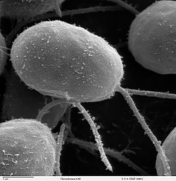
function of flagella
Image credit- FlagellaWikipedia

Function of flagella in eukaryotic cells

  • The flagella in the eukaryotes are much different from that of the prokaryotes in serving its usage. The function of flagella in the eukaryotes strives to be a conserved one and serves for the use of getting transport system for proteins, to serve motility, and works also as a sensory.  
  • The flagella of the eukaryotes like the animals and the plants helps in serving as the motility purpose that shall help in movement and also in chemotaxis. The bacteria have only one flagella and also can have many of them ad can be polar as well. The flagella show a rotationally movement which is counterclockwise that helps in the cell to propel forward and then move.
  • There are not only the flagella but also the cilia that extend on the surface of the eukaryotes. They are commonly in terms associated with locomotion and is by technique linked with the inside of the cell is covered by the extra of the cytoplasmic membrane.
  • The flagella of the eukaryotes unlike the ones that are in the prokaryotes are located in the cytoplasmic membrane. They give the organism the freedom to move back and forth with the flagella spins. There are not found in all eukaryotes but some. While there is a clockwise movement there is a tumble and this helps in getting the movement of organism changed.
  • Both the flagella present in the eukaryotes and the prokaryotes do run in movement being circular in motion of the filaments that helps in propelling the cell or the fluids inside the cell to move past the cell. The movement can also be whip like on addition to being circular as well.
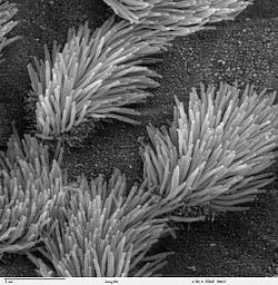
cilia
Image credit- CiliaWikipedia

Function of flagella in algae

  • The flagella is an organelle that helps in the cell to move back and forward On addition to this is also serves uses in organism. In an aqueous surrounding, the flagella mechanism even shows its reaction to the chemical, the mechanical, the light and the gravitational stimulus of then cell. Flagella overall has the same function to perform in any type of cell yet differs in few respect.
  • The flagella on the algae also help in playing a vital role in the sexual fission of the oogamous, the anisogamous and the isogamous species of the algae and mostly the green algae species. They are also called the flagellates for having the whip like flagella. There can be a creation of powers stroke.
  • There is the presence of adenosine triphosphate seen in the algae, the dynein molecule are then activated and the whip like flagella bends as the dynein arms on one side of the dynein tends to cross the bridge and gets them activated and helps in moving up the tube.
  • The species of Chlamydomonas having the flagella has a character that can get converted into the sexual organelle at the time of gametogenesis. They show a specific species adhesion or a reaction of agglutination while in between the cells for the opposite type of mating. This happens due to the presence of a molecule called agglutination that is seen on its surface.

Function of flagella in cells

  • Flagella are said to be a hair like structure that is microscopic and is involved in getting to contribute in the cell movement.
  • The very basic function of flagella is to help the cells or rather any type of cell in their movement. Yet in some of them, the flagella can be seen to serve much other function like as sensory part and more. The definition or types of flagella depend on the usage of them in cells.
  • There are two type of cell seems which the eukaryotic and prokaryotic ones. Both the cells have flagella where the prokaryotes tend to have only one or more and while there are only few eukaryotes that have flagella. There are many types of flagella and are not named after structure but there roles.
  • The flagellum is mostly classified to be a characteristic of the cells of the prokaryotes like the archaea and the bacteria. Along with the prokaryotic cells it is also linked up with the group of protozoan that is also seen in the gametes of the animals, the algae, mosses, the slime molds and the mosses.
  • The motion of the flagella is the cause for the water currents that are needed for the process of circulation and respiration of the sponges and the coelenterates. Most of the bacteria that are considered to be motile are cause of the presence of flagella that helps in the movement.
  • Yet, there is a difference between the pattern of structure for the prokaryotes and the eukaryotes and thus are different. The character of them that is likely is the movement being whip like. The flagella quite resemble that of the cilium in the structure. They have nine pairs of microtubules with each of them having a protein.
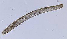
pr
Image credit- ProtozoanWikipedia

Function of flagella in animal cell

  • The flagella are stricture to be defined by their use in the different types of cells rather than their structure.
  • The function of the flagella in the cells of animal is same to that of the purpose of the flagella in the cells of eukaryotes. They helps in locomotion which is the most common function and is then also termed to be a apart of sensory organelle.
  • The animals’ cells do not only have one or more flagella for locomotion but also cilia. They are actually the appendages that are found in almost all the microbes and the animals bit not in the higher level plants. For any of the eukaryotes that have only one cell, both flagella and cilia are needed for movement of them.
  • The use of cilia is to get the water keep moving in relative speed inside the cell concerned with regular process of movement of the cilia. This process can give an end of water the cell moving via the water which is mostly concerned with the single cell species or the second one can be the contents of the moving water across the cell surface.
  • The movement of the flagella for the eukaryotic cells is actually based on the adenosine triphosphate for their energy while in the prokaryotic cells they are based on the energy that is derived from the prokaryotic proton motive force or is termed as the ion gradient across the membrane of the cell.
  • The shape of the flagella is helical in the prokaryotic bacteria and has inclusion like the protein flagella. The flagella base which is called the hook is seen near the surface of cell and is linked with the basal body within the cell envelope. It gets a motion which is circular and is the clockwise way.

Also Read:

The Intricate Functions of the Golgi Apparatus: A Comprehensive Exploration

golgi apparatus function

The Golgi apparatus is a crucial organelle in cellular biology, playing a pivotal role in protein processing, sorting, and lipid biogenesis. This dynamic structure undergoes significant volumetric changes during the interphase of the cell cycle, with its functionality closely linked to the intricate kinetics of vesicular exchange and biochemical conversion. Golgi Apparatus Volume Dynamics during … Read more

Parenchyma Cells Function: In Plants, Xylem, Phloem, Roots

image 2

Parenchyma is a simple yet permanent tissue type that includes a major portion of the ground tissues for the plants with other having vascular ones.

The parenchyma cells are actually said to be alive and these parenchyma cells function is to have-

A parenchyma is a type of tissue that is non-vascular in form and is made of very simple and undifferentiated type of cell that is modifies to play a vital part in for several functions. The other two types of permanent simple tissues are sclerenchyma and collenchyma. The cells in it seem to have very less intercellular space or has no space at all being tightly packed.

The structure of parenchyma cells is that it is a living cell that has a good view of nucleus and the protoplast. The parenchyma cells functions to be isodiametric or is polyhedral in its shape. It can also be seen to be in the shape of oval round, sometimes elongated and polygonal. The cell wall of the parenchyma cells are composed of hemicellulose, cellulose.

parenchyma cells function
Image credit-Parenchyma cellsWikipedia

Plasmodesmata seen to connect the cells of all the tissue of parenchyma. They also have the feature of having many small and tiny vacuoles. In the parenchyma that are older small vacuoles get to link and form a large central vacuole that shall gather tannins or the anthocyanin. The vacuoles are not empty but have water.

Water is found in abundance inside the tiny vacuoles of the parenchyma cells and also serves to be a water reservoir. The parenchyma cells that are for storage might see to have thick walls of xyloglucan which are the endosperm of the date palm. The sugar is held to be used for the purpose of germination and making the wall thinner.

Along with parenchyma cells functions it also has fruits and flowers that have chromoplasts. The cells of parenchyma may have walls that are thick and lignified that helps in act hard to separate from the sclerenchyma. The mechanical strength for the parenchyma cells are taken from the cells property of hydraulic.

The following tend to show the parenchyma cells function-

Energy stored- The parenchyma cells are simple tissue that has cells that are non-specialized with having thin walls. They have cells that are not tight in packaging. It helps in getting the food storage and then provide plant support. The rest part of the part help in storing of nutrients and water. Parenchyma cells perform storage like starch and oils or secretory functions.

Store waste products- The cells of the parenchyma are actually bigger. They also have large vacuoles placed centrally that helps them to have the function of ion regulation and storage along with keeping in waste materials and water. The tissue that is specific for food keeping is made in the parenchyma cells. They keep in resins and gums and form the wood rays that are radially aligned tissues interspersed between the axial elements.

Help in photosynthesis– Some of the parenchyma cells are able to differentiate for phloem and then send special path for the sugars and the products for taking up photosynthesis to travel in the plant. These cells now have the product made from the leaves itself while they make its way via the whole root.

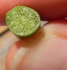
Gaseous exchange- Aerenchym are type of parenchyma cells that help in having the oxygen level maintained via the method of respiration. They are present in continuous form from the roots to shoots and thus help in having the air diffused from the leaves to the roots. They are the cells that are help through maturity.  These store various substances like water, starch, proteins etc. They act as a food and water reservoir. Parenchyma cells may be specialized as a water storage tissue in succulent plants such as Cactus or aloe Vera.

Secretion and transport-Parenchyma with chloroplast that helps in performing photosynthesis is called chlorenchyma. Sclerenchyma tissue, when mature, is composed of dead cells that have heavily thickened walls containing lignin and a high cellulose content with abut 60% to 80%, and serves the function of providing structural support in plants. Sclerenchyma cells possess two types of cell walls: primary and secondary walls.

Types of parenchyma

The parenchyma cells seem to have chloroplasts that are specific to photosynthesis.

The several types of parenchyma are-

Chlorenchyma

These are the parenchyma cells that are inherited with chloroplast and help in photosynthesis by performing the method.

It is seen in the green plants with situated mainly ate the region of sepal or stems. The mesophyll cells of the leaves that differentiate into spongy and palisade cells also have it.

The main criteria for this type of cell is that this type of parenchyma cells functions to repair. In the laves, they create the mesophyll and them are held active for generating photosynthesis and help in exchange of gases. These parenchyma cell types are in the leave mesophyll and are special type of parenchyma cells canned the chlorenchyma.

File:Herbaceous Dicot Stem Collenchyma, Sclerenchyma and Parenchyma in  Cucurbita (37411748021).jpg - Wikimedia Commons
Image credit-ChlorenchymaWikimedia

They are said to be a special type of tissue under parenchyma. It is said to be specific as it has chlorophyll that is green color pigment that helps in getting the process of photosynthesis done. All of the cells of this have same function as it is a permanent and simple tissue and thus all the chlorenchyma cells apart from the one having bulk area works for photosynthesis.

Chlorenchyma are the parenchyma cells which contain chloroplast. The tissue containing chlorenchyma is present in the leaf interiors as well out outer cortex of young stem. It is the tissue which carry out photosynthesis. Chlorenchyma tissues are the types of parenchymatic tissue having chlorophyll. It helps to perform the function of photosynthesis in plants.

Xylem parenchyma

They are the element having the type of complex tissue called to be xylem. The cells of parenchyma of the xylem have functions.

The parenchyma cells functions of the xylem parenchyma are to help in having the fats, the carbohydrates and the water stored for conduction. The plants cells are actually classified based on the functions and its strictures.

Xylem is seen to come from the work xylon that means wood. It was termed by Carl Nageli. It is a type of vascular tissue that is seen in the plants that helps in the primary support of water and also the nutrients from the stem and the leaves. They also provide with mechanical support to the plants. There can be two types of xylem based on its origin.

Xylem - Simple English Wikipedia, the free encyclopedia
Image credit-XylemWikipedia

The first can be primary xylem that generates from procambium and is further staged into metaxylem and the protoxylem. The secondary xylem has its vascular cambium as its origin. Xylem as a whole is made up of four types of elements that are different being vessels, xylem fiber, tracheid and xylem parenchyma.

In simple word, the parenchyma cells that are linked with xylem are called xylem parenchyma. There are two types of the parenchyma inside the secondary structure of xylem. They are the radial parenchyma cells and the axial parenchyma cells. The storage of the protein and fast vary with seasons. There is a migration of nucleus and cytoplasm.

The main attribute of parenchyma cells functions is-

  • Storing of the fats, crystals, starch and fats.
  • The parenchyma xylem cells are linked with the vessels or the tracheid via the outgrowth that are called the tyloses.
  • The parenchyma of xylem is connected with having to maintain the xylem transport tissue.
  • They are help actual for having the vessels restored and the help tracheids function when there is a cavity block as for the air bubble. Cavitation takes place due to the large water tension in the tissues of xylem
  • The parenchyma cells of xylem have the radial conduction going on inside it.

Arenchyma

They are said to be the artificial type of parenchyma Cells. It is made up of the sponge tissues that makes up space for the air.

The space is made in the roots, stems or the leaves of few plants that allot h passing of the gas exchange between the root and the shoot. The space can be said to be the channels and have air in them.

The channels seem to make low resistive for having the gases exchanged like ethylene or oxygen above water and then have the tissues submerged in it. This type of parenchyma cells is mostly common in the wetland and also the aquatic that shall grow in the hypoxic soils. The very word aerenchyma has been taken from a Latin word which means air infusion.

image 3
Image credit-AerenchymaWikipedia

While there is a flood in the soil, hypoxia takes place and the microbes eat up the oxygen fast that the method of diffusion. The presence of the hypoxic in soils is one of the greatest feature of the wetlands. There can be several other reasons for the hypoxia in the soils. In common terms, the lower level of oxygen helps in stimulating the plants and the trees like manganese and iron.

Prosenchyma

These are the type of parenchyma cells that have starch in them with walls made up of lignin mostly seen in Bougainvillea.

They are said to be a special type of cells of the parenchyma ad is also called the transfer cells which is linked in the short path of having the solutes moved or transfer by the cell.

The structure of parenchyma cells is that they are living cells with a clear view of the nucleus and protoplast. The parenchyma cells are either isodiametric or polyhedral in shape. It can also be seen to be oval, round, elongated, and polygonal in shape. The cell walls of parenchyma cells are made up of hemicellulose and cellulose.

A vessel element or vessel member that are also called to be trachea or xylem vessel is one of the cell types found in xylem, the water conducting tissue of plants. Vessel elements are typically found in angiosperms said to be the flowering plants but absent from most gymnosperms such as conifers. Parenchyma is living tissue, and it is a thin-walled and unspecialized structure, and it is adaptable.

They make a good amount of the plant tissue in the ground that helps them specialized in the process of transport, photosynthesis and storage. Parenchyma is an inside part of the vascular tissues that shows a path for having the materials exchanged inside and between the phloem and xylem. They are the ones which have elongated cell type and that interpenetrate the ends. 

Mesophyll parenchyma

Mesophyll is said to be a ground tissue that is seen in between the epidermal layer call of leaves and is made of two kinds if tissue.

The mesophyll is a parenchyma type of tissue that is simple as well as permanent. The palisade parenchyma is placed below the upper surface of the leaf and exactly lower to the epidermis and makes the spongy parenchyma fit the space.

Leaf mesophyll is made up of the parenchyma cells. The elongated parenchyma that is the palisade has several number of large chloroplasts per cycle and is the basic site for photosynthesis in many green plants. Mesophyll is found in both the upper and lower epidermis.

image 4
Image credit-Palisade cellWikipedia

Mesophyll conductance is a vital component of photosynthesis, whose importance for accurate characterization of photosynthetic limitations has increased during the last two decades. Carbon dioxide diffusion across the leaf mesophyll is a complex process implying both biochemical and anatomical factors. Among them, aquaporins, chloroplast distribution, and cell wall thickness are its principal determinants.

The mesophyll is used in having the exchange of gas and also supports in photosynthesis via the chloroplasts. The xylem helps in getting the water transported and also the minerals along the leaves while phloem helps in having the transfer of the products of photosynthesis to the rest part of the green plant. Mesophyll is the internal ground tissue located between the two epidermal cell layers of the leaf.

After the cells of the ground meristem tend to divide and the differentiate, they shall be able to distinguish in several numbers of tissue that consist of the cortex, pith rays and the pith. Inside the leaves, they give rise to many of the parenchyma cells for the mesophyll layers that get involved in for photosynthesis. The palisade layer of mesophyll is quite vital.

Also Read:
7 Chromosome Functions In Animal Cell: Detailed Explanations

1024px Drosophila XY sex determination.svg

Most living cells both plant and animal include chromosomes, which are filamentous structures made of nucleic acids and proteins and that carry genetic information in the form of genes.

A single chromosome is made up of a single DNA molecule wrapped around histone proteins. Information is passed on from one generation to the next via the DNA molecules in the form of genes.

Chromosome functions in animal cell include:

DNA molecules carry specific instructions about every organism. The compact structure of DNA is maintained because the long DNA molecule is wrapped around circular protein molecules called histones, like threads wrapping around a spool. If this arrangement was not maintained, then it would be impossible to fit all the DNA inside the cell nucleus. For example, if not compacted a single DNA molecule in a single human cell is laid out unraveled it would be a massive 6 feet tall.

Detailed discussion of chromosome functions in animal cell:

Chromosomes carry genetic information:

The most basic and vital function that chromosomes perform is to carry the genetic information stored in the DNA from one cellular generation to the next and then from parent to offspring. DNA contains genetic information that is used to perform a variety of biological tasks. These functions are necessary for the organism’s growth, survival, and reproduction.

Chromosomes allow large DNA molecules to be packed inside the cell:

The chromosomal structure makes sure that the DNA molecule is compactly wound around the circular molecules called histones. Without this feature, it would be impossible to compact the huge DNA molecule inside the cell nucleus. Chromosomes can further condense to chromatids during cell division for easier chromosomal distribution.

Chromosome regulates gene action:

Chromosomes not only contain histone proteins but also others that are simply referred to as non-histone proteins. These proteins control gene activity. Cellular molecules that regulate genes can activate or deactivate these proteins at will. The chromosomes can expand and contract in various situations based on the activation and deactivation of these gene regulatory molecules.

Chromosomes carry Mendelian factors:

Considering the chromosomal theory of inheritance, which says that chromosomes carry Mendelian factors. This includes height, hair colour, eye colour, diseases, blood groups and many more. Hence the offspri1ng are equally influenced by the factors coming from both parents

They protect the genetic information:

Chromosomes are protected from chemical (e.g., enzymes) and physical forces by histones and some other proteins. As a result, chromosomes, in turn, safeguard the genetic material i.e. the DNA from damage during cell division when the nuclear membrane dissolves.

Sex chromosomes determine the gender of animals:

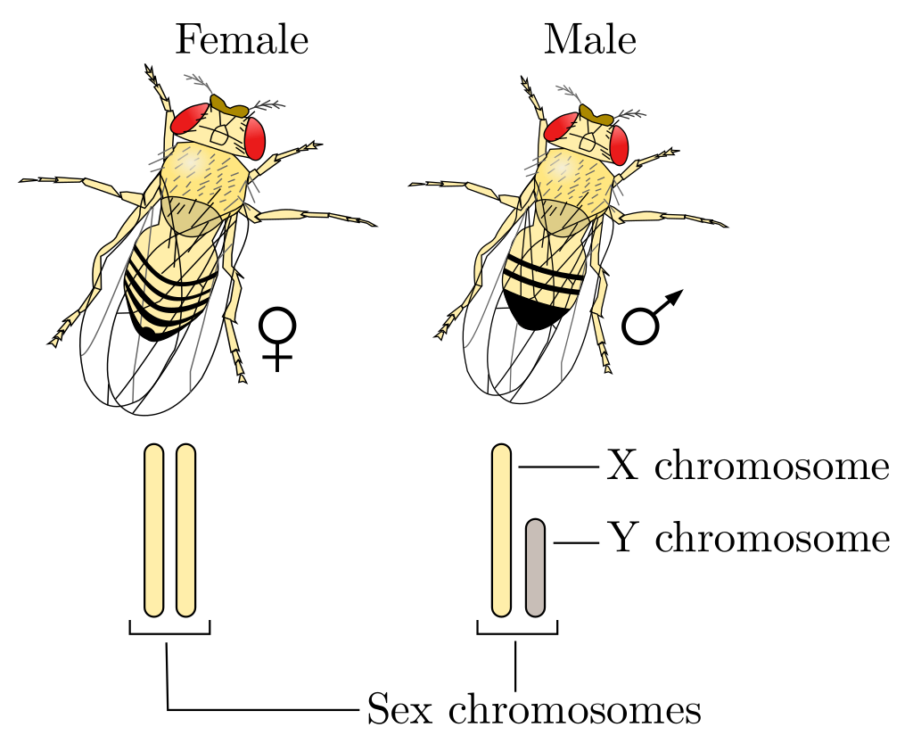
An allosome or simply a  sex chromosome is the type of chromosome that determines a person’s gender. Humans and most other mammals have the X and Y chromosomes. In their cells, males have both X and Y chromosomes, whereas females have two X chromosomes. Hence all egg cells have X chromosomes, whereas sperm cells have either X or Y chromosomes. This system proves that the sex of the offspring after fertilization is dictated by the chromosome of the father rather than the mother.

1024px Drosophila XY sex determination.svg
Drosophila sex chromosomes that work similarly in humans
Image: Wikipedia

Chromosomes allow for the equal distribution of genetic material during cell division:

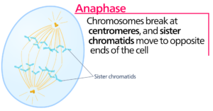
Spindle fibres connect to the centromeres of chromosomes and contract during the anaphase stage of cell division thereby pulling the sister chromatids apart. The contraction spindle fibres ensure that DNA (genetic material) is distributed evenly to daughter nuclei as sister chromatids are separated. The spindle fibres attach to the centromere when they are pulled apart to the sister cells.

1920px Anaphase.svg
How sister chromatids are equally distributed in anaphase due to the contraction of spindle fibers
Image: Wikipedia

How does chromosomal inheritance work?

Technically a child gets one chromosome from either parent to form a pair. Meaning in every pair of chromosomes in a single cell is a sister chromosome derived from the mother and the other from the father. Now it is absolutely luck which of the alleles are more prominently seen in the offspring.

The mitochondrial circular chromosome has a distinct inheritance pattern. Only egg cells, not sperm cells, keep their mitochondria during fertilisation. As a result, a child inherits its mitochondrial DNA from the mother without exception.

 Several human diseases, including various types of hearing loss and diabetes, are thought to be transmitted from the mother via the mitochondrial DNA. Alleles are genetic pairings that influence a variety of our features.

Genetic disorders:

Some common genetic disorders include:

Thalassemia:

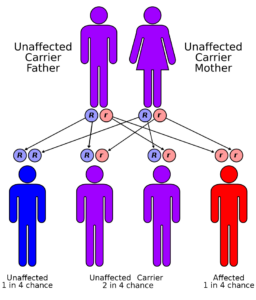
Thalassemia is a group of hereditary genetic disorders that decrease the quantity of haemoglobin a person can make naturally. This disorder prevents oxygen from reaching all parts of the body. Thalassemia is a recessive gene disease. This means if both parents have a thalassemia gene, there is a 25% chance that the child is born with Thalassemia carrying the recessive gene from both parents. There is also a 50%chance of the child being an asymptomatic carrier, which does not affect them greatly but v=can affect their future generation in turn. With any type of Thalassemia, severe anaemia is frequent, demanding specialised care such as regular blood transfusions and chelation therapy.

Sickle cell anemia:

Sickle Cell Disease is another recessive gene hereditary illness that can be passed on to offspring if both parents have the Sickle Cell recessive gene. People having sub-Saharan, Indian, or Mediterranean ancestry are more likely to inherit the characteristic. Red blood cells in Sickle Cell Disease take on a sickle shape instead of the usual concave shape.

chromosome-functions-in-animal-cell
Image showing the autosomal recessive nature of Sickle cell anemia inheritance
Image: Wikipedia

The reason for the shape change is that A single amino acid is mutated in the Hb gene causing the codon to change from GAG to GUC changing the amino acid from Glutamic acid to Valine. This results in the cell changing from its normal concave shape, to a sickle shape, which decreases its surface area and stability. These cells hence end up rupturing and aggregating into lumps clogging blood vessels. Often it can lead to internal bleeding, infections, organ failure or even acute respiratory syndrome.

Down Syndrome:

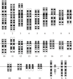
Down syndrome is a disease caused by the abnormality in the chromosome number causing a child to be born with 47 total chromosomes instead of the normally occurring 46. Genes in chromosomes shape and function the body of a baby as it develops throughout pregnancy and after birth. In most cases, a baby is born with 46 chromosomes or 23 pairs.

800px Down Syndrome Karyotype
Image showing karyotype for trisomy 21 in Down Syndrome
Image: Wikipedia

 Down syndrome is caused when one of these chromosomes, chromosome 21, creates an extra copy, resulting in three chromosomes instead of two, resulting in a total count of 47. A chromosome with an extra copy is referred to as a trisomy. Down syndrome is also medically termed trisomy 21. Down’s seriously causes a  baby’s body and brain to develop differently as a result of the additional copy, resulting in physical and metal disruptions to their normal growth.

Also Read:
13 Spliceosome Function:Detailed Facts

images 6 300x290 1

Spliceosome is a multi megadalton ribonucleoprotein complex in eukaryotes, which stimulates RNA splicing mechanisms. Let’s discuss each and every Spliceosome function respectively.

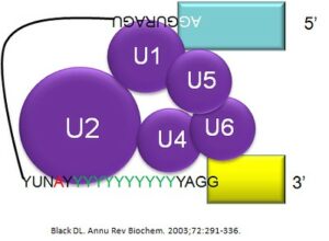
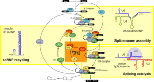
The assembly of Ribonucleic acids and protein elements stimulates the spliceosome function. There are five major Uridine rich  snRNAs known as U-RNAs, such as U1, U2, U4, U5 ,U6 and around 100 protein components are assembled to form spliceosome complexes. Some major functions of spliceosome are mentioned below-

Recognise number of substrates and binds

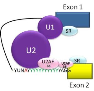
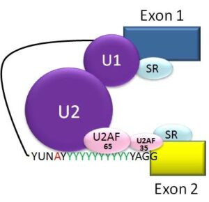
The first function of a spliceosome complex is to recognise the substrate. Here the U1 unit plays a major role and recognises the 5′ splice site of the intron and binds with that. The U2 snRNP recognises the branch point sequence near the 3′ splice site and binds with that, forming A complex. The U4, U5, U6 snRNPs join together and form a B complex. 

Stimulates transesterification

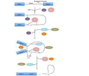
After binding the spliceosome causes two steps transesterification process. In the first transesterification step the 3’ hydroxyl group of adenine nucleotides in the branch point attacks the 5’ splice site of the intron. At second esterification step 3′-hydroxyl of the releasing 5′ exon attacks 3′ splice site and cut the intron between the exons. 

Creates a loop

After the first nucleophilic attack the spliceosome folds the intron structure and causes a loop in the pre-mRNA. This loop-like structure reassembles as the lariat which eliminates after the second nucleophilic attack .

Remove introns

After the nucleophilic attacks the spliceosome causes the intron excision. The two attacks cut each of the splice sites of an intron (non coding sequences of pre-mRNA) and remove it from the structure. 

Ligase exons

After the intron excision process the spliceosome facilities exon ligation. The exon or the coding sequences of pre-mRNA joins together and forms a mature RNA structure.

Involves in different interactive activities within cell

The spliceosome complex is also involved in different kinds of protein -protein interactions, RNA- Protein interactions and RNA-RNA interactions. 

Work as a editor

The spliceosome functions as an editor, thus it edits or removes all the unnecessary area or sequences of precursor RNA and produces a mature RNA.

Finally form a mature RNA from precursor RNA

The main spliceosome function is to produce a mature RNA from the pre-mRNA or hnRNA. It removes all the non-coding regions or introns and joins the exons together. This way the spliceosome produces a mature functional RNA.

Each spliceosome function is facilitated by interactions between the subunits of it. In alternative splicing the exon can be seen. In that case different RNA isoforms are produced through creating exon combinations, from a single transcript.

The spliceosome mediated RNA splicing is only present in eukaryotic cells. In prokaryotes splicing mechanism is not needed. Only tRNA splicing occurs in prokaryotes.

images 6

Spliceosomal A Complex formation from Wikimedia Commons

5514952747 8d57782084 w

Splicesomal B complex formation from Flickr.com

Minor spliceosome function

Minor spliceosome is a ribonucleoprotein complex found in some rare classes of pre-mRNA introns, having U5, U11, U12, U4atac, U6atac and several protein elements as their functional subunits.

The minor spliceosome function is the same as the major spliceosome Function. It forms a  mature RNA from a precursor RNA or hnRNA. Here U11 and U12 snRNPs play crucial roles in recognition of substrates. After that association of U4atac, U6atac and U5 facilitates two step nucleophilic attacks and causes intron excision. After the removal of introns, exons are joined with each other and form a mature RNA.

The minor spliceosome mediated splicing is generally found in some rare classes of eukaryotes such as insects, fungi and plants, etc. 

images 5

Minor spliceosome function from Wikimedia Commons

Spliceosome assembly function

The spliceosome is made up of five major snRNPs such as U1, U2, U4, U5 ,U6 units and around 100 protein subunits. These all subunits assemble to facilitate splicing mechanism and produce a mature RNA structure.

The function of spliceosome assembly is to stimulate splicing and form a mature RNA from a precursor RNA.

The interaction and binding of snRNP U1 to the GU rich 5′ splice site and binding of snRNP U2 to the Branch Point Sequence (BPS) near 3′ splice site, begins the spliceosome assembly functions. It folds the RNA structure and form A complex.

After that U4, U5 and U6 snRNPs interact and bind with the intron structure in close proximity to the Adenine of the branch point, forming B complex. This causes a nucleophilic attack of -OH of the adenine (branch point sequence)  to the 5′ splice site and cut off the end. It gives a loop-like structure called lariat to the intron. After that the newly exposed -OH of 5′ exon attacks on the 3′ splice site. The U5 then removes the intron structure and joins the exons to form a mature RNA.

Spliceosome Function

Spliceosome Function from Wikimedia Commons

Spliceosome components function

Spliceosome contains five major snRNP (U1, U2, U4, U5 ,U6 units)and around 100 protein subunits in its structure. The whole structure is involved actively in the splicing mechanism.

The function of  spliceosomal components like U1 and U2 is to recognise the substrate or the number of substrates in which they have to work. After recognising they bind with the introns at some conserved sequences. The U4, U5, U6 and all other proteins are majorly involved in the intron excision process and exon ligation process.

To know more about RNA splicing steps read our article on RNA Splicing Steps: Detailed Analysis And Facts

Function of spliceosomes in protein synthesis

The spliceosome function in protein synthesis is very crucial. If the non coding sequences or introns are not cut out by spliceosome before the translation, the protein synthesis process is hampered. 

After transcription the precursor RNA has several non coding unnecessary sequences between them. The spliceosome removes all the introns or unessential sequences and joins the exon or the coding sequences together to form a mature mRNA. These mature mRNAs  are then ready to decode specific functional protein elements. This way the spliceosome function is very essential in the protein synthesis process. 

To know more about RNAs read our article on Is RNA Antiparallel: What, Why, Detailed Facts

Small RNAs in spliceosome function

The small RNAs are non coding poly ribonucleotides, having less than 200 nucleotides in their structure. Among several small RNAs, specifically the small nuclear RNAs or snRNAs are used in spliceosome function.

The snRNAs or U-RNAs used in the spliceosomal complex are U1, U2, U4, U5, U6, etc. These snRNAs actively participate and stimulate the whole splicing mechanism. U1 and U2 first recognizes and binds with them. The U4, U5 and U6 stimulate the next splicing mechanism. The U5 is very crucial in the intron removal and exon ligation process.

As a whole we can say that spliceosome function is very important in the RNA translation and protein synthesis process. It produces a mature RNA from precursor RNA. The spliceosome function completely depends on its snRNPs components. Hope this article on spliceosome function will be helpful to you.

Also Read:

Uracil Function in RNA: Detailed Explanations

kj

RNA is said to be a molecule which is similar to that of DNA. Just with the difference of RNA being single stranded.

Uracil function in RNA is just placed to one area which is to pair up with adenine via the hydrogen bond. While the time of base pairing along with adenine, uracil seem to act both as a donor of hydrogen bond and also an acceptor of it. Uracil binds with sugar in RNA.

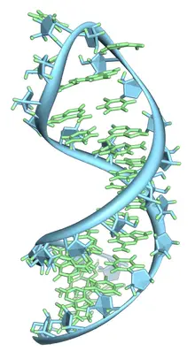
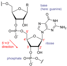
RNA strands do seem to have a backbone and it is formed by sugar called ribose and it placed in alternating manner and also attached to it are the groups of phosphate. There are four bases linked with each of the sugar said to be adenine, guanine, cytosine and uracil.

There are several types of RNA seen in the cell. They are transfer RNA, messenger RNA and ribosomal RNA. In the recent times, there is few tiny RNA that has been seen to be used up in getting the gene expression regulated. RNA is same to that of DNA with just one strand.

As said earlier RNA is quite same to that of a molecule of DNA with just having a variation in its structure. There are several functions of RNA that the cells make it does. One of them can be said to be the messenger RNA or the mRNA. There are also the others serving the goals. The molecules of amino are made to gather into the portion chain.

It is a nucleic acid that informs the molecule to help the conversion of data from the genome to the proteins by the method of translation. Another type of RNA is called the transfer RNA that is the tRNA and they are said to be the non-protein ones that encode the molecules of RNA and helps in getting the amino acid accrued away physically to the site of translation.

RNA as a molecule

RNA is quote of a molecule that is flexible and instructs the protein making industry in the cell of the task that needs to be done.

It helps in storming of the genetic data with making the cell understand the motif of DNA and those acts as a part of helping to start a life. RNA helps in playing a role of a parent in converting the genetic data to the protein in our body.

This is termed to be a good molecule for its helps carry the genetic codes for many of the organism and also it has played its role in making a life start. Along with RNA the DNA makes up he nucleic acid and one of the four or three classes of the major portion of the macromolecules. They are vital for life.

kj
Image credit-
RNAWikipedia

The other parts that make up macromolecule are the lipids and proteins along with a portion of the carbohydrates. Macromolecules are a large part of the molecules and often get to repeat themselves as a subunit. Both the RNA and DNA make up subunits and are called as nucleotides. When taken by mouth- RNA and DNA are quite safe when consumed in the amounts found in food.

The two of the nucleic acids get to team up to make proteins, the process of making up of proteins using up the genetic data in the nucleic acids is vital for the life as said by people. It is called the central dogma of the biology world. The dogma signifies the flow of the genetic data in any organism. Also, RNA is safe for most people when taken along with omega-3 fatty acids and L-arginine.

RNA in simple word can be said to a molecule that links up the DNA and the proteins. The ability of this molecule is to store and get to copy the data depending on the molecule that repeats its nucleotide. The nucleotide is made to organize in specific sequence and can also read the letters in any code. It is used for essential in various biological roles in coding, decoding, regulation and expression of gene.

Uracil structure

Uracil gets to bind with adenine via the hydrogen bond in RNA While in DNA it gets replaced by Thymine.

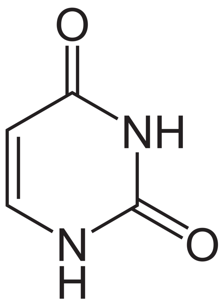
At the time of synthesis, in the RNA strand, the base of uracil inks with adenine and then cytosine pairs up with guanine. The molecular formal for uracil is C4H4N2O2 with it being an organic pyrimidine compound.

Uracil gets to replace thymine as the complementary nucleotide for the base adenine. This means that at the time of elongation process the presence of the base adenine in the template of DNA strand can tell the RNA polymerase to link it with the corresponding place of the growing RNA strand. It is involved with hereditary feature. Found in RNA, it base pairs with adenine and replaces thymine during DNA transcription

uracil function in rna
Image Credit-
Uracil StructureWikipedia

In the molecule of DNA adenine is seen to always pair up with thymine and guanine pairs with cytosine. In RNA, uracil gets to replace with thymine and thus in RNA uracil is said to always pair with adenine. Both uracil and thymine are said to have hydrogen bonds that are two in number and in between them awhile the rest have three. So uracil is the nucleotide that is found almost exclusively in RNA.

As there is a lot similar behavior in structure of purine and pyrimidine it is usually referred to as having double ring with one member adenine and guanine are said to purines. A six member thymine, uracil and cytosine single ring are called to be the pyrimidine. In view of context, uracil is also said to be polar. Uracil is a nucleotide, much like adenine, guanine, thymine, and cytosine, which are the building blocks of DNA, except uracil replaces thymine in RNA.

Uracil replaces thymine as an alternate nucleotide to the base of adenine. This states that the presence or finding of the base for adenine in the DNA strand template during the elongation process or method and can actually instruct RNA polymerase to bind to the appropriate site of the growing RNA strand. It is related to genetic traits. Uracil is a common and naturally occurring pyrimidine nucleobase in which the pyrimidine ring is substituted with two oxo groups at positions 2 and 4.

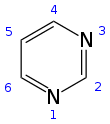
The pyrimidines are the building blocks of DNA and RNA and involved in the formation of active intermediates in carbohydrate and phospholipid metabolism. Pyrimidine synthesis differs from that of purines in that the single pyrimidine ring is first assembled to form orotic acid and then linked to ribose phosphate to form the central pyrimidine nucleotideuridine monophosphate. The pyrimidine bases, uracil and thymine, are catabolized in steps.

jjl
Image credit-Pyrimidine ring-Wikipedia

Uracil vs. thymine

The DNA molecule has in it the bases of adenine, cytosine, guanine and thymine while RNA has in it uracil.

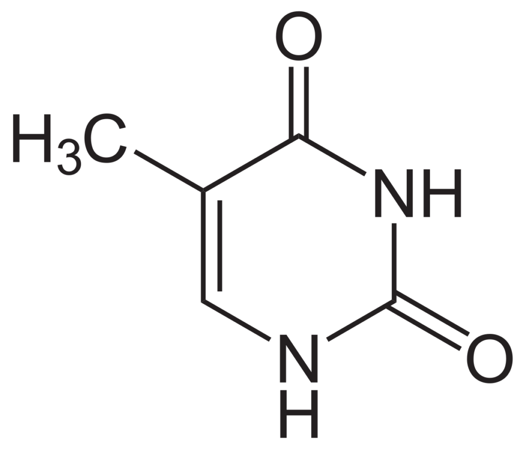
The molecule of RNA has in it uracil whereas the DNA has in it thymine. Thymine is the base that has a group of methyl group at the 5th position of carbon while uracil has the molecule of hydrogen in it at 5th place. Thymine is said to synthesize by uracil.

Uracil as a base is said to be less expensive to make and with less energy and can be responsible for the Uracil function in RNA. Thus having thymine as the normal base can make the detection of general base and repair the incipient sudden changes that are taking place. Uracil is a nucleotide, much like adenine, guanine, thymine, and cytosine, which are the building blocks of DNA, except uracil replaces thymine in RNA

Uracil is a nucleotide, much like adenine, guanine, thymine, and cytosine, which are the building blocks of DNA, except uracil replaces thymine in RNA. So uracil is the nucleotide that is found almost exclusively in RNA. DNA does not use uracil as mostly due to the deamination of cytosine to uracil via hydrolysis-which releases ammonia. When thymine is used in the cell can easily recognized.

tt
Image credit-
ThymineWikipedia

It get seen that the uracil doesn’t belong there and can repair it by substituting it by a cytosine again. DNA uses thymine instead of uracil because thymine has greater resistance to photochemical mutation, making the genetic message more stable. Outside of the nucleus, thymine is quickly destroyed. Uracil is resistant to oxidation and is used in the RNA that must exist outside of the nucleus. In RNA, the thymine is replaced by uracil.

DNA uses up the base of thymine as thymine seem to have better resistance to the mutation that are photochemical and then makes up more specific genetic information and becomes more stable. The difference between both the bases can be-

  • Thymine is seen in the molecule of DNA while Uracil is seen in RNA.
  • In all of the system of biology, thymine is said to be synthesized form the vase of uracil.
  • Thymine has its ribonucleoside as thymidine and uracil has its own as uradine.
  • Thymine has its deoxyriblonucleoside called as deoxythymidine and uracil has the Deoxyribonucleoside as deoxyuridine.
  • The molar mass of uracil is 112.08 g while thymine has its mass as 126.11 g.
  • Uracil serves to be allosteric regulator and also as coenzyme in plants while thymine are derive from the uracil.

Also Read:
Middle Lamella Function: What, Structure FAQs

Plant Cell Wall 300x169 2

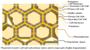
The middle lamella is structurally the last outer layer of the plant cell wall.

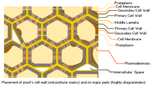
Plant cells wall is made up of three layers in total, among which the middle lamella is the outermost layer lying in contact with other cells or the environment.

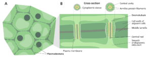
The middle lamella is what connects one adjacent plant cell to another, by forming a cement or jelly-like connective layer. This layer is what allows the cells to communicate with one another and share information and materials by the formation of something called the plasmodesmata.

What is middle lamella?

Essential in plant physiology middle lamella is a special material that joins adjacent plant cells.

Neighboring plant cells adhere or attach to the cells beside or adjacent to them via the middle lamella. The cell wall is composed of three layers among which the middle lamella is the one that is on the outermost edge.

what-is-middle-lamella
Structure of the cell wall showing the middle lamella
Image: Wikipedia

The middle lamella is high in pectin binding the neighboring cells’ main cell walls together. It stabilizes the cells and forms plasmodesmata(small channels that allow adjacent cells to communicate or share materials) between them. The middle lamella is the first layer to be formed from the cell plate splitting the two sister cells, during plant cell cytokinesis.

Middle lamella structure:

  • The plant cell wall is composed of 3 layers-Primary cell wall. Secondary cell wall and the middle lamella. 
  • The primary cell wall is the innermost layer lying in contact with the plasma membrane, while the middle lamella is the outermost layer in contact with adjacent cells or the out environment. The secondary cell wall is sandwiched in between these 2 layers.
  • The middle lamella is essentially composed of peptic polysaccharides just like the cell wall. If the cell is the outermost cell, the middle lamella can also have lignin in its composition for bark formation.
  • The pectins are mainly composed of Calcium and magnesium pectates
  • These polysaccharides are synthesized by the Golgi bodies and transported to the outer layers of the cell wall via exocytosis.
  • Using antibody staining it has been determined that in mature cells the pectins in the middle lamella are partially esterified.
  • The presence of hydroxyproline-rich glycoproteins found only in the cell junctions of the middle lamella is another hallmark of its chemical makeup.

Middle lamella function:

The middle lamella has several functions that can be classified mainly into- mechanical support and cell communication.

Mechanical and structural functions:

  • The intermediate or middle lamella acts as a cementing layer between the major walls of neighbouring cells.
  • The plant cell’s cell wall shields it from mechanical stress. The cell wall offers strength, rigidity, and protection to plant cells, particularly against osmotic lysis.
  • The middle lamella is a portion of the cell wall that connects neighbouring cells. 

Cell communication:

  • Being the outermost layer of the cell wall, they allow the cell to communicate with adjacent cells and the environment.
  • This communication includes the exchange of gases, cellular materials, nutrients and also information.
  • This mainly occurs via plasmodesmata- small pores in the middle lamella that act and channels for the byway transport of materials.
  • As the middle lamella is degraded by enzymes like in the ripening of fruits it allows the cells to separate from each other.
what-is-middle-lamella
Transport of materials via plasmodesmata Image: Wikipedia

How does middle lamella work?

The main job of the middle lamella is to adhere adjacent cells to one another.

The middle lamella’s primary role is to hold neighbouring cells together. Pectin makes up the central lamella, which functions as a gelling agent or glue to keep the plant together.

Plant Cell Wall
Placement of the middle lamella Image: Wikipedia

Adhesion of neighbouring cells is the function of the intermediate lamella in basic words. The middle lamella is a section of the cell wall that links neighbouring (similar or different) cells to form a compact and stable tissue structure. It also facilitates cell communication by forming plasmodesmata between them.

Why middle lamella is optically inactive and amorphous?

The middle lamella of the cell wall is chemically amorphous and optically inactive.

The middle lamella of the cell wall is mainly composed of Calcium pectate, Magnesium pectate, Calcium Carbonate, Calcium Oxalate along with some lignin. The composition of the middle lamella lamella makes it amorphous in nature.

Due to the presence of pectates and proteins, the middle lamella is also optically inactive or isotropic. This is mainly because it is made up of inorganics salts rather than large carbon associated molecules that lack the ability to rotate the plane of plane-polarized light directed at it.

This is very different from the cell wall that is anisotropic or optically active. It may be due to the fact that unlike the middle lamella it is composed of long-chain polysaccharides like cellulose pectin(in the case of bacteria)

Why is the middle lamella of the cell wall important?

The middle lamella is simply an important aspect of plant physiology.

It functions in varied ways to hold the cell compactness together. It helps to keep the cells structure stable by attaching adjacent cells to each other. Not only cells that are side by side but also those above and below the line.

The compact structure of plant tissues are complex and require the cementing property of the middle lamella to keep them stable. A single plant tissue can have more than one type of cell with different structures that must remain attached to each other tightly to allow the passage of water or food molecules from one part of the plant to another.

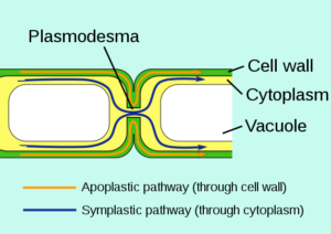
1920px Plasmodesmata en.svg
Structure of plasmodesmata
Image: Wikipedia

 The middle lamella also forms plasmodesmata or small pores that form channels that allow the above transport to function flawlessly. This is because the cell wall is quite rigid which makes it difficult for the exchange of materials possible through it. Without the presence of the plasmodesmata processes like- water transport, food storage and transport or guttation would not be possible in plant systems.

Also Read:
Adenine Function in RNA: Detailed Facts

ij

RNA is a nucleic acid which is same as DNA but RNA is just single stranded. They are used for much purpose.

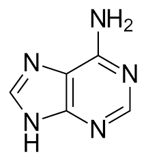
Adenine is one of the double bases that are used in forming of the nucleotides for the nucleic acids structures being the DNA and RNA. The adenine function in RNA is to bind uracil through the two molecules of hydrogen and is a pyrimidine.

Along with the above mentioned adenine function in RNA, adenine is also used up in places within the cell with just not in DNA or RNA, it is a part of the adenosine triphosphate molecule and plays as a source of energy in the cell. It plays a dual role in cell with building RNA and DNA.

Ribonucleic acid is commonly short termed as RNA. It is a molecule which is polymeric and is vital for different biological roles in regulating, coding, expression of genes and decoding. RNA also is vital for synthesis of protein and cellular respiration.

Taking in all the function of adenine in RNA or any other, along with the proteins, the lipids, the carbohydrates, the nucleic acid seems to be of much importance with respect to the four great macromolecules needed for life forms.

The most basic adenine functions in RNA are-

  • It is one of the two based of purines that is used to make nucleotides of the nucleic acids. It helps in bonding with the thiamine in the structure of DNA and also in RNA binds with uracil.
  • They have their role in for cellular respiration in form of NAD, ATP, FAD and synthesis of protein as a chemical for in RNA and DNA.
  • It is an organic and pre base of purine. Thus, have many roles to play in biochemistry mostly in cell to cell respiration. It is also a chemical compounder of RNA.
  • It provides with formation of the bonds by supporting it with energy needed.
  • They link with the phosphate in order to keep energy. When 3P is linked with adenine, it is ATP while in DNA adenine interacts with in the form of AMP. So, in general in DNA the formation of adenine is used as a substrate.
ij
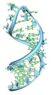
Image Credit- Structure of RNA moleculeWikipedia

Adenine function in RNA as a base

The bases of adenine take up one of the two forms. The purines have a double ring in where a 5 ring atom links to a 6 atom ring.

Pyrimidines are usually a 6 atom ring. The purines in the acids are the adenine and guanine. The pyrimidine refers to uracil, cytosine and the thymine. The chemical formula for adenine is C5H5N5. Most DNA is found inside the nucleus of a cell, where it forms the chromosomes.

The base of adenine binds with thymine in DNA and uracil in RNA. It is a vital base as it is not only supportive in both the nucleic acids strands bit also acts as a source of energy and a carrier for the molecule of ATP, the cofactor for flavin adenine dinucleotide and the factor for nicotinamide adenine dinucleotide. . Chromosomes have proteins called histones that bind to DNA.

The metabolism purines consist of the formation of guanine and adenine. Both of the bases are made for the monophosphate and the present method is still being recognized for getting adenine made in large scale. DNA has two strands that twist into the shape of a spiral ladder called a helix. DNA is made up of four building blocks called nucleotides: adenine (A), thymine (T), guanine (G), and cytosine (C). The nucleotides attach to each other (A with T, and G with C) to form chemical bonds called base pairs.

There are many that tend to confuse both the names of bases which are adenosine and adenine. Both however are not the same. Adenine is the name which is popular to hat if the purine base. Adenine is the great molecule of nucleotide that is made up of pure adenine and the sugar being deoxyribose or ribose and more or one of the phosphate groups.

adenine function in rna
Image Credit- AdenineWikipedia

What is adenine in RNA?

The adenine in RNA is a derivative of purine and is a nucleobase. It is on among the rest three bases.

The rest of the bases except adenine are cytosine, guanine and thymine in DNA. In RNA, the four bases are adenine, cytosine, uracil and guanine. Adenine is represented as A and binds with uracil.

The derivatives of the bases have a good role in biochemistry which consists of getting the protein synthesized and then taking pat is cell respiration. The adenine has its shape complementary to that of wither thymine in DNA or uracil in RNA.

Pure adenine is always said to be a molecule that is independent. When it is connected with DNA there is a formation of covalent bond in between the sugar called the deoxyribose and in the left side bottom nitrogen that gets a help in removing the atom of hydrogen. Genes are short pieces of DNA that carry specific genetic information.

After it the rest of the structures is said to be adenine residue and forms great part of the entire molecule. Adenosine is the adenine that reacts with ribose sugar which is used in RNA and the adenosine triphosphate. The deoxyadenosine is the adenine linked with the deoxyribose and then used to make DNA.

Any of the hard structures are made of smaller blocks, just like a house is made of smaller parts. Living beings are also made in the same way with combining the smaller parts being the molecules made of atoms. Adenine is vital life building block. It is among the major four bases. So adenine plays a dual role in the cell: it’s used for building DNA and RNA, but it’s also used at storing energy in the cell.

The RNA and DNA have the genetic code for all living organism that are the plants, animals, the fungi, humans and other microbes. Adenine helps them to get a stabilized nucleic acid composition for the molecules. It is found in adenosine triphosphate which is a molecule that helps in carrying the enemy needed for life.

hj
Image Credit- RNAWikipedia

Structure of the Adenine RNA molecule

Adenine is one of the rest there molecules found in both of the stands of nucleic acids that is needed for sustaining any of the living being.

Adenines gets top make many types of tautomer and compounds that can fast be inter changed and are generally in common referred to be as same. Yet, in condition being isolated, the adenine tautomer is seen in inert or gas phase.

The metabolism of the purines involves the making of guanine and adenine. Both of the components being guanine and adenine are taken from the nucleotide called the inosine monophosphate that in turn is made to synthesize from the already kept ribose phosphate. Adenine is one of nitrogenous bases utilized in the synthesis of nucleic acids.

This complex is taken as a tunnel and via this pathway of the complex with using the atoms from the amino acids like the aspartic acid, glutamic and glycine atoms and also used up is the coenzyme called the tetrahydrofolate. It also involves the transfer of protein molecules form amino to the ribosomes. Adenine also bonds with Thymine in the DNA structure. Adenine is likewise utilized somewhere else in the cell, in DNA and RNA, however it’s essential for the atom adenosine triphosphate, which is the energy hotspot for the cell.

Some of the molecules of RNA do play a good role in getting the reactions in biology catalyzed by letting the control of gens or communicating with sensing responses and sending back cell signals. One of the vital adenine functions in RNA is getting the synthesis of protein done. In DNA, adenine binds to thymine via two hydrogen bonds to assist in stabilizing the nucleic acid structures. In RNA, which is used in the cytoplasm for protein synthesis, adenine binds to uracil.

Also Read:
Function of plasmid in bacteria: Detailed Facts

aa

Plasmids are the tiny molecule inside the cell which is also an extrachoromosomal DNA and can be separated.

The Function of plasmid in bacteria can differ and serves quite a bit of usage. They help in gene carrying that helps in organism to survive, some are sensory organelle, some help replication.

The genes that they have are for having the organism to survive wither by getting to kill the rest of microbes or also can be by getting to defeat the host cell that is done by generating of toxins.

Some of the bacteria get to work via this process by helping the bacteria in replication of its DNA. As the plasmids are nothing they actually tend to have less genes with some functions. There are provisions for multiple plasmids to exist in the same cell with having different functions.

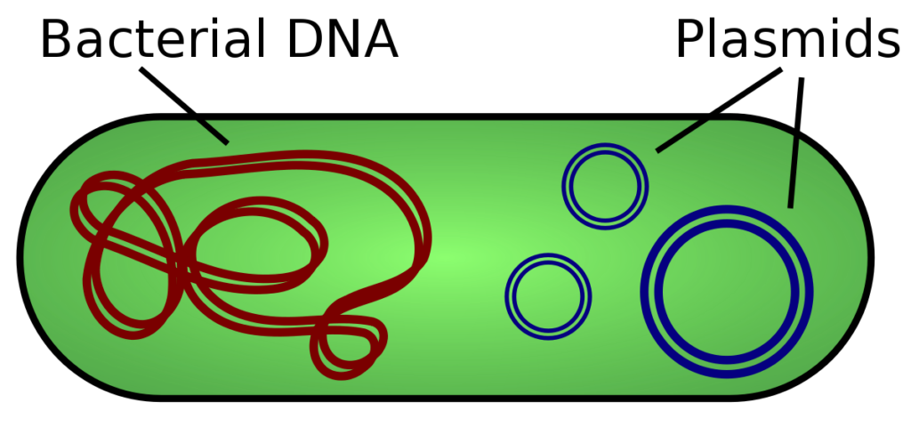
The plasmid can be made to part ways from the chromosomal DNA and can replicate on its own. They are mostly found in as small, being double stranded and in circular shape inside the bacteria with sometimes even showing its presence to archaea and eukaryotes.

The main function of the plasmid in bacteria are-

  • They carry in genes that are antibiotic resistant and then spread them in entire human or the body of animal. This helps the eukaryotes get them treated.  
  • The plasmids on the DNA are also able to produce proteins that are antibacterial.
  • They also are capable of carrying out those genes that are involves in all the metabolic process and let themselves be used u for having the pollutants digest from the outer space.
  • They are also able to let carry genes that are connected with getting the pathogenicity of the bacteria increased that can help cause the diseases like that of tetanus and anthrax.

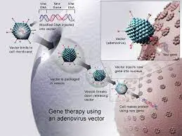
Function of plasmid in bacteria as Gene therapy

Plasmids play a good role in gene therapy. They are most in common to be used for getting the therapeutic genes inserted.

They are also easy to be manipulated and also are easy for the replication in the cells of the bacteria. They have the ability to be efficient to target the cells that shall be defeating and triggering the genes inside them.

They are also no plasmids that are harmful or have any bad effect like that of viral spreaders. The genes is inside the human system for getting them fights against the disease. Gene therapy is the introduction of genes into existing cells to prevent or cure a wide range of diseases.

aa
Image credit- Gene therapyWikipedia

Function of plasmid in bacteria as Recombinant DNA technology

The recombinant DNA technology makes a good use of the plasmids for a lot of purpose.

For the part of the drug delivery, the technology makes a good use of the plasmids to have the likely drugged get inserted in the body. The function of plasmid in Bacteria is much likely and targeted here.

The DNA technology being recombinant which is applied in plasmids for being resistance and can be used up to get the bacteria killed for being harmful to the human cells.

The very first rime when the plasmid was applied inside the human was while insulin was getting inserted inside the human cells. It did give good result and the other application for it was inserting it in the hormone for human growth in the animal’s mammalian cells.

aa 1
Image credit- Recombinant DNA technologyWikipedia

Application of plasmid in bacteria

The humans have been able to develop a lot of uses that can be obtained from plasmid and thus have created software.

The Function of plasmid in bacteria is so diverse that a software has been made that shall be able to record the sequence of DNA of the plasmids for getting it used in many techniques.

The plasmids are sued up in genetic engineering for amplifying or making of many copies of specific genes. In the molecular cloning, a plasmid is considered to be a variable. It is a sequence in DNA that shall help in transferring any foreign genetic particle form one cell to other.

This is done so that the genes can be made to express and also replicated. Plasmids are also used in cloning the tiny segments of the DNA. They can also be made to replicate proteins like that of the protein that are used up to code for insulin in big spaces.

On addition, the plasmids are seen to transfer genes into the human cells as part of the gene therapy. There is investigation or research going on this term. The cells may be lacking few of the proteins if the person has a hereditary disorder that has gene mutation. A plasmid in the DNA shall allow the cells to express a protein that they lack.

Function of plasmid in bacteria
Image credit- PlasmidWikipedia

Characteristic of plasmids

The plasmids can be classifies in many ways with mostly being conjugative and non-conjugative ones.

For the purpose of the plasmids to get into the process of replication on its own, they have to stretch the DNA inside the cell. The DNA that is trenched acts as a self-replicating unit here.

The bacteria replicon is quite typical and may have a number of elements like that of the gene plasmid specific to replication, initiation and protein and the repeating units is called introns along with the DNA boxes and with the adjacent AT-rich area. The ones that integrate are called the episomes.

There are also many small plasmids that make the use of the host replicative enzymes to make themselves copies while the bigger plasmids can carry out genes that is specific to replication with respect to plasmids. There are a few of them that can get inserted into the chromosome which are the host.

The plasmids carry out only one gene. Mostly the genes that they carry on are useful to the host cells like that of the cells that enable them to live in a surrounding that should or else be lethal or restrict their growth. Some genes tend to encode trait for being antibiotic resistance and are unlike toward heavy metals.

Some of the other characteristics of plasmids in bacteria are-

  • They have size ranging from small to being less that 1 kilobase pairs.
  • They are generally circular but can also be linear sometimes.
  • Plasmids vary in different individual with being in range from one to many.
  • The plasmids that are larger in size tend to have less number of copies.
  • The plasmids can be carried on from one cell to other.
tt
Image credit- Self replicating PlasmidWikipedia

Plasmid in bacterial transformation

The bacteria can take in any of the foreign DNA and this is called transformation. It is the key step towards cloning of DNA.

After the very step of transformation, the bacteria are already made to get selected on the plates of antibiotic. The bacteria along with the plasmids are then said to be antibiotic resistant with each making a colony.

The colonies that have the correct plasmids shall be able to grow to make cultures that are bigger if that of the identical bacteria that shall be sued up to make plasmid or making of proteins. The key steps to get the DNA cloned is first transforming and selecting the bacteria.

In any method of general cloning method, the scientist at the basic insert a DNA piece like that of the gene, inside the circle shape DNA and is called the plasmid. The step that is used is restriction enzymes and the DNA ligase and is called the ligation.

After the method of ligation is done, the next way if to get the DNA of travelled inside the bacteria in a way called the transformation. Thus, one can use the process of antibiotic selection and then analyses DNA ways to make sure the bacteria is correct and then contain it in the plasmid.

There are many steps that should be kept in mind while plasmids are being utilized in bacteria transformation. They are-

  • The bacteria that are made special are mixed long with the DNA just like ligation
  • The bacteria are then treated with heat shock that results in some of them to take up on as a plasmid.
  • The plasmids that are used in cloning have a gene that is antibiotic resistance. Thus all the bacteria that are placed on any of the antibiotic plate are selected for the ones that are able to make up on a plasmid.
  • The bacteria that are not able to take on any plasmid are dead. Each of the bacterium having the plasmid produce a cluster that is identical and have plasmid have those bacteria called the colony.
  • There are many colonies that are formed and are checked upon to get them recognized with the correct one like that of PCR or the restriction digestion
  • A colony is made that has all the right plasmids grown in large quantity and is then used to make plasmid or produces proteins.

There always need to be an eye kept on the colonies for a good use of plasmid for transformation. The cause’s are-

  • All the colonies should have plasmids taken up and would be antibiotic resistance.
  • It is however not needed that the plasmid that have the colonies need to be of the same plasmid.
  • When any of the DNA is made to cut and paste, it is possible that the side materials of the products can are formed which was not made to build.
  • The bacteria can be lysed up to release the protein along with gene releasing to express itself having chemical sign.
  • For this reason sometimes the gene can back up by without taking any of the gens and sometimes it can move backwards. It can be complex process depending on the hands.
  • If the gene seems to move backwards, the wrong strand of the DNA shall be transcribed and no protein can be made out of it.
  • In some of the cases, the bacteria can be sued up for building of proteins by using a lot of plasmid DNA. This is also one of the functions of plasmid in bacteria.

Also Read:

Cytosol Function And Structure:Why,How And Detailed Insights

images 300x200 1

Cytosol is one of the major intracellular fluids (ICF), acting as the matrix part of a cell. Here we are going to discuss cytosol functions and structure inside the cell.

As a medium of most of the cellular activities, the cytoplasm performs several functions inside the cell.

  • The cytosol is the cytoplasmic semi-fluid matrix, consisting of several micro and macro bio molecules. With these components of cytosol, it makes a cellular organization and gives structural support to the cell.
  • The cytosol is the main platform for all enzymatic activities within cells. It contains several cellular enzymes and acts as a medium in their activities.
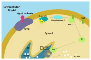
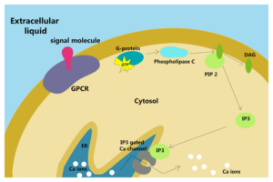
  • In signal transduction procedure, the role of cytosol is immense. The cytosol transport metabolites from their production site to their destinations. 
  • Cytosol performs a significant role in osmoregulation. It contains several inorganic ions which regulate and maintain the osmotic pressure in the cell.
  • The cytosol contains trehalose, an osmoprotectant which helps to survive the cell during adverse situations. It protects all the cellular organelles and biomolecules by turning the cytosol into solid.
  • Cytosol has a major role in generating and maintaining the action potential of the cell.
  • After the division of the membrane, the cytosol performs a significant role in the cytokinesis process and stimulates cell division.
  • In the endocytosis process where metabolites from outside of the cell enters, the cytosol helps to bring them in.
  • Most of the metabolic activities like protein biosynthesis, Pentose phosphate pathway, glycolysis, gluconeogenesis,etc takes place in the cytosol of the cell.
  • Growth and expandation of a cell also depends on the cytosol part of that cell.

Apart from these there are several more cytosol functions included, such as assisting cytoplasm streaming, ion channels maintaining,  etc.

cytosol function,cytosol structure

Cytosol function from Wikimedia

Cytosol Structure

The cytosol is typically a mixture of  ions, small molecules,large water-soluble molecules and water. However, a structural organization can be seen within cytosol. Let’s have a closer look at it.

Concentration Gradient

In this portion of cytosol, small molecular sparks can be produced such as calcium spark, oxygen spark or adenosine triphosphate, etc. These sparks are few micrometers in length and lasts upto only a few milliseconds. These sparks mostly arise from the site near to mitochondria.

Protein complexes

It is an organization of protein associates, where the proteins contain functional similarities. The association of protein complexes allows substrate channeling, important for metabolic pathways. Most enzymes related to the same metabolic pathway are associated to form the protein complexes.

Protein compartments

Protein compartments are a large central association of protein substances. Some common protein compartments are proteasome, containing proteases which can cause degradation of other cytosolic protein elements. Carboxysome compartment contains enzymes necessary for the carbon fixation process.

In case of prokaryotic cells where the organelles lack membrane bound structure, some macromolecules around them undergo  clustering or polymerization and form an organization called biomolecular condensates. 

The cytoskeleton is not considered a part of cytosol but its presence influences the cytosol structure. The cytoskeletal filaments restrict the diffusion of large particles of cytoplasm.

Cytosol Composition

The cytosol is an unequal mixture of several components like water, ions, micro molecules, macromolecules, etc. At different Parts of cytosol the component amounts vary randomly.

Water

The cytosol consists of about 79% of water in it. The pH of the cytosolic water is 7.4. The viscosity of this is the same as the pure water. The cytosolic water influences most metabolic activities within the cytoplasm. 

Inorganic and organic Ions

The cytosol consists of several inorganic ions in it. The amount of these ions differs from in other extracellular fluids. It contains potassium (139-150 millimolar), sodium (12 millimolar), chloride (4 millimolar), bicarbonate (12 millimolar), amino acids (138 millimolar), etc. It also has some amount of magnesium and calcium in it.

Micro And Macro Molecules

About 20%-30% of cytosol consists of protein molecules in it. These protein molecules are mostly enzymes involved in various  metabolic pathways. In case of prokaryotic cells the cytosol also contains the genome of the cell or the nucleoid. Related to these genetic material, replication and transcription enzymes (protein) are also available in the prokaryotic Cytosol.

To know more about DNA replication read our article on DNA Replication Steps and Critical FAQs

Cytosol Location

In case of eukaryotic cells the cytosol is located between the space of cytoplasmic membrane and nuclear membrane. It is the part of cytoplasm avoiding the membrane bound organelles,cytoskeleton and inclusions. 

In case of prokaryotic cells the cytosol is located inside the cytoplasmic membrane. The genome or nucleoid is a situation within the cytosolic matrix of a cell.

To know more about eukaryotic cells read our article on Eukaryotic Cells Examples: Detailed Insights
Cytosol Vs Cytoplasm

The cytosol is a component of cytoplasm itself. Let’s try to find out the difference between them.

TopicCytosolCytoplasmDefinitionThe cytosol is a fluid component of cytoplasm present in cells. The cytoplasm is the component of the cell present inside the cell.
CompositionIt contains ions, water and water soluble proteins. It contains enzymes, lipids, nucleic acids, carbohydrates, ions, etc.
Involved in Metabolic reaction takes place in cytosol. Glycolysis, cell division, etc takes place in cytoplasm.
Main function Transportation of molecules and signal transduction is the main function of Cytosol. The main function of Cytoplasm is to hold all the cellular organelles and other molecules within it, Give protection and serve like a medium of all metabolic reactions.
Components Water, ions and large water soluble molecules are the major components of cytosol. Cytosol, membrane bound organelles and cytoplasmic inclusions are the major components of cytoplasm.
Diversity The diversity is low in case of cytosol. The diversity is high in case of cytoplasm.
Difference between cytosol and cytoplasm
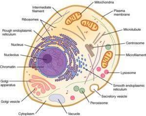
0312 Animal Cell and Components 1

Cytoplasm from Wikimedia

How Does Cytosol Function?

The importance of cytosol in cellular activities is immense. It performs several functions inside the membrane. 

The cytosol is the semifluid gel-like matrix which gives the cellular components structural support. It transports molecules from one to another place of the cell and helps in signal transduction procedure. In prokaryotes it contains the nucleoid and serves as a medium of replication and transcription process.

The components of cytosol are highly related to the efficiency of all metabolic pathways. 

How Many Coenzymes Are Reduced In Cytosol?

During the glycolysis process two acetyl coenzymes are reduced in cytosol.

Here we describe the cytosol function and cytosol structure briefly. We mention the composition of the cytosol. We also state the difference between the cytosol and the cytoplasm of a cell. We also find out how the cytosol functions within the cell. Lastly we can say the role of cytosol in cellular activities is immense.

Also Read: