Is Maltase an Enzyme?

is maltase an enzyme

Maltase is a crucial enzyme involved in the digestion of carbohydrates, specifically the disaccharide maltose. As a hydrolase enzyme, maltase plays a vital role in breaking down maltose into two glucose molecules, which can then be absorbed into the bloodstream and utilized by the body for energy. Understanding Maltase Maltase is found primarily in the … Read more

Is Catalase an Enzyme? A Comprehensive Guide

is catalase an enzyme

Catalase is a ubiquitous enzyme found in the cells of all aerobic organisms, playing a crucial role in protecting cells from oxidative damage caused by the accumulation of hydrogen peroxide (H2O2). This powerful enzyme catalyzes the decomposition of hydrogen peroxide into water and oxygen, making it an essential component of the cellular defense system against … Read more

3 Simple Diffusion Examples: Explanation You Should Know

0305 Simple Diffusion Across Plasma Membrane 1

This article would present a set of simple diffusion examples which would help in the better understanding of types of diffusions in a detailed manner. 

Diffusion is described as the process where the movement of the particles are from the higher concentration level to a lower concentration level. The differences in the concentration of the two areas are termed as the concentration gradient and the process of diffusion continues till the neutralisation of the gradient. 

Passive transport and types 

Passive transport is explained as the movement of the ions or a set of molecular substances within the cells along a specific concentration gradient without the requirement of any form of eternal energy. There are four types of passive transport, simple diffusion, facilitated diffusion, filtration and osmosis.

Simple diffusion

Simple diffusion is the basic movement of the ions or molecular particles from a higher concentration region to a lower concentration region. In cells, the movement is across a semipermeable membrane and occurs without the support of any protein channels. 

For example, diffusion tends to occur in both liquid and gases as the particles have the ability to move freely from one place to the other in a random manner. Similarly, in cells, the movement of the particles occurs in and out through simple diffusion. 
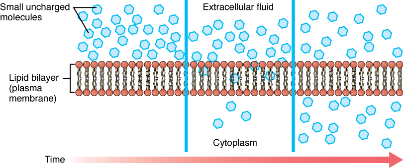
simple diffusion examples
Simple Diffusion from Wikimedia

Facilitated Diffusion

Facilitated diffusion is the type where the transportation of the ions across the semipermeable membrane is done with the support of specific sets of transmembrane integral proteins. These molecules are mostly large in size and are dependent on the carrier substance to pass through the membrane. 

There is no requirement of any external energy for this process. For example, glucose transport or amino acid transport are carried with the help of facilitated diffusion. The cell membrane is permeable to molecules that are non-polar and smaller in size. 

Filtration

The selective absorption of few sets of nutrients within the body is termed as filtration. This process has no requirement of external energy and occurs through the concentration gradient. Here, mostly the solids are filtered from liquids and gases. The substances are mostly soluble in nature and hence can pass easily through the pores.

For example, in our body, the kidney establishes filtration. Here, the blood gets filtered by the glomerulus where the necessary molecules for the body are reabsorbed. 

Osmosis

Osmosis is the process where the water molecules along with other kinds of molecules pass through the semipermeable membrane from a higher concentration to a lower concentration in order to balance the overall contraction level. Osmosis is directly proportional to the concentration gradient and temperature. The greater is the concentration gradient and temperature, the faster would be the process of osmosis. 

For example, there is the absorption of water from the soil by the plants due to the process of osmosis. The concentration of the water is lower in the roots than the soil, hence the flow of water is made into the roots which is further utilised by the plants. 

Simple diffusion examples

Few of the simple diffusion examples in our body are as follows:

Oxygen and Carbon dioxide

It has been identified that movement of gases across the membranes are among the most classic examples for simple diffusion within animals. Both the oxygen and carbon dioxide get dissolved in the blood in order to process the exchange of gases using the simple diffusion method. 

The direction regarding the movement of gases is dependent on the concentration gradient of the gases within the cells. During the time of inhalation, the concentration of oxygen is greater than carbon dioxide within the alveoli than the blood vessels. Hence, the movement of oxygen is into the blood from the alveoli

During the time of exhalation, the concentration of the carbon dioxide is greater than oxygen within the blood vessels than the alveoli. Hence, the movement of oxygen is into the alveoli of the lungs from the blood, which is then exhaled out through windpipe. 

Movement of waste materials

The process of removing waste from the body within animals is processed using simple diffusion. In the liver, the set of waste material called urea is excreted from the blood. In a similar manner, the waste like toxins and chemicals are removed from the kidneys and the absorption of water is processed by the method of simple diffusion. It is often found in collaboration with active transport and osmosis as well. 

Bacteria 

Bacteria are simple organisms which leave them with a limited number of ways to intake the necessary nutrients. Hence, the nutrients are diffused across the cell membrane. Bacteria lack the specialised organelles in transporting substances and hence the reliance is more on simple diffusion. 

Bacteria tend to include facilitated diffusion as well in transporting most nutrients but simple diffusion is important in terms of delivering water, oxygen and few of the small sized nutrients within the cytoplasm. 

Conclusion 

The article concludes that simple diffusion forms an important process in terms of managing life forms on earth which can be noticed through various simple diffusion examples.  

Also Read:

The Intricate Dance of Osmosis and Diffusion: A Comprehensive Guide

is osmosis diffusion

Osmosis and diffusion are fundamental processes in biology that govern the movement of molecules across semi-permeable membranes. These two phenomena are intricately linked, yet distinct, and play a crucial role in maintaining the delicate balance within living organisms. In this comprehensive guide, we will delve into the intricacies of osmosis and diffusion, exploring their mechanisms, factors that influence their rates, and their vital functions in various biological systems.

Understanding Osmosis

Osmosis is the spontaneous movement of water molecules across a semi-permeable membrane from an area of higher water potential (lower solute concentration) to an area of lower water potential (higher solute concentration). This process is driven by the tendency of the system to reach equilibrium, where the water potential on both sides of the membrane is equal.

The water potential (Ψ) of a solution is determined by the equation Ψ = π + P, where π represents the solute potential and P represents the pressure potential. A solution with a higher solute concentration has a lower water potential, and water will move from an area of higher water potential to an area of lower water potential until equilibrium is reached.

The rate of osmosis is influenced by several factors, including:

  1. Concentration Gradient: The greater the difference in solute concentration across the membrane, the faster the rate of osmosis.
  2. Membrane Permeability: The permeability of the semi-permeable membrane to water molecules affects the rate of osmosis. Membranes with higher water permeability allow for faster osmosis.
  3. Temperature: Increased temperature enhances the kinetic energy of water molecules, leading to a higher rate of osmosis.
  4. Pressure: Applying external pressure to the solution can counteract the osmotic flow of water, a phenomenon known as osmotic pressure.

Osmosis plays a crucial role in various biological processes, such as:

  • Plant Cell Turgor: Osmosis helps maintain the rigid structure of plant cells by regulating the water potential and pressure within the cells.
  • Animal Cell Homeostasis: Osmosis is essential for the regulation of water balance and the exchange of nutrients and waste products across cell membranes in animals.
  • Kidney Function: The kidneys rely on osmosis to reabsorb water and essential nutrients from the filtrate, while excreting waste products.
  • Osmotic Shock: Sudden changes in the osmotic environment can lead to cell lysis (bursting) or plasmolysis (cell shrinkage) in both plant and animal cells.

Exploring Diffusion

is osmosis diffusion

Diffusion is the process by which molecules move from an area of higher concentration to an area of lower concentration, driven by the random motion of the molecules and the tendency of the system to reach equilibrium. This movement occurs without the need for external energy input, as the process is driven by the concentration gradient.

The rate of diffusion is influenced by several factors, including:

  1. Concentration Gradient: The greater the difference in concentration between two areas, the faster the rate of diffusion.
  2. Molecular Size and Weight: Smaller and lighter molecules tend to diffuse more rapidly than larger and heavier molecules.
  3. Temperature: Increased temperature enhances the kinetic energy of molecules, leading to a higher rate of diffusion.
  4. Membrane Permeability: The permeability of the membrane to the diffusing molecules affects the rate of diffusion.

Diffusion plays a crucial role in various biological processes, such as:

  • Gas Exchange: Diffusion is the primary mechanism for the exchange of oxygen and carbon dioxide in the lungs and other respiratory systems.
  • Nutrient and Waste Transport: Diffusion facilitates the movement of nutrients, such as glucose and amino acids, into cells, as well as the removal of waste products, such as carbon dioxide and urea, from cells.
  • Cellular Communication: Diffusion of signaling molecules, such as hormones and neurotransmitters, allows for intercellular communication and coordination within an organism.
  • Membrane Transport: Diffusion of small, uncharged molecules, such as oxygen and carbon dioxide, across cell membranes is a crucial aspect of cellular metabolism and homeostasis.

Measuring Osmosis and Diffusion

To quantify the rates of osmosis and diffusion, various experimental techniques have been developed:

  1. Osmosis Measurement:
  2. Dialysis Tubing Experiment: A solution of known concentration is placed inside a dialysis tube, which is then immersed in a solution of different concentration. The change in the volume or concentration of the solution inside the dialysis tube over time can be used to calculate the rate of osmosis.
  3. Plasmolysis Observation: The degree of plasmolysis (cell shrinkage) in plant cells can be observed under a microscope to estimate the rate of osmosis.

  4. Diffusion Measurement:

  5. Fluorescence Recovery After Photobleaching (FRAP): This technique involves selectively bleaching a small region of a fluorescently labeled sample and then measuring the rate at which the fluorescence recovers due to the diffusion of unbleached molecules into the bleached area.
  6. Agar Diffusion Assay: This method involves placing a substance of interest (e.g., an antibiotic) in a well cut into an agar plate and measuring the diameter of the resulting zone of inhibition, which is proportional to the rate of diffusion of the substance.

By understanding the principles and measurement techniques of osmosis and diffusion, researchers and students can gain valuable insights into the fundamental processes that govern the movement of molecules in biological systems.

Conclusion

Osmosis and diffusion are essential processes that underpin the delicate balance and functioning of living organisms. From maintaining the structural integrity of plant cells to facilitating the exchange of gases and nutrients in animals, these phenomena play a crucial role in sustaining life. By exploring the intricacies of osmosis and diffusion, we can better understand the complex mechanisms that govern the movement of molecules across semi-permeable membranes and their vital importance in the natural world.

References:

  1. Biology Junction. (2017). Osmosis Lab Example 2. Retrieved from https://biologyjunction.com/osmosis-lab-example-2/
  2. Southern Biological. (n.d.). Osmosis and Diffusion. Retrieved from https://www.southernbiological.com/osmosis-and-diffusion/
  3. Quizlet. (n.d.). AP Bio Lab Osmosis and Diffusion Flashcards. Retrieved from https://quizlet.com/254765215/ap-bio-lab-osmosis-and-diffusion-flash-cards/
  4. Open Washington Pressbooks. (n.d.). Lab 5. Diffusion and Osmosis. Retrieved from https://openwa.pressbooks.pub/lwtechbiol160/chapter/lab-5-diffusion-and-osmosis/
  5. Alberts, B., Johnson, A., Lewis, J., Raff, M., Roberts, K., & Walter, P. (2002). Molecular Biology of the Cell. Garland Science.
  6. Lodish, H., Berk, A., Zipursky, S. L., Matsudaira, P., Baltimore, D., & Darnell, J. (2000). Molecular Cell Biology. W. H. Freeman.
  7. Campbell, N. A., & Reece, J. B. (2005). Biology. Pearson Education.

Unraveling the Mysteries of Enzyme Denaturation: A Comprehensive Guide

how is enzyme denatured

Enzyme denaturation is a complex and fascinating process that occurs when enzymes are exposed to conditions outside their optimal environment, leading to a loss of their three-dimensional structure and functionality. This comprehensive guide delves into the intricate details of this phenomenon, providing a wealth of information for biology students and enthusiasts alike.

Understanding Enzyme Structure and Function

Enzymes are essential biomolecules that catalyze a wide range of chemical reactions within living organisms. These remarkable proteins are characterized by their unique three-dimensional structures, which are crucial for their ability to recognize and bind to specific substrates, and to facilitate the transformation of those substrates into products.

The structure of an enzyme can be divided into four distinct levels:

  1. Primary Structure: The linear sequence of amino acids that make up the enzyme’s polypeptide chain.
  2. Secondary Structure: The local, regular patterns of hydrogen bonding between the amino acid residues, such as alpha-helices and beta-sheets.
  3. Tertiary Structure: The overall three-dimensional shape of the enzyme, which is determined by the interactions between different parts of the polypeptide chain.
  4. Quaternary Structure: The arrangement of multiple polypeptide chains (subunits) within a single enzyme complex.

These structural features work in harmony to create the enzyme’s active site, a specialized region where the substrate binds and the catalytic reaction takes place. The precise shape and chemical properties of the active site are essential for the enzyme’s ability to perform its function effectively.

Factors Affecting Enzyme Denaturation

how is enzyme denatured

Enzymes are delicate structures, and their three-dimensional conformation can be disrupted by various environmental factors, leading to denaturation. The primary factors that can cause enzyme denaturation include:

  1. Temperature: Enzymes typically function best within a narrow range of temperatures, usually between 20°C and 40°C. Exposing enzymes to temperatures outside this range can disrupt the hydrogen bonds and other non-covalent interactions that stabilize the enzyme’s structure, leading to denaturation.

  2. pH: Enzymes are also sensitive to changes in pH, as the ionization state of the amino acid residues within the enzyme can be altered. Extreme pH values, either too acidic or too basic, can cause the enzyme to denature.

  3. Chemical Agents: Certain chemicals, such as strong acids, bases, organic solvents, and heavy metals, can interact with the enzyme’s structure and disrupt the non-covalent interactions that maintain its three-dimensional shape, leading to denaturation.

  4. Mechanical Forces: Physical stress, such as shear forces or high-pressure conditions, can also cause enzymes to denature by disrupting the delicate balance of interactions that stabilize the enzyme’s structure.

Quantifying Enzyme Denaturation

The degree of enzyme denaturation can be quantified using various techniques, including:

  1. Enzyme Assays: By measuring the rate of reaction between an enzyme and its substrate over time, researchers can determine the enzyme’s activity and how it changes under different conditions. Denatured enzymes will exhibit a decreased rate of reaction compared to their native, functional counterparts.

  2. Spectrophotometry: This technique measures the amount of light absorbed by a solution containing the enzyme, which can provide information about the concentration of native enzyme remaining after exposure to denaturing conditions.

  3. Circular Dichroism (CD) Spectroscopy: CD spectroscopy can be used to detect changes in the enzyme’s secondary and tertiary structure, as denaturation often leads to a loss of the characteristic CD signal associated with the native enzyme.

  4. Differential Scanning Calorimetry (DSC): DSC measures the heat absorbed or released by an enzyme as it undergoes denaturation, providing insights into the thermodynamics of the process and the stability of the enzyme’s structure.

  5. X-ray Crystallography and Electron Microscopy: These structural techniques can be used to visualize the three-dimensional structure of an enzyme before and after denaturation, revealing the specific changes that occur in the enzyme’s conformation.

Factors Influencing the Degree of Enzyme Denaturation

The extent of enzyme denaturation can be influenced by a variety of factors, including:

  1. Enzyme Structure: Enzymes with more complex and fragile tertiary and quaternary structures are generally more susceptible to denaturation than those with simpler, more stable structures.

  2. Enzyme Source: Enzymes from different organisms may exhibit varying degrees of stability and resistance to denaturation, depending on the evolutionary adaptations of the source organism to its environment.

  3. Presence of Cofactors or Stabilizing Agents: Certain ions, small molecules, or other biomolecules can bind to enzymes and help stabilize their structure, making them more resistant to denaturation.

  4. Duration and Intensity of Denaturing Conditions: The longer an enzyme is exposed to denaturing conditions, or the more extreme the conditions, the greater the degree of denaturation that will occur.

  5. Reversibility of Denaturation: In some cases, the denaturation of an enzyme may be reversible, meaning that the enzyme can regain its native structure and function if the denaturing conditions are removed or mitigated.

Practical Applications of Enzyme Denaturation

Understanding the process of enzyme denaturation has numerous practical applications in various fields, including:

  1. Biotechnology and Enzyme Engineering: By studying the factors that influence enzyme denaturation, researchers can design more stable and robust enzymes for use in industrial processes, pharmaceuticals, and other applications.

  2. Food Science and Preservation: Enzyme denaturation is a key factor in food processing and preservation, as it can be used to control the activity of enzymes that contribute to spoilage or undesirable changes in food quality.

  3. Diagnostic and Analytical Techniques: The quantification of enzyme denaturation is essential for the development and optimization of various analytical techniques, such as enzyme-linked immunosorbent assays (ELISA) and enzyme activity assays.

  4. Protein Structure and Folding Studies: Investigations into the denaturation of enzymes and other proteins can provide valuable insights into the fundamental principles of protein structure, stability, and folding mechanisms.

  5. Environmental Monitoring and Bioremediation: Enzymes can be used as sensitive indicators of environmental stress or pollution, as their denaturation can be a marker of the presence of harmful substances or conditions.

Conclusion

Enzyme denaturation is a complex and multifaceted process that is crucial to our understanding of enzyme structure, function, and stability. By delving into the various factors that influence enzyme denaturation and the techniques used to quantify it, this comprehensive guide has provided a wealth of information for biology students and researchers alike. With this knowledge, we can continue to unlock the secrets of these remarkable biomolecules and harness their power for a wide range of practical applications.

References:

  1. Fersht, A. (1999). Structure and Mechanism in Protein Science: A Guide to Enzyme Catalysis and Protein Folding. W. H. Freeman.
  2. Voet, D., Voet, J. G., & Pratt, C. W. (2016). Fundamentals of Biochemistry: Life at the Molecular Level. Wiley.
  3. Whitford, D. (2005). Proteins: Structure and Function. Wiley.
  4. Segel, I. H. (1993). Enzyme Kinetics: Behavior and Analysis of Rapid Equilibrium and Steady-State Enzyme Systems. Wiley-Interscience.
  5. Pace, C. N., Shirley, B. A., McNutt, M., & Gajiwala, K. (1996). Forces contributing to the conformational stability of proteins. FASEB Journal, 10(1), 75-83.

A Comprehensive Guide to Plant Cell Wall Formation: Unraveling the Intricate Biosynthesis and Assembly Process

plant cell wall formation

Plant cell wall formation is a complex and dynamic process that involves the coordinated synthesis, transport, and assembly of various polysaccharides, proteins, and other molecules. This intricate process is crucial for plant growth, development, and adaptation to environmental stresses. In this comprehensive guide, we will delve into the key aspects of plant cell wall formation, providing a detailed and data-driven exploration of the underlying mechanisms.

Quantifying Plant Cell Wall Precursors

The biosynthesis of plant cell wall components begins with the production of various precursor molecules. One of the most critical precursors is UDP-glucose, which serves as a building block for the synthesis of cellulose, hemicelluloses, and pectins – the major structural components of the plant cell wall.

To understand the regulation of cell wall biosynthesis, researchers have employed advanced analytical techniques to quantify the levels of UDP-glucose and other nucleotide sugars. A study by Verbančič et al. (2017) utilized liquid chromatography-tandem mass spectrometry (LC-MS/MS) to measure the levels of these precursors in Arabidopsis thaliana. The researchers found that the concentrations of UDP-glucose and other nucleotide sugars varied significantly depending on the plant’s carbon status, indicating a tight coupling between cell wall biosynthesis and carbon metabolism.

For example, the study reported that the levels of UDP-glucose ranged from 0.5 to 5.0 nmol/g fresh weight in Arabidopsis leaves, with higher concentrations observed in plants grown under high-carbon conditions. This data provides valuable insights into the regulatory mechanisms that govern the availability of cell wall precursors and, consequently, the rate of cell wall formation.

Measuring Cell Wall Polysaccharide Synthesis

plant cell wall formation

In addition to quantifying the precursor molecules, researchers have also developed methods to directly measure the rate of cell wall polysaccharide synthesis. One widely used approach is radioisotope labeling, where plants are fed with radioactive precursors, such as [^14^C]glucose, and the incorporation of the label into cell wall polymers is monitored.

A study by Zabotina et al. (2008) employed this technique to investigate the synthesis of cell wall polysaccharides in Arabidopsis thaliana. The researchers found that the rate of cell wall polysaccharide synthesis varied significantly during plant development and in response to changes in carbon availability. For instance, the rate of cellulose synthesis was highest during the early stages of seedling development, reaching up to 2.5 μg/mg dry weight per hour, and decreased as the plants matured.

Furthermore, the study revealed that the availability of carbon had a profound impact on the synthesis of cell wall polysaccharides. When Arabidopsis plants were grown under high-carbon conditions, the rate of cell wall polysaccharide synthesis increased by as much as 50% compared to plants grown under low-carbon conditions. This data highlights the intricate relationship between carbon metabolism and cell wall formation, underscoring the importance of understanding these processes in the context of plant growth and development.

Imaging Cell Wall Formation

Visualizing the distribution and dynamics of cell wall components is another powerful approach to studying plant cell wall formation. Confocal laser scanning microscopy (CLSM) has emerged as a valuable tool for this purpose, allowing researchers to observe the organization and rearrangement of cell wall structures in living cells.

A study by Xiao et al. (2016) utilized CLSM to investigate the orientation of cellulose microfibrils, the fundamental building blocks of the plant cell wall, in Arabidopsis thaliana. The researchers found that the orientation of cellulose microfibrils underwent dynamic changes during cell elongation, with the microfibrils initially aligning perpendicular to the direction of growth and then gradually reorienting to a more longitudinal arrangement.

Quantitative analysis of the CLSM images revealed that the angle of cellulose microfibril orientation shifted from approximately 90 degrees at the start of cell elongation to around 30 degrees by the end of the process. This data provides valuable insights into the mechanisms that govern the assembly and reorganization of the cell wall during plant growth and development.

Modeling Cell Wall Formation

To further understand the complex process of plant cell wall formation, researchers have developed mathematical models that integrate various biophysical and biochemical factors. One such model, proposed by Somerville et al. (2004), describes the synthesis and assembly of cellulose microfibrils in the plant cell wall.

The model takes into account the physical properties of cellulose microfibrils, such as their bending stiffness and persistence length, as well as the mechanical forces acting on the cell wall. By incorporating these parameters, the model predicts that the orientation of cellulose microfibrils is determined by the balance between the bending stiffness of the microfibrils and the mechanical forces exerted on the cell wall.

Furthermore, the model suggests that the rate of cell elongation is directly linked to the rate of cellulose microfibril synthesis and assembly. Specifically, the model predicts that a higher rate of cellulose microfibril deposition leads to a faster rate of cell elongation, as the newly synthesized microfibrils provide the necessary structural support and reinforcement for the expanding cell wall.

By integrating these quantitative data and modeling approaches, researchers can gain a deeper understanding of the complex and dynamic process of plant cell wall formation, paving the way for advancements in areas such as crop improvement, renewable resource development, and plant growth optimization.

Conclusion

Plant cell wall formation is a multifaceted process that involves the coordinated synthesis, transport, and assembly of a diverse array of polysaccharides, proteins, and other molecules. Through the use of advanced analytical techniques, imaging methods, and mathematical modeling, researchers have been able to quantify and visualize the various aspects of this intricate process.

By understanding the regulation of cell wall precursor levels, the dynamics of cell wall polysaccharide synthesis, the organization and rearrangement of cell wall components, and the biophysical factors that govern cell wall formation, we can gain valuable insights into the mechanisms that underlie plant growth, development, and adaptation. This knowledge can be leveraged to enhance crop productivity, develop sustainable biomaterials, and unravel the complex interplay between plant physiology and the cell wall.

As our understanding of plant cell wall formation continues to evolve, the integration of these diverse approaches will undoubtedly lead to groundbreaking discoveries and innovative applications in the field of plant biology and beyond.

References

  1. Verbančič, J., Lunn, J. E., Stitt, M., & Persson, S. (2018). Carbon Supply and the Regulation of Cell Wall Synthesis. Trends in Plant Science, 23(10), 854-869. https://doi.org/10.1016/j.tplants.2018.06.004
  2. Zabotina, O. A., Avci, U., Cavalier, D., Pattathil, S., Chou, Y. H., Eberhard, S., … & Hahn, M. G. (2008). Mutations in multiple XXT genes of Arabidopsis reveal the complexity of xyloglucan biosynthesis. Plant Physiology, 147(4), 1763-1771. https://doi.org/10.1104/pp.108.119982
  3. Xiao, C., Zhang, T., Zheng, Y., Cosgrove, D. J., & Anderson, C. T. (2016). Xyloglucan deficiency disrupts microfibril organization and cell expansion in Arabidopsis thaliana seedlings. Plant Physiology, 171(1), 1-14. https://doi.org/10.1104/pp.15.01395
  4. Somerville, C., Bauer, S., Brininstool, G., Facette, M., Hamann, T., Milne, J., … & Youngs, H. (2004). Toward a systems approach to understanding plant cell walls. Science, 306(5705), 2206-2211. https://doi.org/10.1126/science.1102765

Globular Protein Functions: A Comprehensive Guide

globular protein functions

Globular proteins are a class of proteins that have a compact, spherical three-dimensional structure, which is crucial for their diverse range of functions, including storage, transport, defense, muscle contraction, and biological catalysis. Understanding the intricate relationship between the structure and function of globular proteins is essential for unraveling the molecular mechanisms underlying various biological processes.

Storage and Transport Functions of Globular Proteins

Globular proteins play a vital role in the storage and transport of various molecules, ions, and small molecules within the body. One prime example is the globular protein hemoglobin, which is responsible for transporting oxygen from the lungs to the tissues throughout the body.

Hemoglobin: Oxygen Transport

Hemoglobin is a tetrameric globular protein found in red blood cells, composed of four polypeptide chains, each containing a heme group. The unique three-dimensional structure of hemoglobin allows it to bind reversibly to oxygen molecules, facilitating their transport from the lungs to the tissues where they are needed for cellular respiration.

  • The heme group in each subunit of hemoglobin contains an iron atom that can bind to an oxygen molecule, forming oxyhemoglobin.
  • The binding of oxygen to hemoglobin is cooperative, meaning that the binding of one oxygen molecule increases the affinity of the remaining binding sites for additional oxygen molecules.
  • This cooperative binding is achieved through the quaternary structure of hemoglobin, which undergoes conformational changes upon oxygen binding, further enhancing the protein’s ability to transport oxygen efficiently.

Ferritin: Iron Storage

Ferritin is another globular protein that plays a crucial role in the storage and transport of iron within the body. Ferritin is composed of 24 subunits that assemble into a hollow, spherical structure, capable of storing up to 4,500 iron atoms.

  • The iron atoms are stored in the form of ferric hydroxide phosphate, which is a non-toxic and readily available form of iron for the body’s needs.
  • Ferritin is primarily found in the liver, spleen, and bone marrow, where it serves as a reservoir for iron, releasing it when the body’s demand for iron increases.
  • The globular structure of ferritin allows for the efficient storage and release of iron, maintaining homeostasis and preventing the toxic accumulation of free iron in the body.

Immune Defense Functions of Globular Proteins

globular protein functions

Globular proteins play a crucial role in the immune system’s defense against pathogens, such as bacteria, viruses, and other foreign invaders. Antibodies, which are globular proteins produced by the immune system, are a prime example of this function.

Antibodies: Pathogen Neutralization

Antibodies are Y-shaped globular proteins produced by B cells in response to the presence of specific antigens, which are molecules or structures found on the surface of pathogens.

  • The variable region of the antibody’s two antigen-binding sites allows it to recognize and bind to a specific antigen, effectively neutralizing the pathogen.
  • The constant region of the antibody can then interact with other components of the immune system, such as complement proteins or phagocytic cells, to initiate the destruction or removal of the bound pathogen.
  • The unique three-dimensional structure of antibodies, with their variable and constant regions, is essential for their ability to recognize and neutralize a wide range of pathogens.

Complement Proteins: Pathogen Lysis

Complement proteins are another class of globular proteins that play a crucial role in the immune system’s defense against pathogens. These proteins are activated in a cascade-like manner upon the recognition of foreign invaders, ultimately leading to the lysis (rupture) of the pathogen’s cell membrane.

  • The activation of the complement system involves the sequential binding and conformational changes of various complement proteins, which are facilitated by their globular structures.
  • The final stage of the complement cascade involves the formation of the membrane attack complex (MAC), a pore-like structure that inserts into the pathogen’s cell membrane, causing it to rupture and leading to the pathogen’s destruction.
  • The intricate three-dimensional structures of the individual complement proteins and their coordinated assembly into the MAC are essential for the efficient and effective elimination of pathogens.

Muscle Contraction Functions of Globular Proteins

Globular proteins also play a crucial role in the process of muscle contraction, which is essential for various bodily functions, such as locomotion, circulation, and respiration.

Myosin: Force Generation

Myosin is a globular protein found in muscle cells that is responsible for generating the force needed for muscle contraction. Myosin is composed of two heavy chains and two pairs of light chains, which assemble into a complex three-dimensional structure.

  • The globular head domain of myosin contains the actin-binding site and the ATPase activity, which allows it to convert the chemical energy of ATP hydrolysis into mechanical force.
  • The tail domain of myosin interacts with other myosin molecules, forming thick filaments that slide past thin filaments composed of the globular protein actin during muscle contraction.
  • The precise three-dimensional structure of myosin, with its head and tail domains, is essential for its ability to generate the necessary force for muscle contraction.

Tropomyosin and Troponin: Regulation of Muscle Contraction

Tropomyosin and troponin are two other globular proteins that play a crucial role in the regulation of muscle contraction. Tropomyosin is a rod-like protein that binds to the actin filaments, while troponin is a complex of three globular proteins that interact with both actin and tropomyosin.

  • In the resting state, tropomyosin blocks the myosin-binding sites on actin, preventing muscle contraction.
  • When calcium ions are released into the muscle cell during the excitation-contraction coupling process, they bind to troponin, causing a conformational change that moves tropomyosin away from the myosin-binding sites on actin.
  • This allows myosin to bind to actin, initiating the sliding of the thick and thin filaments and resulting in muscle contraction.
  • The precise three-dimensional structures of tropomyosin and the troponin complex are essential for their regulatory functions in muscle contraction.

Biological Catalysis Functions of Globular Proteins

Globular proteins also play a crucial role in biological catalysis, serving as enzymes that accelerate the rate of chemical reactions within the body.

Enzymes: Biological Catalysts

Enzymes are globular proteins that function as biological catalysts, increasing the rate of chemical reactions without being consumed in the process. Enzymes have a specific three-dimensional structure that allows them to bind to their substrates and facilitate the chemical transformation.

  • The active site of an enzyme, where the substrate binds and the reaction occurs, is a pocket or cleft in the enzyme’s three-dimensional structure, formed by the specific arrangement of amino acid residues.
  • The shape and chemical properties of the active site are complementary to the substrate, allowing the enzyme to bind and orient the substrate in a way that lowers the activation energy barrier for the reaction.
  • Enzymes can increase the rate of chemical reactions by several orders of magnitude, making them essential for the efficient and regulated functioning of various metabolic pathways within the body.

Enzyme Specificity and Regulation

The specific three-dimensional structure of an enzyme is not only crucial for its catalytic function but also for its specificity and regulation.

  • Enzymes can be highly specific, recognizing and binding to only a single substrate or a small group of structurally related substrates, due to the unique shape and chemical properties of their active sites.
  • Enzyme activity can be regulated through various mechanisms, such as allosteric regulation, where the binding of a regulatory molecule to a site other than the active site can induce conformational changes that affect the enzyme’s catalytic activity.
  • The precise three-dimensional structure of enzymes, including the active site and regulatory sites, is essential for their ability to catalyze specific reactions and be regulated in response to the cell’s needs.

Conclusion

Globular proteins are a diverse class of proteins that play a crucial role in a wide range of biological functions, including storage, transport, immune defense, muscle contraction, and biological catalysis. The unique three-dimensional structure of globular proteins, determined by the specific spatial organization of their amino acid side chains, is the key to their functional versatility.

Understanding the structure-function relationship of globular proteins is essential for unraveling the molecular mechanisms underlying various physiological processes and for developing targeted therapeutic interventions for diseases associated with the dysfunction of these proteins.

References:
– Quizlet, Chapter 5 – Function of Globular Proteins Flashcards, https://quizlet.com/94621129/chapter-5-function-of-globular-proteins-flash-cards/
– ScienceDirect, Globular Protein – an overview, https://www.sciencedirect.com/topics/medicine-and-dentistry/globular-protein
– Khan Academy, Globular proteins structure and function (article), https://www.khanacademy.org/test-prep/mcat/biomolecules/amino-acids-and-proteins1/a/the-structure-and-function-of-globular-proteins
– NCBI, Residue level quantification of protein stability in living cells, https://www.ncbi.nlm.nih.gov/pmc/articles/PMC4128145/
– PMC, Quantitative analysis of visual codewords of a protein distance matrix, https://www.ncbi.nlm.nih.gov/pmc/articles/PMC8815937/

Are Algae Multicellular? A Comprehensive Guide

are algae multicellular

Algae are a diverse group of photosynthetic organisms that can be found in a wide range of aquatic and terrestrial environments. While some algae are unicellular, others have evolved to become multicellular, forming complex structures with specialized cells and tissues. In this comprehensive guide, we will explore the fascinating world of multicellular algae, their characteristics, growth rates, and techniques used to assess their culture quality.

Multicellular Algae: Complexity and Diversity

Multicellular algae can range from simple filamentous structures to highly complex, spherical colonies like Volvox carteri. These algae are composed of multiple cells that work together to perform various functions, such as photosynthesis, reproduction, and structural support.

One of the most well-studied examples of multicellular algae is Volvox carteri, a freshwater green alga that forms a spherical colony. V. carteri is composed of two distinct cell types: small, biflagellate somatic cells near the surface, and larger reproductive cells located inside the sphere. The spheroid of V. carteri is made up of over 95% extracellular matrix, which provides structural support and allows for the coordinated movement of the colony.

Another fascinating example of multicellular algae is the marine alga Ulva meridionalis, which has been found to have the highest growth rate ever reported for a multicellular autotrophic plant. This alga can achieve a fourfold daily growth rate, as measured using the relative growth rate (RGR) formula, which expresses the continuously accelerating growth of algae during the exponential phase.

Assessing Microalgae Culture Quality

are algae multicellular

Maintaining the quality and health of microalgae cultures is crucial for various applications, such as biofuel production, wastewater treatment, and the production of high-value compounds. To assess the quality of microalgae cultures, researchers and industry professionals can employ a variety of techniques, including ATP assays and chlorophyll quantification.

ATP Assays

ATP (Adenosine Triphosphate) assays are a common method used to measure cell viability in microalgae cultures. ATP is a crucial energy-carrying molecule in living cells, and its presence can be used as an indicator of cell activity and overall culture health. However, it’s important to note that ATP assays can overestimate the amount of ATP if exogenous ATP is present in the medium. To overcome this limitation, commercial kits like CheckLite (Kikkoman, Japan) can be used to enzymatically remove exogenous ATP, providing a more accurate assessment of the cellular ATP levels.

Chlorophyll Quantification

Chlorophyll, the green pigment responsible for photosynthesis in algae, can also be used to evaluate the status of the photosynthetic apparatus. Chlorophyll levels are sensitive to changes in environmental factors, such as temperature and pH, making it a useful indicator of the overall health and productivity of the microalgae culture.

Recent studies have employed advanced techniques, such as image-based cell counters with fluorescent filters, to quantify chlorophyll levels in microalgae populations. This approach allows for the rapid and accurate assessment of chlorophyll integrity, providing valuable insights into the physiological state of the culture.

Factors Influencing Multicellular Algae Growth

The growth and development of multicellular algae are influenced by a variety of environmental and biological factors. Understanding these factors is crucial for optimizing the cultivation and production of these organisms.

Nutrient Availability

Multicellular algae, like their unicellular counterparts, require a balanced supply of essential nutrients, such as nitrogen, phosphorus, and trace elements, to support their growth and metabolism. Imbalances or deficiencies in these nutrients can lead to reduced growth rates, altered cell morphology, and decreased overall productivity.

Light Intensity and Photoperiod

Light is a critical factor for the growth and development of multicellular algae, as it drives the process of photosynthesis. The intensity and duration of light exposure can significantly impact the growth rate, pigment production, and cellular composition of these organisms.

Temperature

Temperature is another important environmental factor that can influence the growth and physiology of multicellular algae. Different species of algae have varying temperature optima, and deviations from these optimal ranges can lead to stress responses, reduced growth, and even cell death.

Salinity and pH

The salinity and pH of the growth medium can also have a profound impact on the performance of multicellular algae. Optimal salinity and pH levels can vary depending on the species, and maintaining these parameters within the appropriate range is crucial for maintaining a healthy and productive culture.

Conclusion

Algae can be both unicellular and multicellular, and the study of multicellular algae has revealed fascinating insights into the evolution of complex life forms. From the intricate structure of Volvox carteri to the remarkable growth rate of Ulva meridionalis, multicellular algae continue to captivate researchers and inspire new avenues of exploration.

By understanding the factors that influence the growth and development of these organisms, as well as the techniques used to assess their culture quality, we can unlock the full potential of multicellular algae in various applications, from biofuel production to environmental remediation.

As the field of algal research continues to evolve, the insights gained from the study of multicellular algae will undoubtedly contribute to our understanding of the natural world and the development of innovative solutions to global challenges.

References:

  1. Unicellular to multicellular: What can the green alga Volvox tell us about the evolution of multicellularity and cellular differentiation? (2017, November 28). Retrieved July 9, 2024, from https://blogs.biomedcentral.com/on-biology/2017/11/28/unicellular-to-multicellular-what-can-the-green-alga-volvox-tell-us-about-the-evolution-of-multicellularity-and-cellular-differentiation/
  2. Hiraoka, M., Kinoshita, Y., Higa, M., Mo.H., S.T., A.O., … & A.D. (2020). Fourfold daily growth rate in multicellular marine alga Ulva meridionalis. Scientific Reports, 10(1), 12606.
  3. Microalgae culture quality indicators: a review (2020). Retrieved July 9, 2024, from https://www.tandfonline.com/doi/full/10.1080/07388551.2020.1854672

Do Red Blood Cells Have a Nucleus?

do red blood cells have a nucleus

Summary

Red blood cells, also known as erythrocytes, are the most abundant type of blood cells in the human body. One of the defining characteristics of mature red blood cells is the absence of a nucleus. This unique feature is crucial for their primary function of transporting oxygen throughout the body. During the maturation process, red blood cell precursors, called erythroid cells, undergo a series of transformations that ultimately lead to the extrusion of the nucleus, resulting in the formation of nucleus-free reticulocytes and, eventually, mature erythrocytes.

The Maturation Process of Red Blood Cells

do red blood cells have a nucleus

The maturation of red blood cells, or erythropoiesis, is a complex process that involves several stages. Here’s a detailed overview of how red blood cells lose their nucleus:

  1. Erythroid Precursor Cells: The process begins with the differentiation of hematopoietic stem cells in the bone marrow into erythroid precursor cells, also known as proerythroblasts.

  2. Chromatin Condensation: As the erythroid precursor cells mature, they undergo a process called chromatin condensation. During this stage, the genetic material within the cell’s nucleus becomes more compact and dense.

  3. Nuclear Polarization: The condensed chromatin in the nucleus then polarizes to one side of the cell, a phenomenon known as nuclear polarization.

  4. Nuclear Extrusion: With the help of mitochondria, the polarized nucleus is then extruded from the cell, leaving behind a nucleus-free reticulocyte.

  5. Reticulocyte Maturation: The reticulocytes continue to mature in the bone marrow, gradually losing their remaining organelles, such as mitochondria, to become fully mature red blood cells, or erythrocytes.

This process of nuclear extrusion is a defining characteristic of mammalian red blood cell development and is crucial for the cells’ ability to efficiently transport oxygen throughout the body.

Presence of DNA Fragments in Mature Red Blood Cells

While mature red blood cells do not have a nucleus, recent studies have revealed that they can contain large DNA fragments. These DNA fragments are believed to be derived from tumor cells, both in vivo and in vitro. This discovery has opened up new avenues for the diagnosis and research of tumors and other diseases.

  1. Tumor Cell-Derived DNA: Mature red blood cells have been found to contain long DNA fragments that can be obtained from tumor cells. This finding suggests that red blood cells may serve as a novel liquid biopsy platform for early cancer screening and diagnosis.

  2. Health Status Differentiation: The presence of DNA in red blood cells has also been shown to be useful in distinguishing samples with different health statuses. Researchers have observed significant differences in the gaps and copy number variations of DNA regions between samples from individuals with different health conditions.

  3. Potential Diagnostic Applications: The ability to detect and analyze the DNA fragments present in red blood cells could lead to the development of new diagnostic tools and techniques for early cancer detection and monitoring disease progression.

Immunoregulatory Functions of Red Blood Cells

In addition to their role in oxygen transport, red blood cells have also been found to play a role in regulating the body’s immune function. This is primarily achieved through their interaction with cell-free mitochondrial DNA (mtDNA) and pathogenic DNA.

  1. Binding of Cell-free mtDNA: Red blood cells have the ability to bind to cell-free mtDNA, which can be released from damaged or dying cells. This binding helps to regulate the immune response by preventing the activation of Toll-like receptor 9 (TLR9), a key receptor involved in the recognition of foreign DNA.

  2. Interaction with Pathogenic DNA: Red blood cells can also interact with pathogenic DNA, such as bacterial or viral DNA, through the expression of TLR9 on their surface. This interaction may contribute to the modulation of the immune system’s response to infections.

  3. Role in Tumor Progression and Metastasis: Red blood cells have been found to interact with cancer cells through a protein called galectin-4. This interaction plays an essential role in tumor progression and metastasis, highlighting the multifaceted functions of red blood cells beyond their primary role in oxygen transport.

Conclusion

In summary, while mature red blood cells do not possess a nucleus, they have been found to contain large DNA fragments, which can be derived from tumor cells. This discovery has opened up new possibilities for the development of novel diagnostic tools and techniques for early cancer detection and disease monitoring. Additionally, red blood cells have been shown to play a role in regulating the body’s immune function, further underscoring their importance in maintaining overall health and homeostasis.

References:

  1. Lichtman, M. A., & Graham, R. C. (2012). Lichtman and Graham’s human hematology and clinical hematology. Elsevier Health Sciences.
  2. Zhang, X., Li, Y., Li, J., Xu, Y., Li, Y., Zhang, X., … & Zhang, J. (2023). Mature Red Blood Cells Contain Long DNA Fragments and Could Be Used as a Novel Liquid Biopsy Technology Platform for Early Cancer Screening and Diagnosis of Malignancy. Frontiers in Oncology, 13, 972254.
  3. Socratic. (2016). Do red blood cells have nucleus? Retrieved from https://socratic.org/questions/do-red-blood-cells-have-nucleus
  4. Mohanty, S., & Agrawal, A. (2021). Extracellular Vesicles in Cancer: Biogenesis, Composition, and Role in Metastasis. In Cancer Metastasis (pp. 1-25). Springer, Cham.
  5. Zhang, Y., Zhang, H., Li, Y., Li, Y., & Li, X. (2021). Galectin-4 promotes tumor progression and metastasis by regulating the interaction between red blood cells and cancer cells. Oncogene, 40(37), 5203-5216.

Comprehensive Guide to Animal Cell Mitochondria Functions

animal cell mitochondria functions

Mitochondria are the powerhouses of animal cells, playing a crucial role in energy production, calcium homeostasis, and cellular signaling. These organelles are responsible for the majority of the cell’s ATP generation through the process of oxidative phosphorylation. Understanding the various functions of mitochondria in animal cells is essential for researchers and students alike, as it provides insights into cellular metabolism, disease pathogenesis, and potential therapeutic interventions.

Mitochondrial DNA (mtDNA) Content

Mitochondria possess their own genetic material, known as mitochondrial DNA (mtDNA), which is a circular, double-stranded DNA molecule. The number of mtDNA copies per cell can vary significantly, ranging from a few hundred to several thousand, depending on the cell type and energy demands. Quantifying the mtDNA content is an important indicator of mitochondrial content and can be measured using real-time PCR (qPCR) techniques. For example, a study on mouse embryonic fibroblasts found an average of 5,000 to 10,000 mtDNA copies per cell, with variations observed between different cell lines and culture conditions.

Mitochondrial Membrane Potential (ΔΨm)

animal cell mitochondria functions

The inner mitochondrial membrane is highly impermeable, creating an electrochemical gradient, known as the mitochondrial membrane potential (ΔΨm). This potential is essential for the production of ATP and the regulation of various mitochondrial functions. The ΔΨm can be measured using fluorescent dyes, such as tetramethylrhodamine methyl ester (TMRM) or JC-1, and flow cytometry or fluorescence microscopy. A decrease in ΔΨm is often indicative of mitochondrial dysfunction and can be observed in various pathological conditions, such as neurodegenerative diseases and cancer. For instance, a study on human neuroblastoma cells reported a significant reduction in ΔΨm upon exposure to oxidative stress, leading to apoptosis.

Reactive Oxygen Species (ROS) Production

Mitochondria are a major source of cellular reactive oxygen species (ROS), such as superoxide (O2•-) and hydrogen peroxide (H2O2), which are byproducts of the electron transport chain. Excessive ROS production can lead to oxidative stress and damage to cellular components, including proteins, lipids, and DNA. ROS levels can be measured using fluorescent probes, such as 2′,7′-dichlorodihydrofluorescein diacetate (H2DCFDA), or by assessing the oxidation of specific substrates, such as NAD(P)H. For example, a study on mouse cardiomyocytes found that exposure to high glucose levels increased mitochondrial ROS production, leading to impaired mitochondrial function and cell death.

Bioenergetic Capacity

Mitochondria are responsible for the majority of cellular ATP production through the process of oxidative phosphorylation. The bioenergetic capacity of mitochondria can be measured using techniques such as high-resolution respirometry, which quantifies the oxygen consumption rate (OCR), or the Clark electrode, which measures oxygen tension. These methods provide insights into the overall mitochondrial function and can be used to assess the impact of various interventions or disease states on cellular energetics. For instance, a study on human skeletal muscle cells found that exercise training increased mitochondrial respiratory capacity and improved overall bioenergetic function.

Calcium Retention

Mitochondria play a crucial role in the regulation of cellular calcium (Ca2+) homeostasis. They can sequester and release calcium ions, which can influence various cellular processes, such as signaling pathways and apoptosis. Calcium retention can be measured using fluorescent probes, such as Fura-2, or by assessing the uptake and release of calcium ions. Alterations in mitochondrial calcium handling have been associated with various pathological conditions, including neurodegenerative diseases and cancer. For example, a study on mouse cortical neurons reported that mitochondrial calcium overload led to the activation of apoptotic pathways and neuronal cell death.

Mitochondrial Morphology and Dynamics

Mitochondria are highly dynamic organelles that can undergo fusion, fission, and movement within the cell. These morphological changes and dynamic behaviors are essential for maintaining mitochondrial function and cellular homeostasis. Mitochondrial morphology and dynamics can be visualized using microscopy techniques, such as widefield fluorescence and confocal microscopy, which can provide information on mitochondrial shape, size, and movement. For instance, a study on human fibroblasts found that the disruption of mitochondrial dynamics, leading to fragmented and dysfunctional mitochondria, was associated with the development of Parkinson’s disease.

Mitochondrial Transport

The movement of mitochondria within the cell, along microtubules, is crucial for the distribution of these organelles to areas with high energy demands, such as synapses in neurons. Mitochondrial transport can be analyzed using live-cell imaging techniques, which allow the tracking of individual mitochondria and the quantification of their movement. Alterations in mitochondrial transport have been linked to various neurological disorders, as well as cancer metastasis. For example, a study on mouse hippocampal neurons reported that the disruption of mitochondrial transport led to synaptic dysfunction and neurodegeneration.

Mitochondrial Biogenesis

Mitochondrial biogenesis, the process of generating new mitochondria, is essential for maintaining cellular energy homeostasis and adapting to changing energy demands. The rate of mitochondrial biogenesis can be measured by assessing the synthesis rate of individual mitochondrial proteins using mass spectrometry. This approach provides insights into the dynamic regulation of mitochondrial content and function in response to various stimuli or disease states. For instance, a study on human skeletal muscle cells found that exercise training increased the expression of key regulators of mitochondrial biogenesis, leading to an expansion of the mitochondrial network and improved metabolic capacity.

In conclusion, the functions of mitochondria in animal cells are multifaceted and crucial for cellular homeostasis. By understanding the various parameters that can be used to assess mitochondrial structure, function, and dynamics, researchers and students can gain valuable insights into the role of these organelles in health and disease. This comprehensive guide provides a detailed overview of the key aspects of animal cell mitochondria functions, equipping you with the necessary knowledge to explore this fascinating field of study.

References:

  1. Picard, M., Taivassalo, T., Ritchie, D., Wright, K. J., Thomas, M. M., Romestaing, C., & Hepple, R. T. (2011). Mitochondrial structure and function are disrupted by standard isolation methods. PLoS One, 6(3), e18317. doi:10.1371/journal.pone.0018317
  2. Pham, A. H., McCaffery, J. M., & Chan, D. C. (2012). Mouse lines with photo-activatable mitochondria to study mitochondrial dynamics. Genesis, 50(11), 833-843. doi:10.1002/dvg.22050
  3. Mishra, P., & Chan, D. C. (2016). Metabolic regulation of mitochondrial dynamics. The Journal of Cell Biology, 212(4), 379-387. doi:10.1083/jcb.201511036
  4. Wai, T., & Langer, T. (2016). Mitochondrial dynamics and metabolic regulation. Trends in Endocrinology and Metabolism, 27(2), 105-117. doi:10.1016/j.tem.2015.12.001
  5. Nunnari, J., & Suomalainen, A. (2012). Mitochondria: in sickness and in health. Cell, 148(6), 1145-1159. doi:10.1016/j.cell.2012.02.035

Reference Links:

  1. Methods to Evaluate Changes in Mitochondrial Structure and Function in Oncology Applications
  2. Guidelines on experimental methods to assess mitochondrial function in cellular models of neurodegenerative diseases
  3. Mitochondrial Metabolic Function Assessed In Vivo and In Vitro
  4. Cell-Based Measurement of Mitochondrial Function in Human Airway Smooth Muscle Cells
  5. Mitochondrial Dysfunction in Cancer: From Mechanisms to Therapeutic Opportunities
  6. Mitochondrial Dynamics in Health and Disease
  7. Mitochondrial Quality Control in Health and Disease
  8. Mitochondrial Metabolism in Cancer: From Biology to Therapy
  9. Mitochondrial Dysfunction in Neurodegenerative Disorders
  10. Mitochondrial Dynamics and Metabolism in Cancer