Sialic acids are found in animal tissues and other microorganisms, and human brain cells contain that in high amount.
For the different linkage, different verities of Sialic acid structure can be seen which are part of glycoproteins, glycolipids, produces different biochemical functions which are described in this article.
Structure of Sialic acid:
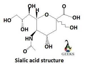
Sialic acid structure is an alpha- keto acid sugar with a skeletal of nine Carbon atoms, which is obtained from a molecule, neuraminic acid by replacing with amino group of one of its hydroxyl group (-OH).
Nearly 50 types of derivatives of Sialic acid structure are known now. In the heterocyclic ring, at the position of Carbon-5, amino group is present and at Carbon-1 a carboxyl group is present which contains negative charge, makes Sialic acid strong organic acid.
The amino group is usually acetylated means replacing one Hydrogen atom with acetyl group, which produces N-acetylneuraminic acid, the common form of Sialic acid.

Numbering of the Carbon atom of Sialic acid structure:
The numbering of the Carbon atoms of the Sialic acid structure starts from the Carboxylate Carbon atom means it is C-1 and then continues along with the chain.
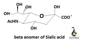
There are two structural configurations, one is alpha (α) anomer and another is beta (β) anomer. If the Carboxylate group at C-1 is in the axial position then the configuration is called σ-anomer and if it is on equatorial position then called β-anomer.

Mainly over 90% the beta anomer is formed.

Diversity in the linkage:
Different types of alpha (α)- linkages are formed Carbon-2 of Sialic acid and sugers, beside this common linkages are occurs to the Carbon-3 or Carbon-6 positions of the galactose residues.
Sialic acids can cover the internal positions within glycans, among which most common is Sialic acid is when attached to another, usually at the Carbon-8 position. The Carbon-5 position can be attached with N-acetyl group or a hydroxyl group.
The Carbon-1 which is a carboxyl group generally remains as ionized form at physiological pH, but it also can be condensed in the lactone form which has hydroxyl group or in the lactam form with a free amino group at Carbon-5 position.
Chemical diversity of Sialic acid structures contribute in the formation of the variety of glycan structures on the cell surface, results in the formation of different cell types.
Biosynthesis of Sialic acid:
Sialic acid is synthesized from glucosamine 6 phosohate and acetyl coenzyme A (deliver acetyl group) through a transferase, enzyme which catalyses the transfer of a specific functional group; results N-acetylglucosamine- 6-P formation.
This product becomes N-acetylmannosamine-6-P through epimerization process, where epimer is transformed to its diastereomeric other part. This reacts with phosphoenolpyruvate (ester derivative from enol of pyruvate and phosphate).
The above reaction produce N-acetylneuraminic-9-P compound which is called Sialic acid. To turn it active for biosynthesis process a monophosphate nucleoside is added with the Sialic acid, results of the formation of cytidine monophosphate-sialic acid.
Function of Sialic acid:
The functions of Sialic acids are generated due to the strong electro- negative charge over it, as it can bind and transport ions (cation) through electrostatic attraction and also stabilize the conformation of proteins including enzymes also.
For the charge Sialic acid carrying, it can increase the viscosity (the resistance power to deformation) of mucin, high molecular weight proteins that can form gel (main component of mucus), protects molecules and cells from attack by glycosidases, increases lifetime.
It controls the affinity of the receptors, protein structures which can bind with chemical messengers and cause change in electrical activity of cell and can report to the modulate processes involve membrane signaling, growth etc.
Hydrophilic nature of Sialic acid structure:
On the surface membranes glycoconjugates are found, containing Sialic acid rich oligosaccharides which help to keep water molecule at the surface of the cell.
As the carboxylate group of the Carbon-1 remains as negatively charged and the Sialic acid remains in the terminal position of carbohydrate chain, so the Sialic acid rich regions create a negative charge on the cell surface.
Water is polar molecule which has partial positive charge on the Hydrogen atoms, so it is attracted to the negatively charged cell surface and membrane.
Acts as masking component:
Erythrocytes and other blood cells are covered by a dense layer of Sialic acid during their normal life time and is removed step wise by hydrolysis. Finally the unmasked cell is destroyed by phagpcytosis, so Sialic acid protects them.
Acts as recognition sites:
Sialic acid structures (components of glycan ligands) are recognized by molecules which belong to Lectin.
Plants don’t have Sialic acids, so these Lectins can help to defense against the microorganisms which contain Sialic acids. In Sialic acid structure the Carbon-1 which contains Carboxylate group (negatively charged) is proven critical for recognition.
Conclusion
For the carboxylate group present in the Sialic acid structure, the negative charge density generated which is one of the cause of the different functions.

Hi…I am Triyasha Mondal, pursuing M.Sc in Chemistry. I am an enthusiastic learner. My specialization is in physical chemistry.
Let’s connect through LinkedIn: