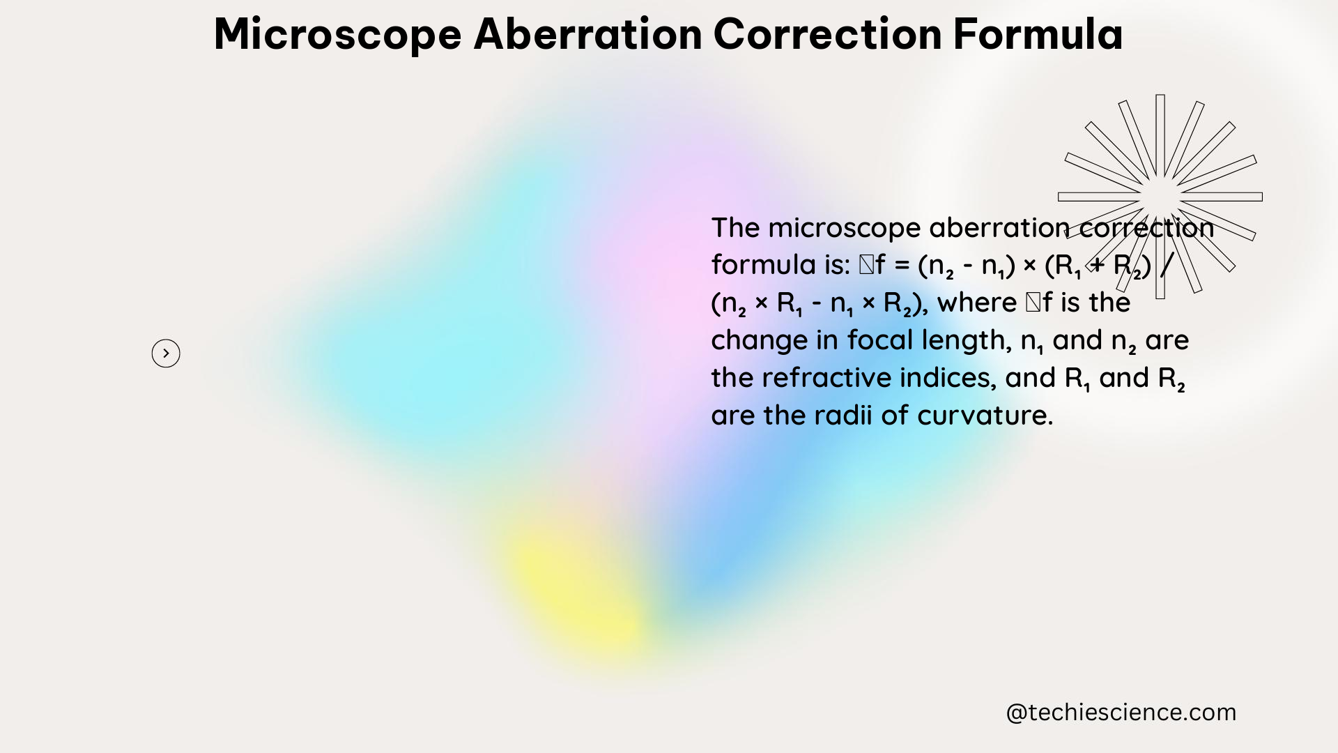
Microscope aberration correction is a critical aspect of microscopy that aims to minimize or eliminate optical aberrations, which can significantly degrade image quality. This comprehensive guide delves into the intricacies of microscope aberration correction, providing a detailed exploration of the underlying principles, mathematical formulations, and practical applications.
Understanding Microscope Aberrations
Microscope aberrations are optical imperfections that can distort the image formed by the microscope’s objective lens. The most common types of aberrations include:
- Spherical Aberration: Occurs when the lens fails to focus all rays of light to a single point, resulting in a blurred image.
- Coma: Causes off-axis points to appear distorted, with a comet-like tail.
- Astigmatism: Occurs when the lens has different focal lengths in different planes, leading to a non-uniform focus.
- Field Curvature: Causes the image plane to be curved rather than flat, resulting in some areas being out of focus.
These aberrations can be caused by various factors, such as the refractive index mismatch between the sample and the immersion medium, cover glass thickness variations, and the objective’s design and quality.
Zernike Polynomial and Aberration Measurement

The Zernike polynomial is a widely used mathematical tool for describing and measuring microscope aberrations. This set of orthogonal functions can represent various types of aberrations, including spherical aberration, coma, and astigmatism.
The Zernike polynomial can be expressed as:
Z(ρ, θ) = Σ a_n^m * R_n^m(ρ) * cos(mθ)
where:
– ρ and θ are the normalized polar coordinates of the pupil plane
– a_n^m are the Zernike coefficients, which represent the magnitude of each aberration term
– R_n^m(ρ) are the radial polynomials
By fitting the wavefront of the microscope’s objective to the Zernike polynomial, the coefficients of each aberration term can be obtained, which can then be used to correct the aberrations.
Aberration Correction Techniques
There are several methods for correcting microscope aberrations, including:
-
Objective Correction Collars: These allow for the adjustment of the lens elements to accommodate cover glass thickness variations and minimize spherical aberration.
-
Computational Aberration Determination and Correction: This approach involves quantifying 4D-STEM data and applying phase retrieval algorithms to correct aberrations.
-
Adaptive Optics: Uses deformable mirrors or spatial light modulators to dynamically correct aberrations in real-time.
-
Pupil Engineering: Employs specialized pupil filters or apertures to selectively block or modify the wavefront and correct aberrations.
Each of these techniques has its own advantages and limitations, and the choice of method depends on the specific requirements of the microscopy application.
Practical Applications and Case Studies
Microscope aberration correction has been successfully applied in various research fields, including:
-
In vivo Sample Aberration Correction in Nonlinear Microscopy: A study used the Zernike polynomial to measure and correct spherical aberration, demonstrating improved image quality.
-
Spectrometer Aberration Correction in Electron Energy Loss Spectroscopy (EELS): Researchers used the Zernike polynomial to measure and correct geometric aberrations and distortions in EELS.
-
Adaptive Optics for Confocal Microscopy: Deformable mirrors were used to dynamically correct aberrations in confocal microscopy, enhancing resolution and contrast.
-
Pupil Engineering for Super-Resolution Microscopy: Specialized pupil filters were employed to improve the resolution and signal-to-noise ratio in structured illumination microscopy.
These case studies highlight the importance of microscope aberration correction and the diverse range of applications where it can be effectively utilized.
Conclusion
Microscope aberration correction is a crucial aspect of modern microscopy, enabling researchers to obtain high-quality, artifact-free images. By understanding the underlying principles, mastering the mathematical tools, and applying the appropriate correction techniques, scientists can unlock the full potential of their microscopy setups and push the boundaries of scientific discovery.
References
- Booth, M. J. (2007). Adaptive optics in microscopy. Philosophical Transactions of the Royal Society A: Mathematical, Physical and Engineering Sciences, 365(1861), 2829-2843.
- Deng, Y., Cao, P., & Zhu, X. (2020). Measurement and correction of in vivo sample aberrations in a nonlinear microscope. Biomedical Optics Express, 11(1), 262-275.
- Krivanek, O. L., Dellby, N., & Lupini, A. R. (1999). Towards sub-Å electron beams. Ultramicroscopy, 78(1-4), 1-11.
- Sheppard, C. J. (2015). Pupil filters for high-resolution microscopy. Journal of the Optical Society of America A, 32(10), 1855-1869.
- Zernike, F. (1934). Beugungstheorie des Schneidenverfahrens und seiner verbesserten Form, der Phasenkontrastmethode. Physica, 1(8), 689-704.

The lambdageeks.com Core SME Team is a group of experienced subject matter experts from diverse scientific and technical fields including Physics, Chemistry, Technology,Electronics & Electrical Engineering, Automotive, Mechanical Engineering. Our team collaborates to create high-quality, well-researched articles on a wide range of science and technology topics for the lambdageeks.com website.
All Our Senior SME are having more than 7 Years of experience in the respective fields . They are either Working Industry Professionals or assocaited With different Universities. Refer Our Authors Page to get to know About our Core SMEs.