The Vital Role of Amino Acids in the Cytoplasm: A Comprehensive Guide

amino acids in cytoplasm

Amino acids in the cytoplasm play a crucial role in various cellular processes, including protein synthesis, energy metabolism, and signaling pathways. The concentration and composition of cytoplasmic amino acids are tightly regulated and can provide valuable insights into cellular physiology and pathology. Understanding the Cytoplasmic Amino Acid Pool The cytoplasmic pool of amino acids is … Read more

When Does the Cytoplasm Divide: A Comprehensive Guide

when does the cytoplasm divide

The cytoplasm, the jelly-like substance that fills the interior of a cell, undergoes a crucial division process called cytokinesis, which occurs after the nuclear DNA has been replicated and segregated during the mitotic phase of the cell cycle. This intricate process ensures the equal distribution of cellular contents into two daughter cells, enabling the continuation … Read more

Can RNA Leave the Nucleus? A Comprehensive Guide

can rna leave the nucleus

RNA, specifically messenger RNA (mRNA), can indeed leave the nucleus through specialized structures called nuclear pores. The process of mRNA export from the nucleus is a complex and tightly regulated event that involves several steps, including nuclear processing, docking at the nuclear pore complex, translocation through the central channel, and cytoplasmic release. This intricate process … Read more

Does Glycolysis Occur in the Mitochondria?

does glycolysis occur in the mitochondria

Glycolysis is a fundamental metabolic pathway that occurs in the cytoplasm of cells, where it breaks down glucose into two molecules of pyruvate, producing a net gain of two ATP molecules and two NADH molecules. However, recent studies have revealed that some eukaryotic organisms, particularly within the stramenopiles group, exhibit a unique form of glycolysis … Read more

Are Ribosomes Found in the Nucleus?

are ribosomes found in nucleus

Ribosomes, the cellular organelles responsible for protein synthesis, are not found within the nucleus of eukaryotic cells. Instead, they reside in the cytoplasm and on the rough endoplasmic reticulum (RER). However, the process of ribosome synthesis, including the production of ribosomal RNA (rRNA) and the assembly of ribosomal subunits, does occur within the nucleus. The … Read more

Is Protease an Enzyme? A Comprehensive Guide

is protease an enzyme

Proteases are a class of enzymes that play a crucial role in various biological processes, including development, differentiation, and the progression of several pathological conditions such as cancer, neurodegeneration, and infectious diseases. Understanding the nature and function of proteases is essential for elucidating their biological roles and harnessing them as diagnostic and therapeutic targets.

What are Proteases?

Proteases, also known as peptidases or proteinases, are enzymes that catalyze the hydrolysis of peptide bonds within proteins. They are responsible for the breakdown and processing of proteins, which is essential for a wide range of biological functions. Proteases can be classified into different families based on their catalytic mechanism, substrate specificity, and structural features.

Importance of Protease Activity

is protease an enzyme

Protease activity is crucial for various biological processes, including:

  1. Protein Degradation and Turnover: Proteases play a central role in the degradation and recycling of proteins, which is essential for maintaining cellular homeostasis and regulating protein levels.

  2. Signaling and Regulatory Pathways: Proteases are involved in the activation and inactivation of signaling molecules, transcription factors, and other regulatory proteins, thereby modulating various cellular processes.

  3. Immune Response: Proteases are involved in the activation of the complement system, the processing of antigens, and the regulation of inflammatory responses.

  4. Tissue Remodeling and Wound Healing: Proteases are essential for the breakdown and reorganization of the extracellular matrix, which is crucial for tissue repair and regeneration.

  5. Pathological Conditions: Dysregulation of protease activity has been implicated in the development and progression of various diseases, such as cancer, neurodegeneration, and infectious diseases.

Measuring Protease Activity

Protease activity can be measured using a variety of molecular tools and techniques, including:

  1. Activity-Based Probes (ABPs): ABPs are designed to covalently bind to the active site of proteases, allowing for the detection and quantification of protease activity in complex biological samples.

  2. Synthetic Peptide Substrates: Short synthetic peptides that mimic the natural substrates of proteases can be used to measure protease activity in vitro and in vivo.

  3. Fluorogenic and Chromogenic Substrates: These substrates undergo a change in fluorescence or color upon cleavage by proteases, enabling the real-time monitoring of protease activity.

  4. Noninvasive Enzyme Activity Sensors: These sensors, such as those based on bioluminescence or fluorescence, can be used to measure protease activity in living organisms without the need for invasive procedures.

  5. Computational Tools: Bioinformatics tools and algorithms have been developed to identify protease substrates and cleavage sites, which can aid in the design of new activity-based probes and synthetic peptide substrates.

Proteases as Diagnostic and Therapeutic Targets

The ability to measure and manipulate protease activity has led to the development of various diagnostic and therapeutic applications:

  1. Disease Biomarkers: Protease activity can be used as a biomarker for the early detection and monitoring of various diseases, such as cancer, neurodegeneration, and infectious diseases.

  2. Biological Imaging: Protease-activated probes and sensors can be used for the in vivo imaging of protease activity, which can provide valuable information about disease progression and treatment response.

  3. Drug Discovery: Proteases are important targets for the development of new therapeutic agents, and the ability to measure their activity is crucial for the screening and optimization of drug candidates.

  4. Activity-Based Diagnostics and Therapeutics: Engineered protease-activated probes and sensors can be used to trigger the specific activation of diagnostic or therapeutic agents in response to disease-associated protease activity.

Challenges and Future Directions

While significant progress has been made in the field of protease research, there are still several challenges and areas for further development:

  1. Rapid Identification and Characterization of New Substrates and Sensors: Developing efficient methods for the identification, design, and characterization of new peptide substrates and activity-based sensors remains a major bottleneck in advancing protease research and applications.

  2. Improving Computational Tools: Current protease databases and analytic tools focus primarily on endogenous substrates and cleavage sites, and there is a need to expand these resources to include synthetic activity-based sensors and large-scale libraries of synthetic peptides.

  3. Translating In Vitro Findings to In Vivo Applications: Bridging the gap between in vitro protease activity measurements and their relevance in complex in vivo systems is crucial for the successful development of diagnostic and therapeutic applications.

  4. Addressing Specificity and Selectivity Challenges: Developing highly specific and selective protease activity probes and sensors is essential for accurate measurements and targeted therapeutic interventions.

  5. Integrating Protease Activity Data with Other Biological Datasets: Combining protease activity data with other omics data, such as transcriptomics, proteomics, and metabolomics, can provide a more comprehensive understanding of the biological roles and regulation of proteases.

By addressing these challenges and continuing to advance the field of protease research, scientists and clinicians can unlock the full potential of proteases as diagnostic and therapeutic targets, ultimately leading to improved patient outcomes and advancements in various areas of biomedicine.

References:

  1. Sigma’s Non-specific Protease Activity Assay – Casein as a Substrate. (2023-02-19). Retrieved from https://www.youtube.com/watch?v=YS6EUxQsn1k
  2. Protease Activity Analysis: A Toolkit for Analyzing Enzyme Activity Data. (2022-03-08). Retrieved from https://www.biorxiv.org/content/10.1101/2022.03.07.483375v1.full.pdf
  3. Protease Activity Analysis: A Toolkit for Analyzing Enzyme Activity Data. (2022-07-06). Retrieved from https://www.ncbi.nlm.nih.gov/pmc/articles/PMC9301967/
  4. Novel Method for the Quantitative Analysis of Protease Activity – NCBI. (2021-01-25). Retrieved from https://www.ncbi.nlm.nih.gov/pmc/articles/PMC7876679/
Is Lactase an Enzyme? A Comprehensive Guide

is lactase an enzyme

Summary

Lactase is a crucial enzyme responsible for the digestion of lactose, a disaccharide found in milk and dairy products. This enzyme is produced in the small intestine of humans and other mammals, allowing them to break down lactose into its constituent monosaccharides, glucose and galactose. However, many adults experience lactose intolerance due to a decline in lactase production, making it difficult for them to properly digest lactose. In a teaching lab setting, the activity of lactase can be measured using various methods, providing valuable insights into the field of enzyme biology and the importance of this enzyme in human health and nutrition.

Understanding Lactase: The Enzyme that Breaks Down Lactose

is lactase an enzyme

Lactase is a member of the glycoside hydrolase family of enzymes, specifically classified as a disaccharidase. Its primary function is to catalyze the hydrolysis of lactose, a disaccharide composed of glucose and galactose, into these two monosaccharides. This process is essential for the proper digestion and absorption of lactose in the human body.

The Structure and Function of Lactase

Lactase is a large, globular protein composed of approximately 1,024 amino acids. Its structure consists of two distinct domains: a catalytic domain and a lectin-like domain. The catalytic domain contains the active site where the hydrolysis of lactose takes place, while the lectin-like domain is responsible for binding to the lactose substrate.

The mechanism of action for lactase involves the following steps:

  1. Lactose binds to the active site of the enzyme, where the glycosidic bond between the glucose and galactose moieties is positioned.
  2. A water molecule is activated by a catalytic amino acid residue, typically a glutamic acid, which acts as a nucleophile and attacks the anomeric carbon of the glucose moiety.
  3. This results in the formation of a covalent intermediate, with the glucose moiety remaining bound to the enzyme.
  4. A second catalytic amino acid, typically an aspartic acid, acts as a general base and facilitates the hydrolysis of the covalent intermediate, releasing the glucose and galactose products.

The hydrolysis of lactose by lactase is a crucial step in the digestion and absorption of dairy products, as it allows the body to utilize the energy and nutrients provided by the glucose and galactose molecules.

Lactase Production and Regulation

Lactase is primarily produced by the cells lining the small intestine, known as enterocytes. The expression of the lactase gene (LCT) is regulated by various transcription factors and epigenetic mechanisms, which can influence the level of lactase production throughout an individual’s lifespan.

In humans, lactase production is typically highest during infancy and early childhood, when the body requires a reliable source of energy and nutrients from milk. However, as individuals age, the expression of the lactase gene often decreases, leading to a decline in lactase production and the development of lactose intolerance in many adults.

The regulation of lactase production is a complex process that involves both genetic and environmental factors. Certain genetic variants in the LCT gene can lead to persistent lactase production (lactase persistence) or early cessation of lactase production (lactase non-persistence), contributing to the varying prevalence of lactose intolerance across different populations and ethnic groups.

Measuring Lactase Activity in the Teaching Lab

In a teaching lab setting, the activity of lactase can be measured using various methods, providing students with hands-on experience in the field of enzymology and enzyme kinetics.

Colorimetric Assay for Lactase Activity

One common method for measuring lactase activity is the colorimetric assay, which utilizes an artificial substrate and a color-based detection system. The protocol typically involves the following steps:

  1. Prepare a solution containing the artificial substrate, such as o-nitrophenyl-β-D-galactopyranoside (ONPG), which is a colorless compound.
  2. Add the lactase enzyme to the substrate solution and incubate for a specific duration, allowing the enzyme to catalyze the hydrolysis of the substrate.
  3. During the hydrolysis, the ONPG is converted into o-nitrophenol, a yellow-colored compound.
  4. Measure the absorbance of the solution at a specific wavelength (typically around 420 nm) using a spectrophotometer.
  5. The absorbance reading is directly proportional to the amount of o-nitrophenol produced, which in turn reflects the activity of the lactase enzyme.

By varying the experimental conditions, such as substrate concentration, temperature, or the presence of inhibitors, students can explore the kinetic properties of lactase and gain a deeper understanding of enzyme behavior.

Glucose Detection Strips for Lactase Activity

Another method for measuring lactase activity involves the use of glucose detection strips, which contain the enzyme glucose oxidase and a color-changing dye, such as toluidine blue.

The protocol for this method is as follows:

  1. Prepare a solution containing the lactose substrate and the sample containing the lactase enzyme.
  2. Incubate the solution for a specific duration, allowing the lactase to hydrolyze the lactose into glucose and galactose.
  3. Dip a glucose detection strip into the solution and observe the color change.
  4. The glucose oxidase enzyme within the strip will convert the glucose into gluconic acid and hydrogen peroxide, which will then interact with the toluidine blue dye, causing a color change from blue to green, yellow, and eventually brown.
  5. The intensity of the color change can be used to estimate the activity of the lactase enzyme, as it is directly proportional to the amount of glucose produced.

This method provides a simple and visual way for students to assess lactase activity, while also introducing them to the principles of enzymatic reactions and glucose detection.

Extending the Experiments

The basic protocols for measuring lactase activity can be further extended and adapted to suit the specific needs and interests of the student cohort. Some possible extensions include:

  1. Repeated measurements at different temperatures to investigate the effect of temperature on lactase activity and enzyme kinetics.
  2. Varying the substrate (lactose) concentration to determine the Michaelis-Menten kinetic parameters, such as the maximum velocity (Vmax) and the Michaelis constant (Km).
  3. Assessing the impact of heat denaturation on lactase activity by exposing the enzyme to different temperatures and durations.
  4. Carrying out competition experiments by adding lactose or other potential inhibitors to the reaction mixture to study the effects on lactase activity.
  5. Exploring the role of cofactors, such as calcium or magnesium ions, in the catalytic activity of lactase.

By engaging in these hands-on experiments, students can develop a deeper understanding of the structure, function, and regulation of lactase, as well as the broader principles of enzyme biology and their relevance in human health and nutrition.

Conclusion

Lactase is a crucial enzyme that plays a vital role in the digestion and absorption of lactose, a disaccharide found in milk and dairy products. Its activity can be measured and quantified in a teaching lab setting using various methods, providing valuable insights into the field of enzyme biology and the importance of this enzyme in human health and nutrition.

Through the exploration of lactase activity, students can gain a deeper understanding of the structure and function of enzymes, the mechanisms of enzymatic reactions, and the factors that influence enzyme kinetics. These hands-on experiments not only enhance the students’ knowledge but also develop their critical thinking and problem-solving skills, preparing them for future endeavors in the field of biology and beyond.

References:

  1. Got Lactase? Quiz Flashcards | Quizlet. (n.d.). Retrieved from https://quizlet.com/362178906/got-lactase-quiz-flashcards/
  2. Measuring Lactase Enzymatic Activity in the Teaching Lab – PMC. (2018, August 6). Retrieved from https://www.ncbi.nlm.nih.gov/pmc/articles/PMC6126642/
  3. Measuring the Enzymatic Activity of Lactase – LOUIS Pressbooks. (n.d.). Retrieved from https://louis.pressbooks.pub/generalbiology1lab/chapter/measuring-the-enzymatic-activity-of-lactase/
  4. Lactase – an overview | ScienceDirect Topics. (n.d.). Retrieved from https://www.sciencedirect.com/topics/biochemistry-genetics-and-molecular-biology/lactase
  5. Lactase Persistence and Lactose Intolerance – PMC. (2010, January 1). Retrieved from https://www.ncbi.nlm.nih.gov/pmc/articles/PMC2879868/
  6. Lactase – Structure, Function and Regulation – NCBI Bookshelf. (n.d.). Retrieved from https://www.ncbi.nlm.nih.gov/books/NBK279225/

9 Monophyletic Group Example:With Their Characteristics

Monophyletic group or Monophyletic group example also called as the clade is the naturally occurring group of organisms that have a single or the common ancestor.

  • Mammals
  • Birds
  • Angiosperms
  • Insects
  • Lions
  • Echidnas
  • Komodo dragons
  • Crocodiles
  • Humans and Great apes

Mammals:

In evolutionary terms, all marine mammals descend from a single and common ancestor and hippos are the closest living relatives of whales, dolphins.

Birds:

Birds are one of the best examples that fall under the monophyletic group as all the birds have a common and single ancestor. Right from the shape, their body morphology till their level of intelligence and swift movement, all the birds descend from a single ancestor.

Angiosperms:

When asked about plants, each and every one of us would definitely relate to them with the bright, radiant and supremely blooming flowers. So, yes the flowering plants or the technical term, angiosperms are a great example of a monophyletic group as they are under Magnoliophyta, or Anthophyta.

Insects:

All the arthropods certainly fall under the monophyletic group and the reason is similar to that of birds (shape, their body morphology etc).

Lions:

A good example of monophyletic group, an adult male lion is larger and has a longer tail than a female and has a rougher coat than the female. Lions are found in Africa and India, where they have a muscular body with a broad chest, a short rounded head, and round ears. 

Echidnas:

A solitary mammal of medium size covered in coarse hair and spines, echidnas are medium-sized and solitary. Same like in few animals which have fur (hair), claws (clamp like nails), nails, and sheaths of horn, spines are made of a protein called the keratin, the same fibrous amino acid linked poly-peptide protein that builds up animal fur, claws, and nails.

Komodo dragon:

Using mitochondrial DNA, genetic analysis reveals that the Komodo dragon is the a very close relative (sister taxon) of the lace monitor (V. varius), with their common ancestor distinguishing from the lineage that gave rise to the crocodile monitor (Varanus salvadorii) of New Guinea.

Crocodile:

This study ( Are crocodiles really monophyletic? Evidence for subdivisions from sequence and morphological data-L. Rex McAliley, United States Department of Agriculture) uses molecular and morphological techniques to address the phylogenetic placement of the African slender snouted crocodile, Crocodylus cataphractus.

Cataphractus is often considered a “basal” form of Crocodylus, but morphological studies have traditionally placed it within the genus Crocodylus, while molecular studies suggest that it is very different from other Crocodylus.

Humans and Great apes:

Based on a cladogram, humans and chimpanzees show a common ancestor with the tribe called hominini. The Gorillini tribe is where gorillas share a common ancestor. It is believed that both tribes, Hominini and Gorillini, share a common ancestor in the subfamily Homininae.

Based on analysis, humans, bonobos, chimpanzees, and gorillas would be considered into the monophyletic group because they share the same features, DNA, and a common ancestor.

What is a monophyle?

This is a portion that comes in evolutionary biology.

In cladistics, a monophyle is a group of organisms that share only one common ancestor (or, more precisely, one ancestral population) with all of its descendants.

What is Paraphyly and how are they different from monophyle?

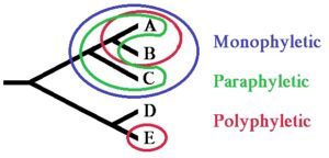
  • In the terms of evolutionary biology, a paraphyletic group is one that has a common ancestor and some descendants, but not completely each and everyone.
  • Example of Paraphyly: Reptilia
monophyletic group example
Monophyletic group
Image credits- Wikimedia
  •  In biology, monophyletic refers to, relates to, or affects a single phylum (or other taxon) of organisms, whereas paraphyletic refers to a defined group of taxa, without including all descendants of the common ancestor of all members.  
  • Example of monophyletic group: Mammals, Birds, Angiosperms, Insects, Lions, Echidnas, Komodo dragons, Crocodiles, Humans and Great apes

Summary:

This article summarizes the monophyletic examples and their characteristics. They are the group of organisms which have a common and single ancestor.

Also Read:

Is Endocytosis Active? A Comprehensive Exploration

is endocytosis active

Endocytosis is a fundamental cellular process that involves the internalization of extracellular materials, including nutrients, signaling molecules, and even entire cells, into the interior of a cell. This process is indeed an active one, requiring a significant amount of energy input from the cell to drive the complex machinery involved. In this comprehensive blog post, we will delve into the intricacies of endocytosis, exploring the energy requirements, the molecular mechanisms, and the crucial role it plays in various cellular functions.

Understanding the Energy Requirements of Endocytosis

Endocytosis is an energy-dependent process, in contrast to passive transport mechanisms like simple diffusion and osmosis. The cell invests a substantial amount of energy, primarily in the form of ATP, to power the various steps involved in endocytosis.

Quantifying the Energy Consumption

A study published in the Journal of Cell Biology provides precise data on the energy requirements of endocytosis. The researchers measured the ATP consumption during endocytic events in mammalian cells and found that it is approximately 2.5 x 10^6 ATP molecules per endocytic event. This staggering energy investment highlights the active nature of endocytosis and the significant resources the cell must allocate to this process.

The Role of Actin and Actin-Binding Proteins

The active nature of endocytosis is further underscored by the crucial involvement of the actin cytoskeleton and its associated proteins. A 2019 review in the journal Traffic discusses the pivotal role of actin and actin-binding proteins in the scission and departure of endocytic vesicles from the plasma membrane.

Actin filaments undergo dynamic rearrangements, driven by the energy-dependent activities of actin-binding proteins, to facilitate the formation and internalization of endocytic vesicles. This dynamic interplay between the actin cytoskeleton and its regulatory proteins is a hallmark of the active nature of endocytosis.

The Energetic Cost of Membrane Deformation

The process of endocytosis also requires significant energy input to overcome the energetic barriers associated with the deformation of the cell membrane. As the cell membrane invaginates to form the endocytic vesicle, it must overcome the inherent resistance to bending and curvature. This energy-intensive process is facilitated by the recruitment and activation of specialized proteins, such as clathrin and its associated adaptor proteins, which help to shape the membrane and drive the formation of the endocytic vesicle.

The Molecular Mechanisms of Active Endocytosis

is endocytosis active

Endocytosis is a complex, multi-step process that involves the coordinated action of numerous molecular players. Understanding the active nature of endocytosis requires a closer look at the specific mechanisms involved.

Clathrin-Mediated Endocytosis

One of the best-studied forms of active endocytosis is clathrin-mediated endocytosis. In this process, the cell membrane invaginates to form a clathrin-coated pit, which then pinches off to form a clathrin-coated vesicle. This process is driven by the energy-dependent assembly and disassembly of the clathrin coat, as well as the recruitment and activation of accessory proteins that facilitate membrane deformation and vesicle scission.

Caveolae-Mediated Endocytosis

Another form of active endocytosis is caveolae-mediated endocytosis, which involves the formation of flask-shaped invaginations in the cell membrane called caveolae. These structures are stabilized by the presence of the protein caveolin, and their internalization is also an energy-dependent process, involving the dynamic rearrangement of the actin cytoskeleton and the recruitment of regulatory proteins.

Phagocytosis and Macropinocytosis

Endocytosis can also occur on a larger scale, with the cell engulfing entire particles or even other cells. This process, known as phagocytosis, is a highly energy-intensive form of endocytosis that is crucial for immune function, tissue remodeling, and nutrient acquisition. Similarly, macropinocytosis, the internalization of large volumes of extracellular fluid, is also an active process that requires significant energy input.

The Role of GTPases and Kinases

The regulation of endocytic processes is mediated by a complex network of signaling molecules, including small GTPases and protein kinases. These energy-dependent enzymes play a crucial role in coordinating the various steps of endocytosis, from the initial membrane deformation to the final scission and internalization of the endocytic vesicle.

The Functional Importance of Active Endocytosis

Endocytosis is a fundamental cellular process that is essential for a wide range of physiological functions. The active nature of endocytosis underscores its importance in maintaining cellular homeostasis and facilitating critical cellular processes.

Nutrient Uptake and Signaling

One of the primary functions of endocytosis is the internalization of nutrients, such as vitamins, minerals, and macromolecules, from the extracellular environment. This active process allows the cell to acquire the necessary building blocks and energy sources for growth, metabolism, and signaling.

Immune Function and Cell-Cell Communication

Endocytosis also plays a crucial role in immune function, as it allows immune cells to internalize and process foreign particles, pathogens, and even other cells for antigen presentation and immune response coordination. Additionally, endocytosis facilitates cell-cell communication by enabling the internalization of signaling molecules and the subsequent activation of intracellular signaling cascades.

Waste Removal and Membrane Recycling

Endocytosis is also responsible for the internalization and recycling of membrane components, as well as the removal of waste and damaged materials from the cell. This active process helps to maintain the integrity and function of the cell membrane and the overall cellular homeostasis.

Conclusion

In conclusion, endocytosis is an undoubtedly active process that requires a significant amount of energy input from the cell. The quantifiable data on ATP consumption, the crucial involvement of the actin cytoskeleton and its regulatory proteins, and the energy-intensive nature of membrane deformation all highlight the active nature of this fundamental cellular process.

Understanding the active nature of endocytosis is crucial for understanding the complex mechanisms that govern cellular function, from nutrient uptake and signaling to immune response and waste removal. This knowledge can have far-reaching implications in fields such as cell biology, physiology, and medicine, as researchers continue to explore the intricacies of this energy-dependent process.

References:

  1. Mettlen, M., Chen, P. H., Srinivasan, S., Danuser, G., & Schmid, S. L. (2018). Regulation of Clathrin-Mediated Endocytosis. Annual Review of Biochemistry, 87, 871-896. https://www.ncbi.nlm.nih.gov/pmc/articles/PMC6714026/
  2. Doherty, G. J., & McMahon, H. T. (2009). Mechanisms of endocytosis. Annual Review of Biochemistry, 78, 857-902. https://onlinelibrary.wiley.com/doi/full/10.1111/tra.12618
  3. Kaksonen, M., & Roux, A. (2018). Mechanisms of clathrin-mediated endocytosis. Nature Reviews Molecular Cell Biology, 19(5), 313-326. https://www.ncbi.nlm.nih.gov/pmc/articles/PMC8108533/
  4. Lumenlearning. (n.d.). Endocytosis and Exocytosis. Retrieved from https://courses.lumenlearning.com/suny-wmopen-biology1/chapter/endocytosis-and-exocytosis/
  5. Doherty, G. J., & McMahon, H. T. (2009). Mechanisms of endocytosis. Biochemical Journal, 422(1), 16-59. https://www.ncbi.nlm.nih.gov/pmc/articles/PMC5614474/
  6. MRG Science. (n.d.). Topic 14: Membrane Transport. Retrieved from https://www.mrgscience.com/topic-14-membrane-transport.html

13 Single Cell Protein Example: Explanation You Should Know

1200px Aspergillus niger 2 1024x768 1

Single cell protein example is the dried and dead cells of microorganisms like yeast, bacteria, fungi and algae that can be used as a food or feed supplement due to high protein source. 

  • Aspergillus niger
  • Rhizopus cyclopean
  • Saccharomyces cerevisiae
  • Candida tropicalis
  • Spirulina
  • Aspergillus fumigatus
  • Chlorella pyrenoidosa
  • Chondrus crispus
  • Pseudomonas fluorescens
  • Candida utilis
  • Lactobacillus
  • Bacillus megaterium
  • Methylophilus methylotrophus

Aspergillus niger:

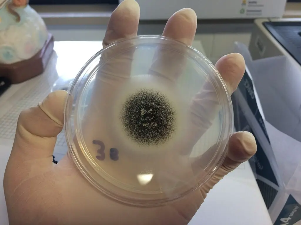
Aspergillus niger is a member from the group of fungi (Aspergillus niger AS-101)

single cell protein example
Aspergillus niger
Image credits: Wikimedia

Aspergillus niger is a cellulolytic mold, which is efficiently used to produce single cell protein by using the raw materials like alkalic corn cobs.

Rhizopus cyclopean:

Rhizopus cyclopean is also a member from the group of fungi.

1487022229 cdde217da0 c
Rhizopus cyclopean
Image credits: Flickr

Rhizopus cyclopean are rich in protein that are truly used in the production of single cell protein.

Saccharomyces cerevisiae:

Saccharomyces cerevisiae is referred to as the king of microbes in the case of biotechnology, microbiology and genetic manipulative techniques.

Saccharomyces cerevisiae is often prefered for single cell protein as it has high rich content of protein about 44.6% in recent research studies.

Candida tropicalis:

The candida tropilis is a fungi with division- ascomycota and class-Saccharomycetes, order- Saccharomycetales, family- Saccharomycetaceae and genus- Candida species- C. tropicalis

Sugarcane bagasse hemicellulosic hydrolysate (SBHH) was found to be a suitable substrate for yeast single cell protein production in Candida tropicalis. 

Spirulina:

The word, Single cell protein directly refers to spirulina as it is one of the most common and flexible available single cell proteins in the world market.

Spirulina culturing is one of the best businesses in the market as the yield is rich protein content.

Aspergillus fumigatus

Aspergillus fumigatus is a member from the group of fungi.

1487022229 cdde217da0 c 1
Aspergillus fumigatus
Image credits: Wikimedia

Aspergillus fumigatus is a cellulolytic mold, which is efficiently used to produce single cell protein by using the raw materials like alkalic corn cobs which is similar to aspergillus niger.

Chlorella pyrenoidosa

Chlorella is a uni-celled, green fresh water algae utilized as a single-cell protein source.

Chlorella pyrenoidosa is one of the best examples for the single protein as it is grown in the tofu waste medium for the yield.

Chondrus crispus:

Irish moss, or chondrus crispus, is a popular edible red seaweed found on rocky coasts in the Northern Atlantic.

Research are being conducted in which this species can be used to produce more single cell protein and yield higher.

Read more about Protein

Pseudomonas fluorescens:

Pseudomonas fluorescens is commonly present in the soil or any surface, when grown in a culture medium the typical green color appears so radiantly.

Pseudomonas fluorescens is also one of the examples in the single cell production for its high protein content and low maintenance.

Candida utilis:

Candida utilis is referred to as the edible yeast as it is very safe to consume.

These cells are rich in protein and also their doubling time is less so fast yield can be expected.

Lactobacillus:

Often the term good bacteria comes, when we come to know about Lactobacillus. They are very much useful for the gut.

1200px Aspergillus niger 2 1
Lactobacillus
Image credits: Wikimedia

Lactobacillus is similar to the other examples of single cell protein as they have good protein content and also gut friendly.

Bacillus megaterium:

Bacillus megaterium commonly found in the soil, and they grow really well on the collagen derived subtract for protein synthesis.

Bacillus megaterium is very easy to cultivate and fast growing as well.

Read more about Monomer Of Protein Example

Methylophilus methylotrophus

Methylophilus methylotrophus, from the name we could know it has significant use in the industrial field for the production of methanol

Methylophilus methylotrophus is used as a single cell protein by using substrate friendly medium for higher yield.

Summary:

This article exclusively has about 13 examples of the single cell protein which are now a blooming field in the area of biotechnology, microbiology, genetic engineering.

Also Read: