Start Codon Example: 5 Facts You Should Know!

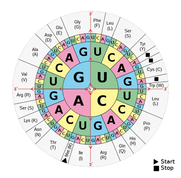
Aminoacids table.svg

Codons are a set of three nucleotides that code for amino acids. Let us see examples of the start codon and a few facts about it. The start codon is the first codon in the mRNA, that is transcripted into amino acid in the ribosomes. Ribosomes are made up of two subunits and are present … Read more

Unraveling the Secrets of Trilobite Characteristics: A Comprehensive Exploration

trilobite characteristics

Trilobites, the iconic arthropods that dominated the ancient seas, have long captivated the minds of paleontologists and enthusiasts alike. These remarkable creatures, with their distinct segmented bodies and compound eyes, offer a wealth of information about the evolution and ecology of the Paleozoic era. In this comprehensive blog post, we delve into the intricate details … Read more

9 Trilobite Examples & Types: Facts That You Should Know!

574px Trilobite Walliserops trifurcatus 1 287x300 1

Trilobites are extinct marine arthropods that are divided into three lobes; head, body, and thorax with jointed legs. Let us see some of its types below. Proetida Redlichiida Corynexochida Harpetida Agnostida Asaphida Odontopleurida Lichida Phacopida Ptychopardiia Types of Trilobite 1. Proetida Proetida are small trilobites made up of an exoskeleton with small pits called tubercles. … Read more

Stop Codon Example: 7 Facts You Should Know!

A codon is a three-letter code that is a combination of the four nucleotides of DNA or RNA. Every single codon codes to specific amino acids. Let us discuss. The stop codon is a sequence of three nucleotides-trinucleotide that signals to stop the protein synthesis that takes place in the cytoplasm of the cell. There … Read more

Does Cytoplasm Have Protein?

does cytoplasm have protein

The cytoplasm, the material inside a cell excluding the nucleus, is a complex and dynamic environment that plays a crucial role in cellular function. At the heart of this cytoplasmic landscape lies a diverse array of proteins, which contribute to a wide range of cellular processes. Understanding the presence and role of proteins within the … Read more

Can DNA Leave the Nucleus? A Comprehensive Exploration

can dna leave the nucleus

DNA, the fundamental genetic material that carries the instructions for life, is typically confined within the nucleus of eukaryotic cells. This strict compartmentalization is essential for the proper functioning and regulation of cellular processes. In this comprehensive guide, we will delve into the intricate details of DNA’s relationship with the nucleus, exploring the reasons why … Read more

Is Vitamin A a Coenzyme?

is vitamin a coenzyme

Vitamin A is an essential nutrient that plays a crucial role in various physiological processes in the human body. While it is not a coenzyme in the strict sense, it can act as a cofactor for certain enzymes, particularly in the visual system. In this comprehensive blog post, we will delve into the intricacies of … Read more

Enzymes and Metabolism: A Comprehensive Guide to Understanding Cellular Physiology

enzymes and metabolism

Enzymes and metabolism are the fundamental pillars of cellular physiology, orchestrating the intricate chemical reactions and energy production that sustain life. This comprehensive guide delves into the intricacies of enzymes, their regulation, and the pivotal role they play in the complex metabolic pathways that power cellular processes.

Understanding Enzymes: The Catalysts of Life

Enzymes are specialized proteins that act as catalysts, accelerating the rate of chemical reactions within cells. These remarkable biomolecules are responsible for catalyzing nearly all vital cellular processes, from energy production and nutrient breakdown to DNA replication and cellular signaling.

Enzyme Structure and Function

Enzymes are composed of intricate three-dimensional structures, with specific active sites that bind to and facilitate the transformation of substrates into products. The unique shape and chemical properties of an enzyme’s active site allow it to selectively bind and stabilize the transition state of a reaction, dramatically increasing the reaction rate.

Enzyme Kinetics and Regulation

The activity of enzymes is tightly regulated at multiple levels, including:

  1. Enzyme Concentration: The amount of an enzyme present in a cell or tissue can significantly impact its overall activity.
  2. Oligomeric State: Enzymes can exist in different oligomeric states, which can affect their catalytic efficiency and regulation.
  3. Allosteric Regulation: Binding of regulatory molecules to specific sites on an enzyme can induce conformational changes that modulate its activity.
  4. Covalent Modification: Post-translational modifications, such as phosphorylation or acetylation, can alter an enzyme’s structure and function.
  5. Compartmentalization: The localization of enzymes within specific cellular compartments can influence their accessibility to substrates and regulatory factors.

Understanding these regulatory mechanisms is crucial for deciphering the complex interplay between enzymes and their role in cellular metabolism.

Metabolic Mapping: Visualizing Enzyme Activity
enzymes and metabolism

Quantitative enzyme histochemistry and cytochemistry, also known as metabolic mapping, are powerful techniques used to study the activity and distribution of enzymes within cells and tissues.

Dehydrogenase Activity Mapping

Dehydrogenases are a class of enzymes that catalyze redox reactions, transferring electrons from one molecule (the substrate) to another (the cofactor). The activity of dehydrogenases can be visualized and quantified using metabolic mapping techniques.

  1. Tetrazolium Salt Reduction: Intact cells or cryostat tissue sections are incubated in a reaction medium containing the substrate, cofactors, and a tetrazolium salt. The dehydrogenase enzyme reduces the tetrazolium salt, forming a water-insoluble blue formazan precipitate at the site of enzyme activity.
  2. Quantification of Formazan Absorbance: The absorbance of the precipitated formazan can be measured using monochromatic light microscopy and image analysis, providing a direct quantitative measure of the local dehydrogenase activity.

Kinetic Parameter Determination

The quantitative nature of metabolic mapping enables researchers to determine in situ kinetic parameters, such as the maximal enzyme activity (Vmax) and the affinity of a substrate for an enzyme (Km). These parameters provide valuable insights into the regulation and efficiency of enzymes within their native cellular environment.

Enzyme Concentration and Reaction Rates

In vivo, the activity of enzymes can be influenced by their concentration within the cell or tissue. Interestingly, studies have shown that as enzyme concentration increases, the apparent enzymatic efficiency (Vmax/Km) can decrease, and the Km can increase.

Substrate Flux Limitation

Simulations have revealed that in situations with attenuated diffusion, the substrate flux can become the rate-limiting factor, rather than the enzyme concentration. This explains why reaction rates in vivo can sometimes be independent of enzyme concentrations, as the availability and transport of substrates to the enzyme active sites become the limiting factor.

Advances in Enzyme Activity Prediction

Recent developments in computational biology have enabled the prediction of Michaelis constants (Km) from structural features of enzymes. Deep learning algorithms have been trained on large datasets of enzyme structures and kinetic parameters, allowing for genome-scale predictions of Km values.

This powerful approach can provide valuable insights into the intrinsic properties of enzymes and their catalytic efficiency, further enhancing our understanding of cellular metabolism and the role of enzymes in various biological processes.

Conclusion

Enzymes and metabolism are the cornerstones of cellular physiology, driving the intricate chemical reactions and energy production that sustain life. By understanding the structure, function, and regulation of enzymes, as well as the techniques used to study their activity, we can gain deeper insights into the complex metabolic pathways that underpin cellular processes.

This comprehensive guide has explored the key concepts and advancements in the field of enzymes and metabolism, equipping you with the knowledge to navigate the intricacies of this fascinating area of biology. As research continues to unveil new discoveries, the understanding of enzymes and their role in metabolism will undoubtedly continue to evolve, shaping our understanding of the fundamental mechanisms that sustain life.

References:

  1. Molenaar RJ, Khurshed M, Hira VVV, Van Noorden CJF. Metabolic Mapping: Quantitative Enzyme Cytochemistry and Histochemistry to Determine the Activity of Dehydrogenases in Cells and Tissues. Frontiers in Physiology. 2018;9:561.
  2. Zotter A, Bäuerle F, Dey D, Kiss V, Schreiber G. Quantifying enzyme activity in living cells. Journal of Biological Chemistry. 2018;293(4):1303-1312.
  3. Kroll A, et al. Deep learning allows genome-scale prediction of Michaelis constants from structural features. PLoS Biology. 2021;19(10):e3001402.
  4. Cornish-Bowden A. Fundamentals of Enzyme Kinetics. Wiley-Blackwell; 2012.
  5. Nelson DL, Cox MM. Lehninger Principles of Biochemistry. W.H. Freeman; 2017.
  6. Berg JM, Tymoczko JL, Stryer L. Biochemistry. W.H. Freeman; 2015.
Bacteria Cell Wall Formation: A Comprehensive Guide

bacteria cell wall formation

Bacteria cell wall formation is a complex and intricate process that involves the synthesis and remodeling of peptidoglycan (PG), a crucial component of the bacterial cell wall. The thickness, organization, and composition of PG vary among different bacterial species, and its synthesis is essential for bacterial viability, growth, and survival.

Understanding the Bacterial Cell Wall Structure

The bacterial cell wall is a multi-layered structure that provides structural integrity, protection, and shape to the bacterial cell. It is composed of several key components, including:

  1. Peptidoglycan (PG): A polymer of N-acetylglucosamine (GlcNAc) and N-acetylmuramic acid (MurNAc) cross-linked by short peptide chains. PG is the primary structural component of the bacterial cell wall.
  2. Teichoic acids: Anionic polymers found in the cell walls of Gram-positive bacteria, which play a role in cell wall integrity, cell division, and cell surface properties.
  3. Lipopolysaccharides (LPS): A complex glycolipid found in the outer membrane of Gram-negative bacteria, which contributes to the structural integrity and permeability of the cell wall.
  4. Proteins: Various proteins embedded in the cell wall, such as porins, enzymes, and surface receptors, which perform essential functions.

The thickness and organization of the cell wall can vary significantly between Gram-positive and Gram-negative bacteria. Gram-positive bacteria typically have a thicker PG layer (20-80 nm), while Gram-negative bacteria have a thinner PG layer (2-7 nm) and an additional outer membrane.

Peptidoglycan Synthesis and Remodeling

bacteria cell wall formation

The synthesis and remodeling of PG are central to bacterial cell wall formation. This process involves several key steps:

  1. Precursor Synthesis: The PG precursor, UDP-N-acetylmuramyl-pentapeptide, is synthesized in the cytoplasm through a series of enzymatic reactions.
  2. Translocation: The PG precursor is then translocated across the cytoplasmic membrane by a flippase enzyme, such as MurJ or Amj, to the periplasmic space.
  3. Polymerization: In the periplasmic space, the PG precursor is polymerized by glycosyltransferases, which catalyze the formation of the glycan backbone.
  4. Cross-linking: The glycan strands are then cross-linked by transpeptidases, which form peptide bonds between the peptide side chains, creating a rigid and stable PG network.
  5. Remodeling: During cell growth and division, the PG layer is continuously remodeled by various hydrolytic enzymes, such as autolysins and endopeptidases, which cleave and rearrange the PG structure.

The regulation and coordination of these PG synthesis and remodeling processes are crucial for maintaining the integrity and shape of the bacterial cell wall.

Visualization and Characterization Techniques

Advances in microscopy and analytical techniques have provided valuable insights into the structure and dynamics of bacterial cell walls. Some of the key techniques used to study PG synthesis and remodeling include:

  1. Cryogenic Electron Tomography (cryo-ET): This technique allows for the high-resolution, three-dimensional visualization of the division site architecture and PG biogenesis dynamics in live bacterial cells, as demonstrated in the study on Escherichia coli.
  2. Fluorescence Microscopy: Fluorescent labeling of PG precursors or cell wall-associated proteins enables the real-time monitoring of PG synthesis and remodeling processes during bacterial growth and division.
  3. Atomic Force Microscopy (AFM): AFM can be used to measure the thickness and organization of the PG layer, as well as to visualize the surface topography of live bacterial cells, as shown in the study on Staphylococcus aureus and Bacillus subtilis.
  4. Chemical and Biochemical Techniques: Techniques such as isotopic labeling, metabolic probes, and the isolation of critical intermediates have provided valuable insights into the biochemistry of cell wall biogenesis, as discussed in the Chemical Biology review.

These advanced techniques have been instrumental in elucidating the complex mechanisms underlying bacterial cell wall formation and have opened new avenues for understanding the role of the cell wall in bacterial physiology, growth, and division.

Factors Influencing Bacterial Cell Wall Formation

Several factors can influence the synthesis and remodeling of the bacterial cell wall, including:

  1. Bacterial Species and Strain: The thickness, composition, and organization of the PG layer can vary significantly between different bacterial species and even between strains within the same species.
  2. Growth Conditions: Environmental factors, such as nutrient availability, pH, temperature, and osmotic pressure, can affect the rate and regulation of PG synthesis and remodeling.
  3. Cell Cycle and Division: The cell wall must be continuously remodeled to accommodate cell growth and division, with the division apparatus playing a crucial role in coordinating these processes.
  4. Genetic Regulation: The expression and activity of genes involved in PG synthesis, translocation, and remodeling are tightly regulated to ensure the proper formation and maintenance of the cell wall.
  5. Cell Wall-Targeting Antibiotics: Certain antibiotics, such as β-lactams and glycopeptides, target and disrupt specific steps in the PG synthesis and remodeling pathways, leading to cell wall weakening and ultimately cell death.

Understanding these factors and their interplay is essential for developing effective strategies to manipulate or inhibit bacterial cell wall formation, which has important implications in the fields of microbiology, biotechnology, and antimicrobial drug development.

Conclusion

Bacterial cell wall formation is a complex and dynamic process that involves the synthesis and remodeling of peptidoglycan, a crucial structural component of the bacterial cell wall. The thickness, organization, and composition of the cell wall can vary significantly among different bacterial species and strains, and its formation is essential for bacterial viability, growth, and survival.

Advances in microscopy and analytical techniques have provided valuable insights into the mechanisms underlying cell wall biogenesis, enabling researchers to visualize and characterize the structure and dynamics of the bacterial cell wall. Understanding the factors that influence cell wall formation is crucial for developing effective strategies to manipulate or inhibit this process, with important implications in various fields, including microbiology, biotechnology, and antimicrobial drug development.

References:

  1. Brown, A. R., Gordon, R. A., Hyland, S. N., & Grimes, C. L. (2020). Chemical biology tools for examining the bacterial cell wall. Chemical biology, 17(8), 1217-1232.
  2. Navarro, P. P., Vettiger, A., Ananda, V. Y., … & VanNieuwenhze, M. S. (2022). Cell wall synthesis and remodelling dynamics determine division site architecture and cell shape in Escherichia coli. Nature microbiology, 7(11), 1621-1634.
  3. Roux, A., … & Armitage, J. P. (2020). The Architecture of the Gram Positive Bacterial Cell Wall. PMC, 7308169.

How Are Traits Made: A Comprehensive Guide

how are traits made

Traits, the observable characteristics of an organism, are the result of a complex interplay between genes and the environment. Understanding the mechanisms behind trait formation is crucial for fields such as genetics, evolutionary biology, and agriculture. In this comprehensive guide, we will delve into the intricate details of how traits are made, exploring the role of genes, the environment, and the various processes that shape the expression of phenotypic traits.

The Genetic Basis of Traits

At the core of trait formation are genes, the fundamental units of heredity. Genes are sequences of DNA that encode the instructions for the production of proteins, which in turn, play a crucial role in determining an organism’s physical and behavioral characteristics.

Genotype and Phenotype

The genotype refers to the genetic makeup of an individual, the specific combination of alleles (different versions of a gene) that an organism inherits from its parents. The phenotype, on the other hand, is the observable physical or behavioral characteristic of an organism, which is the result of the interaction between the genotype and the environment.

Polygenic Traits

Many traits are controlled by multiple genes, known as polygenic traits. These traits exhibit continuous variation within a population, with individuals displaying a range of phenotypic values. Examples of polygenic traits include height, skin color, and intelligence.

Additive Genetic Effects

The expression of polygenic traits is influenced by the cumulative, additive effects of multiple genes. Each gene contributes a small, incremental effect to the overall phenotype, and the combined effects of these genes result in the continuous variation observed in the population.

Heritability

The degree to which a trait is influenced by genetic factors versus environmental factors is measured by heritability. Heritability is expressed as a value between 0 and 1, where a value closer to 1 indicates a stronger genetic influence, and a value closer to 0 indicates a stronger environmental influence.

Quantitative Trait Loci (QTLs)

Quantitative trait loci (QTLs) are regions of the genome that contain genes or genetic markers associated with the variation of a quantitative trait. By identifying and studying QTLs, researchers can gain insights into the genetic architecture of complex traits and the specific genes involved in their expression.

The Role of the Environment

how are traits made

While genes provide the blueprint for trait formation, the environment plays a crucial role in shaping the final phenotype. Environmental factors, such as temperature, nutrition, and social interactions, can influence the expression of genes and the development of traits.

Phenotypic Plasticity

Phenotypic plasticity refers to the ability of a single genotype to produce different phenotypes in response to varying environmental conditions. This flexibility allows organisms to adapt to changing environments and optimize their chances of survival and reproduction.

Epigenetic Modifications

Epigenetic modifications are changes in gene expression that do not involve alterations to the DNA sequence. These modifications, such as DNA methylation and histone modifications, can be influenced by environmental factors and can be passed on to subsequent generations, contributing to the inheritance of certain traits.

Trait Development and Expression

The process of trait development and expression involves a complex interplay between genetic and environmental factors, as well as various developmental and regulatory mechanisms.

Developmental Pathways

During an organism’s development, genes are expressed in a coordinated manner, activating specific developmental pathways that guide the formation of various tissues, organs, and structures. Disruptions or variations in these pathways can lead to the emergence of different phenotypic traits.

Gene Regulation

The expression of genes is tightly regulated by various mechanisms, such as transcription factors, epigenetic modifications, and post-transcriptional processes. These regulatory mechanisms ensure that genes are expressed at the right time, in the right place, and at the appropriate levels, contributing to the overall development and expression of traits.

Phenotypic Variation

Phenotypic variation within a population is the result of the combined effects of genetic and environmental factors. This variation is essential for the process of natural selection, as it provides the raw material for evolution to act upon.

Quantitative Genetics and Trait Prediction

Quantitative genetics is the field of study that focuses on the genetic basis of quantitative traits, those that exhibit continuous variation within a population. Researchers in this field use various tools and techniques to understand the genetic architecture of complex traits and predict their evolutionary responses.

F2 Crossing Experiments

One of the classic methods used in quantitative genetics is the F2 crossing experiment. By crossing two parental lines with known phenotypes and genotypes, researchers can observe the segregation and recombination of alleles in the F2 generation, revealing the underlying genetic basis of the trait.

Additive Genetic Variance

The degree to which a population can respond to selection depends on the amount of additive genetic variance present for a particular trait. Researchers can estimate the additive variance through quantitative genetic breeding experiments, which provide insights into the evolutionary potential of a trait.

Predicting Evolutionary Responses

Understanding the additive genetic variance for traits under selection is crucial for predicting how populations might respond to environmental changes, such as climate change. By estimating the additive variance, researchers can make informed predictions about the evolutionary trajectories of these traits.

Conclusion

Traits are the result of a complex interplay between genes and the environment, shaped by various genetic, developmental, and regulatory mechanisms. By understanding the intricate details of how traits are made, researchers can gain valuable insights into the genetic architecture of complex traits, the mechanisms of phenotypic plasticity, and the evolutionary potential of populations. This knowledge is essential for advancing fields such as genetics, evolutionary biology, and agriculture, ultimately contributing to our understanding of the natural world and our ability to address pressing challenges.

References:

  • The Evolution of Quantitative Traits: https://www.nature.com/articles/nrg3426
  • Quantitative Genetics: https://www.nature.com/articles/nrg3426
  • Inheritance of Quantitative Traits: https://www.nature.com/articles/nrg3426