Is Mitochondria The Powerhouse Of The Cell? 5+ Facts

350px Mitochondrion structure.svg 300x210 1

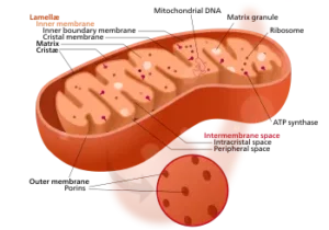
Mitochondria are subcellular organelles derived from initial endosymbiosis in eukaryotes. Let us explore if mitochondria is the powerhouse of the cell. Mitochondria are referred to as the “powerhouse of the cell” because they produce the energy needed for other cell organelles to function. The production of ATP, or adenosine triphosphate, takes place in an area … Read more

Do Prokaryotes Have a Nucleus? A Comprehensive Guide

do prokaryotes have a nucleus

Prokaryotes, which include bacteria and archaea, are single-celled organisms that lack a true nucleus or membrane-bound organelles, a fundamental characteristic that distinguishes them from eukaryotic cells. While the absence of a nucleus may seem like a limitation, prokaryotes have evolved unique mechanisms to efficiently carry out essential cellular processes. The Nucleoid Region: Prokaryotic DNA Storage … Read more

Are Endonucleases Restriction Enzymes: A Comprehensive Guide

are endonucleases restriction enzymes

Endonucleases, a class of restriction enzymes, are essential tools in the field of molecular biology, renowned for their ability to recognize and cleave specific DNA sequences. These remarkable enzymes have revolutionized various applications, from genetic engineering to forensic analysis, earning their discoverers the prestigious Nobel Prize in Medicine and Physiology in 1978.

Understanding Endonucleases: The Molecular Workhorses

Endonucleases are enzymes that can recognize and cut DNA molecules at specific nucleotide sequences, known as recognition sites. These recognition sites are typically 4 to 8 base pairs long and are often palindromic, meaning they read the same forward and backward. When an endonuclease binds to its recognition site, it creates a double-strand break in the DNA, either leaving blunt ends or generating staggered cuts that result in single-stranded overhangs.

The discovery of these remarkable enzymes is credited to the pioneering work of three scientists: Werner Arber, Hamilton O. Smith, and Daniel Nathans. Their groundbreaking research on the mechanisms of bacterial defense systems against viral infections led to the identification and characterization of these DNA-cleaving enzymes, earning them the 1978 Nobel Prize.

Types of Endonucleases: Diversity and Specificity

are endonucleases restriction enzymes

Endonucleases can be classified into several types based on their recognition sequences, cleavage patterns, and cofactor requirements. Some of the most common types include:

  1. Type I Endonucleases: These enzymes recognize and cleave DNA at sites distant from their recognition sequence, requiring ATP and a cofactor, such as S-adenosylmethionine (SAM), for their activity.

  2. Type II Endonucleases: This is the most widely used class of endonucleases in molecular biology. They recognize and cleave DNA within or adjacent to their specific recognition sequences, typically 4 to 8 base pairs long. Examples include EcoRI, BamHI, and HindIII.

  3. Type III Endonucleases: These enzymes recognize and cleave DNA at a fixed distance from their asymmetric recognition sites, requiring ATP and a cofactor for their activity.

  4. Type IV Endonucleases: This class of enzymes recognizes and cleaves modified DNA, such as methylated or hydroxymethylated DNA, as a defense mechanism against foreign genetic material.

The high specificity of endonucleases is a crucial feature that makes them invaluable in various applications. Each enzyme recognizes and cleaves a unique DNA sequence, allowing for precise manipulation and analysis of genetic material.

Applications of Endonucleases: Revolutionizing Molecular Biology

Endonucleases have become indispensable tools in the field of molecular biology, enabling a wide range of applications:

  1. Genetic Engineering: Endonucleases are essential for DNA cloning, gene insertion, and genetic modification. They allow for the precise excision and insertion of DNA fragments, facilitating the creation of recombinant DNA molecules.

  2. DNA Mapping and Sequencing: Endonucleases are used to generate DNA fragments of specific sizes, which can then be analyzed to create detailed genetic maps and facilitate DNA sequencing.

  3. Diagnostic and Forensic Analysis: Endonucleases are employed in DNA fingerprinting and forensic investigations, where they are used to generate unique DNA profiles for identification and comparison purposes.

  4. Gene Expression Studies: Endonucleases are utilized in techniques like Northern blotting and RNase protection assays to analyze gene expression patterns and quantify specific RNA transcripts.

  5. Epigenetic Studies: Certain endonucleases, such as those that recognize and cleave methylated DNA, are employed in the study of epigenetic modifications and their impact on gene regulation.

  6. Genome Editing: The development of CRISPR-Cas9 technology, which utilizes a guide RNA and the Cas9 endonuclease, has revolutionized genome editing, allowing for precise and targeted modifications of genetic sequences.

  7. Molecular Diagnostics: Endonucleases are used in various molecular diagnostic techniques, such as the Direct Restriction Assay (DRA), which enables the detection and quantification of specific DNA targets with high sensitivity and specificity.

The versatility and precision of endonucleases have made them indispensable tools in the fields of genetics, genomics, and biotechnology, driving advancements in our understanding of the genetic code and enabling groundbreaking applications in medicine, agriculture, and beyond.

Endonucleases in Action: Techniques and Protocols

Researchers and scientists have developed a wide range of techniques and protocols to harness the power of endonucleases effectively. Some of the commonly used methods include:

  1. Restriction Fragment Length Polymorphism (RFLP): This technique utilizes endonucleases to generate DNA fragments of varying lengths, which can be separated and analyzed to identify genetic variations or polymorphisms.

  2. Southern Blotting: Endonucleases are used to cleave DNA samples, and the resulting fragments are separated by gel electrophoresis, transferred to a membrane, and probed with labeled DNA sequences to detect specific genetic targets.

  3. DNA Fingerprinting: Endonucleases are employed in DNA profiling techniques, such as Amplified Fragment Length Polymorphism (AFLP) and Variable Number Tandem Repeat (VNTR) analysis, to generate unique DNA patterns for identification and forensic applications.

  4. Methylation-Sensitive Restriction Enzyme (MSRE) Analysis: Certain endonucleases, such as HpaII and MspI, are used to detect and analyze DNA methylation patterns, which play a crucial role in epigenetic regulation.

  5. Direct Restriction Assay (DRA): This technique utilizes endonucleases to cleave target-probe hybrids, releasing a molecular marker that can be detected and quantified, enabling the sensitive and specific detection of target DNA sequences.

  6. Genome Editing with CRISPR-Cas9: The Cas9 endonuclease, guided by a specific RNA sequence, is used to precisely target and cleave desired DNA sequences, allowing for efficient genome editing and modification.

These are just a few examples of the numerous techniques and protocols that leverage the unique properties of endonucleases to advance scientific research and applications.

Endonucleases: Pushing the Boundaries of Molecular Biology

As the field of molecular biology continues to evolve, the role of endonucleases is expected to expand even further. Ongoing research and technological advancements are unlocking new frontiers in areas such as:

  1. Improved Enzyme Engineering: Scientists are exploring ways to engineer endonucleases with enhanced specificity, activity, and stability, expanding their utility in various applications.

  2. Multiplexed Genome Editing: The development of CRISPR-Cas9 systems with multiple guide RNAs allows for the simultaneous targeting and modification of multiple genetic loci, revolutionizing genome engineering.

  3. Epigenetic Regulation: Endonucleases that recognize and cleave specific epigenetic modifications, such as DNA methylation or histone modifications, are enabling deeper insights into the complex mechanisms of gene regulation.

  4. Diagnostic and Therapeutic Applications: The high specificity and sensitivity of endonucleases are being leveraged in the development of advanced molecular diagnostics and targeted therapeutic approaches, such as gene therapy.

  5. Synthetic Biology: Endonucleases are playing a crucial role in the design and construction of synthetic genetic circuits, paving the way for the creation of novel biological systems and applications.

As the scientific community continues to push the boundaries of molecular biology, the versatility and precision of endonucleases will undoubtedly remain at the forefront of groundbreaking discoveries and technological advancements.

Conclusion

Endonucleases, the remarkable restriction enzymes, have revolutionized the field of molecular biology, enabling unprecedented control and manipulation of genetic material. From their discovery to their widespread applications, these molecular workhorses have become indispensable tools in the hands of researchers and scientists worldwide.

As the understanding of these enzymes continues to deepen, and new applications emerge, the future of endonucleases in advancing scientific knowledge and addressing global challenges remains bright and full of promise.

References

  1. Promega Corporation. (n.d.). Restriction Enzyme Resource. Retrieved from https://www.promega.com/resources/guides/nucleic-acid-analysis/restriction-enzyme-resource/
  2. Guo, J., Ju, J., & Turro, N. J. (2012). Fluorescent hybridization probes for nucleic acid detection. Analytical and Bioanalytical Chemistry, 402(10), 3115-3125. https://doi.org/10.1007/s00216-011-5528-z
  3. Quizlet. (n.d.). Forensic Analysis: Restriction Enzymes. Retrieved from https://quizlet.com/93432607/forensic-analysisrestriction-enzymes-flash-cards/
  4. Nature Education. (n.d.). Restriction Enzymes. Retrieved from https://www.nature.com/scitable/topicpage/restriction-enzymes-545/
  5. ScienceDirect. (n.d.). Restriction Enzyme. Retrieved from https://www.sciencedirect.com/topics/biochemistry-genetics-and-molecular-biology/restriction-enzyme
  6. Xu, S. Y., Zhu, Z., Zhang, P., Chan, S. H., Samuelson, J. C., Xiao, J., … & Guan, S. (2007). Discovery of natural nicking endonucleases Nb. BbvCI and Nb. BtsI and engineering of top-strand nicking variants from BbvCI and BtsI. Nucleic Acids Research, 35(14), 4608-4618. https://doi.org/10.1093/nar/gkm496
  7. Pingoud, A., Fuxreiter, M., Pingoud, V., & Wende, W. (2005). Type II restriction endonucleases: structure and mechanism. Cellular and Molecular Life Sciences CMLS, 62(6), 685-707. https://doi.org/10.1007/s00018-004-4513-1
Is Amylase an Enzyme? A Comprehensive Guide

is amylase an enzyme

Amylase is a crucial enzyme found in various organisms, including plants, animals, and microorganisms. It plays a vital role in the breakdown of starch, a complex carbohydrate, into smaller, more easily digestible molecules. In this comprehensive guide, we will delve into the intricacies of amylase, its structure, function, and the methods used to measure its activity.

What is Amylase?

Amylase is a group of hydrolase enzymes that catalyze the hydrolysis of 1,4-α-glucosidic linkages in starch, glycogen, and related polysaccharides. This process breaks down these complex carbohydrates into smaller, more easily digestible units, such as maltose and glucose. Amylases are classified into two main types: α-amylase and β-amylase, which differ in their specific mode of action and the products they generate.

Structure and Function of Amylase

is amylase an enzyme

Amylase enzymes are typically composed of a single polypeptide chain, with a molecular weight ranging from 50 to 100 kDa. The structure of amylase can be divided into several domains, each with a specific function:

  1. Catalytic Domain: This domain contains the active site of the enzyme, where the hydrolysis of the 1,4-α-glucosidic linkages occurs. The active site is typically composed of a number of amino acid residues, such as aspartic acid, glutamic acid, and histidine, which are responsible for the enzyme’s catalytic activity.

  2. Substrate-Binding Domain: This domain is responsible for the recognition and binding of the starch substrate to the enzyme, allowing the catalytic domain to effectively hydrolyze the 1,4-α-glucosidic linkages.

  3. Calcium-Binding Domain: Some amylases, particularly those found in plants and microorganisms, require the presence of calcium ions (Ca2+) for their optimal activity. This domain is responsible for the binding and coordination of the calcium ions, which are essential for the enzyme’s structural integrity and catalytic function.

The specific function of amylase is to break down starch into smaller, more easily digestible units, such as maltose and glucose. This process is crucial for the metabolism and energy production in living organisms, as it allows them to extract the necessary nutrients from their food.

Measuring Amylase Activity

The activity of amylase can be measured using a variety of methods, with the most common being the standard iodine assay. This assay is based on the decrease in the absorbance of the iodine-starch complex at 580 nm, which occurs as the starch is hydrolyzed by the amylase enzyme.

The standard reaction mixture for the iodine assay typically contains:

  • Enzyme solution
  • 1% (w/v) potato starch solution in 20% NaCl
  • Phosphate buffer (50 mM, pH 7, 20% NaCl)

The reaction mixture is incubated at 50°C for 30 minutes, and the reaction is then stopped by cooling on ice. A 100 μL aliquot of the reaction mixture is then mixed with commercial Lugol’s iodine solution, and the absorbance at 580 nm is measured after adding 1 mL of distilled water.

One unit of amylase activity is defined as the amount of enzyme that degrades one microgram of starch per minute under the specified assay conditions. This measurement of enzyme activity is crucial for understanding the efficiency and effectiveness of amylase in various applications, such as in the food and pharmaceutical industries.

Factors Affecting Amylase Activity

The activity of amylase can be influenced by various factors, including:

  1. Temperature: Amylase activity typically increases with temperature up to an optimal point, after which it begins to decrease due to enzyme denaturation.

  2. pH: Amylase enzymes have an optimal pH range, usually between 6.0 and 7.0, where their activity is highest. Deviations from this range can lead to a decrease in enzyme activity.

  3. Substrate Concentration: The activity of amylase is directly proportional to the concentration of the starch substrate, up to a certain saturation point. Beyond this point, the enzyme activity may plateau or even decrease due to substrate inhibition.

  4. Enzyme Concentration: The activity of amylase is also dependent on the concentration of the enzyme itself. Increasing the enzyme concentration generally leads to a corresponding increase in the rate of starch hydrolysis.

  5. Inhibitors and Activators: Certain molecules, such as metal ions, can act as inhibitors or activators of amylase activity. For example, calcium ions (Ca2+) are known to activate some amylases, while heavy metal ions, such as mercury (Hg2+) and lead (Pb2+), can inhibit enzyme activity.

Understanding these factors is crucial for optimizing the performance of amylase in various applications, such as in the food industry, where it is used for the production of syrups and the processing of starch-based products.

Applications of Amylase

Amylase enzymes have a wide range of applications in various industries, including:

  1. Food Industry: Amylase is used in the production of syrups, the processing of starch-based products, and the baking industry, where it helps improve the texture and shelf-life of bread and other baked goods.

  2. Pharmaceutical Industry: Amylase is used in the production of certain medications, such as those used to treat digestive disorders, as well as in the development of diagnostic tests.

  3. Textile Industry: Amylase is used in the desizing of textile fabrics, where it helps remove the starch-based sizing agents used during the weaving process.

  4. Paper Industry: Amylase is used in the production of paper, where it helps in the removal of starch-based coatings and adhesives.

  5. Biofuel Production: Amylase is used in the production of biofuels, such as bioethanol, where it helps in the hydrolysis of starch-based feedstocks.

The versatility and importance of amylase in various industries highlight the crucial role this enzyme plays in our daily lives and the ongoing research and development efforts to further optimize its applications.

Conclusion

In conclusion, amylase is a crucial enzyme that plays a vital role in the breakdown of starch, a complex carbohydrate, into smaller, more easily digestible molecules. This enzyme is found in a wide variety of organisms, including plants, animals, and microorganisms, and its activity can be measured using standard iodine assays. Understanding the structure, function, and factors affecting amylase activity is essential for optimizing its use in various industries, from food production to biofuel development. As research in this field continues to evolve, the importance of amylase is likely to grow, making it an increasingly valuable tool in the pursuit of sustainable and efficient solutions.

References:

  1. Biochemical Characterization of the Amylase Activity from the Halophilic Archaeon Haloarcula sp. HS. https://www.ncbi.nlm.nih.gov/pmc/articles/PMC8073556/
  2. Amylase Activity Experiment. https://chem.beloit.edu/classes/Chem260/Labs/Amylase/Amylase%20Activity.pdf
  3. Amylase – from Molecules to Systems. https://www.ableweb.org/volumes/vol-35/?art=+47
  4. Colorimetric Measurements of Amylase Activity. https://edisciplinas.usp.br/pluginfile.php/7605277/mod_folder/content/0/Projeto%202/Supporting%20information_Projeto_2.pdf
  5. Amylase – an overview. https://www.sciencedirect.com/topics/materials-science/amylase

Enzymes and Respiratory: A Comprehensive Guide to Understanding the Intricate Relationship

enzymes and respiratory

Enzymes and respiratory processes are fundamental to the survival and functioning of all living organisms. Enzymes act as catalysts, accelerating chemical reactions, while respiration is the process by which cells convert the energy stored in organic molecules into a form that can be used by the cell, known as ATP. This comprehensive guide will delve into the intricate relationship between enzymes and respiratory processes, providing a detailed exploration of the various techniques used to measure and quantify their activities.

Enzymes in the Electron Transport Chain

The electron transport chain (ETC) is a crucial component of the respiratory process, responsible for the generation of the proton gradient that drives the production of ATP. Enzymes play a vital role in this process, acting as catalysts to facilitate the transfer of electrons through a series of complexes.

The ETC is composed of four main complexes: Complex I (NADH dehydrogenase), Complex II (succinate dehydrogenase), Complex III (cytochrome bc1 complex), and Complex IV (cytochrome c oxidase). Each of these complexes contains multiple enzymes that work together to transfer electrons and generate the proton gradient.

Measuring Respiratory Chain Enzyme Activity (RCEA)

One technique used to assess the activity of these enzymes is the measurement of respiratory chain enzyme activity (RCEA) in human renal biopsy specimens. This method, as described in a study published in the Journal of the American Society of Nephrology, involves measuring the activities of Complexes I, II/III, and IV of the ETC, expressed as a ratio of citrate synthase activity.

The study found a range of RCEA values for each complex:
– Complex I: 0.161 to 0.866
– Complex II/III: 0.021 to 0.318
– Complex IV: 0.001 to 0.025

These data provide a baseline for assessing mitochondrial function in kidney tissue and can aid in the diagnosis of mitochondrial disorders, which often involve defects in the respiratory chain.

Measuring Mitochondrial Respiratory Chain Enzymatic Activities in Drosophila

enzymes and respiratory

Another study, published in the journal Mitochondrion, describes a protocol for measuring mitochondrial respiratory chain enzymatic activities in Drosophila melanogaster samples. This method involves the isolation of mitochondria from Drosophila tissues, followed by the measurement of the activity of MRC complexes I–V, as well as citrate synthase (CS), using UV-VIS spectrophotometry.

The study provides a verified and reliable protocol for this analysis, which can be useful for researchers studying mitochondrial function in this model organism. The authors also reference various biochemical assays that can be used to assess mitochondrial function, such as the measurement of oxygen consumption, ATP production, and the activities of specific enzymes.

Quantifying Enzyme Activity

In addition to the techniques used to measure respiratory chain enzyme activity, enzyme activity can also be quantified by measuring the amount of substrate converted or product formed in the presence or absence of an enzyme. This approach can be applied to a wide range of enzymes, including those involved in respiratory processes.

One example is the measurement of the activity of the enzyme catalase, which breaks down hydrogen peroxide. This can be done by monitoring the rate of oxygen production in the presence of varying concentrations of hydrogen peroxide, as described in a practical biology resource.

By quantifying the activity of enzymes involved in respiratory processes, researchers can gain valuable insights into the underlying mechanisms of cellular respiration and how they may be affected by various factors, such as environmental conditions, genetic mutations, or disease states.

Factors Affecting Enzyme Activity

The activity of enzymes can be influenced by a variety of factors, including temperature, pH, substrate concentration, and the presence of inhibitors or activators. Understanding how these factors affect enzyme activity is crucial for interpreting the results of enzyme activity measurements and for designing experiments to study respiratory processes.

For example, temperature can have a significant impact on enzyme activity, with most enzymes exhibiting an optimal temperature range for their activity. Similarly, pH can affect the structure and function of enzymes, with most enzymes having an optimal pH range for their activity.

Substrate concentration is another important factor, as the rate of an enzyme-catalyzed reaction is directly proportional to the concentration of the substrate, up to a certain point. Beyond this point, the enzyme becomes saturated, and the rate of the reaction reaches a maximum.

Inhibitors and activators can also modulate enzyme activity, either by binding to the enzyme and preventing substrate binding (competitive inhibition) or by binding to a different site on the enzyme and altering its conformation (allosteric regulation).

Conclusion

Enzymes and respiratory processes are intricately linked, with enzymes playing a crucial role in the electron transport chain and the production of ATP. Understanding the activity of these enzymes and the factors that influence their function is essential for researchers and clinicians working in the field of biology and medicine.

The techniques described in this guide, including the measurement of respiratory chain enzyme activity, the quantification of enzyme activity, and the study of factors affecting enzyme activity, provide valuable tools for investigating the complex relationships between enzymes and respiratory processes. By applying these methods, researchers can gain deeper insights into the underlying mechanisms of cellular respiration and develop new strategies for the diagnosis and treatment of mitochondrial disorders and other respiratory-related diseases.

References:
Respiratory Chain Enzyme Activity in Human Renal Biopsy Specimens
Measuring Mitochondrial Respiratory Chain Enzymatic Activities in Drosophila
Factors Affecting Enzyme Activity
AP Bio Unit 3: Enzymes and Cellular Respiration
BIOL 1011 Midterm

Do Fungi Have Enzymes?

do fungi have enzymes

Fungi are remarkable organisms that play a crucial role in various ecosystems and industries. One of the key features that make fungi so versatile is their ability to produce a wide range of enzymes. These enzymes are essential for the growth, reproduction, and survival of fungi, as well as their ability to break down complex organic matter and utilize it as a source of nutrients.

The Importance of Enzymes in Fungi

Enzymes are biological catalysts that accelerate chemical reactions within living organisms. In the case of fungi, these enzymes are responsible for a variety of functions, including:

  1. Nutrient Acquisition: Fungi produce a diverse array of enzymes that allow them to break down complex organic compounds, such as cellulose, lignin, and chitin, into simpler molecules that can be absorbed and utilized as nutrients. This enables fungi to thrive in a wide range of environments, including decomposing plant matter, soil, and even the human body.

  2. Fungal Growth and Development: Enzymes play a crucial role in the growth and development of fungal cells. For example, cell wall-degrading enzymes are essential for the penetration and expansion of fungal hyphae, while enzymes involved in the synthesis of cell wall components are necessary for maintaining the structural integrity of the fungal cell.

  3. Reproduction: Certain enzymes are involved in the production and release of fungal spores, which are the reproductive units of fungi. These enzymes facilitate the formation and dispersal of spores, ensuring the continued propagation of the fungal species.

  4. Defense Mechanisms: Fungi also produce enzymes that help them defend against predators, competitors, and environmental stressors. For instance, some fungi produce lytic enzymes that can degrade the cell walls of other microorganisms, giving them a competitive advantage in their ecological niche.

Diversity of Fungal Enzymes

do fungi have enzymes

Fungi are known to produce a vast array of enzymes, with estimates suggesting that they can synthesize over 30,000 different types of enzymes. These enzymes can be broadly classified into several categories based on their function and substrate specificity:

  1. Carbohydrate-Active Enzymes (CAZymes): Fungi are renowned for their ability to produce a diverse range of CAZymes, which are essential for the breakdown of complex carbohydrates, such as cellulose, hemicellulose, and chitin. These enzymes include cellulases, hemicellulases, and chitinases, and they play a crucial role in the decomposition of plant biomass.

  2. Lignin-Modifying Enzymes: Certain fungi, particularly white-rot fungi, are capable of producing enzymes that can degrade the recalcitrant lignin polymer, which is a major component of plant cell walls. These enzymes, such as laccases and peroxidases, are of great interest in the biofuel and pulp and paper industries.

  3. Proteases: Fungi produce a variety of proteolytic enzymes, or proteases, that can break down proteins into smaller peptides and amino acids. These enzymes are important for fungal nutrition, as well as in various industrial applications, such as the production of detergents and pharmaceuticals.

  4. Lipases: Fungal lipases are enzymes that catalyze the hydrolysis of lipids, such as fats and oils. These enzymes have numerous applications in the food, detergent, and biofuel industries, as well as in the production of fine chemicals and pharmaceuticals.

  5. Oxidoreductases: Fungi produce a range of oxidoreductase enzymes, which are involved in the oxidation and reduction of various substrates. These enzymes, such as laccases and peroxidases, play crucial roles in the degradation of recalcitrant compounds, bioremediation, and the production of fine chemicals.

Fungal Enzyme Production and Applications

Fungi have become an increasingly important source of enzymes for various industrial and biotechnological applications. The ability of fungi to produce large quantities of enzymes, coupled with their versatility in utilizing a wide range of substrates, has made them a valuable resource in numerous industries:

  1. Pharmaceutical Industry: Fungal enzymes are used in the production of various drugs and pharmaceuticals, such as antibiotics, anti-inflammatory agents, and immunosuppressants.

  2. Agricultural Industry: Fungal enzymes are employed in the production of biopesticides, biofertilizers, and feed additives, which can improve crop yields and animal health.

  3. Food Industry: Fungal enzymes are used in the production of cheese, bread, wine, and other food products, where they enhance flavor, texture, and nutritional value.

  4. Paper and Textile Industry: Fungal enzymes, such as cellulases and xylanases, are used in the pulp and paper industry for the biobleaching and deinking of paper, as well as in the textile industry for the bioscouring and biopolishing of fabrics.

  5. Biofuel Production: Fungal enzymes, particularly cellulases and hemicellulases, play a crucial role in the conversion of lignocellulosic biomass into biofuels, such as ethanol and biodiesel.

  6. Bioremediation: Certain fungal enzymes, such as laccases and peroxidases, have the ability to degrade a wide range of environmental pollutants, making them valuable in bioremediation efforts.

The global market for fungal enzymes is expected to continue growing, driven by the increasing demand for sustainable and eco-friendly solutions in various industries. As our understanding of fungal enzyme diversity and production continues to expand, the potential applications of these remarkable biomolecules will only continue to grow.

Conclusion

Fungi are remarkable organisms that possess a vast array of enzymes, which play crucial roles in their growth, development, and survival. These enzymes also have numerous applications in various industries, from pharmaceuticals to biofuels, highlighting the importance of fungi as a valuable natural resource. As we continue to explore and harness the enzymatic potential of fungi, we can expect to see even more innovative and sustainable solutions emerge in the years to come.

References:

  1. Zhao, Z., Liu, H., Wang, C., & Xu, J. R. (2014). Comparative analysis of fungal genomes reveals different plant cell wall degrading capacity in fungi. BMC genomics, 15(1), 1-14.
  2. Sánchez, C. (2009). Lignocellulosic residues: biodegradation and bioconversion by fungi. Biotechnology advances, 27(2), 185-194.
  3. Polizeli, M. L. T. M., Rizzatti, A. C. S., Monti, R., Terenzi, H. F., Jorge, J. A., & Amorim, D. S. (2005). Xylanases from fungi: properties and industrial applications. Applied microbiology and biotechnology, 67(5), 577-591.
  4. Kües, U., & Rühl, M. (2011). Multiple multi-copper oxidase gene families in basidiomycetes–what for?. Current genomics, 12(2), 72-94.
  5. Martínez, Á. T., Speranza, M., Ruiz-Dueñas, F. J., Ferreira, P., Camarero, S., Guillén, F., … & Martínez, M. J. (2005). Biodegradation of lignocellulosics: microbial, chemical, and enzymatic aspects of the fungal attack of lignin. International microbiology, 8(3), 195-204.

The Intricate Structure of the Cell Membrane in Plant Cells

cell membrane structure in plant cell

The cell membrane, also known as the plasma membrane, is a crucial component of plant cells that serves as a barrier between the internal and external environments. This thin, flexible layer is composed of a phospholipid bilayer, with hydrophilic (water-loving) heads facing outward and hydrophobic (water-fearing) tails lining up in the bilayer’s interior. This unique structure allows the cell membrane to be semi-permeable, regulating the passage of specific materials while blocking larger and charged substances.

The Phospholipid Bilayer: The Foundation of the Cell Membrane

The foundation of the cell membrane in plant cells is the phospholipid bilayer, which is made up of two layers of phospholipid molecules. Each phospholipid molecule consists of a hydrophilic head and two hydrophobic fatty acid tails. The hydrophilic heads face outward, interacting with the aqueous environments on both sides of the membrane, while the hydrophobic tails are oriented inward, forming a barrier that prevents the passage of water-soluble molecules.

The phospholipid bilayer is not a static structure; it is a dynamic and fluid-like arrangement, with the phospholipid molecules constantly moving and rearranging within the membrane. This fluidity is essential for the proper functioning of the cell membrane, as it allows for the movement of proteins and the passage of materials across the membrane.

Membrane Proteins: Gatekeepers and Signaling Hubs

cell membrane structure in plant cell

In addition to the phospholipid bilayer, the cell membrane in plant cells also contains a variety of proteins that serve crucial functions. These membrane proteins can be embedded within the bilayer or associated with the outer surface of the membrane.

Integral Membrane Proteins

Integral membrane proteins are those that are embedded within the phospholipid bilayer, with portions of the protein extending into the extracellular and intracellular environments. These proteins play a vital role in various cellular processes, such as:

  1. Transport Channels: These proteins act as gateways, allowing the selective passage of specific molecules, ions, and nutrients across the membrane. Examples include aquaporins, which facilitate the movement of water, and ion channels, which regulate the flow of charged particles.

  2. Membrane Receptors: These proteins act as sensors, detecting and responding to various chemical signals or environmental cues from the extracellular space. They play a crucial role in signal transduction, allowing the cell to perceive and respond to changes in its surroundings.

  3. Enzymatic Proteins: Some integral membrane proteins are enzymes that catalyze specific chemical reactions, either on the extracellular or intracellular side of the membrane.

Peripheral Membrane Proteins

Peripheral membrane proteins are associated with the outer surface of the phospholipid bilayer, either through interactions with integral membrane proteins or by directly binding to the lipid heads. These proteins often serve as:

  1. Structural Proteins: Peripheral membrane proteins can help maintain the shape and integrity of the cell membrane, providing a scaffold for the overall structure.

  2. Signaling Proteins: Some peripheral membrane proteins are involved in signal transduction, relaying information from the extracellular environment to the cell’s interior.

  3. Anchoring Proteins: These proteins can serve as attachment points for the cytoskeleton, linking the cell membrane to the internal structures of the cell.

The combination of the phospholipid bilayer and the diverse array of membrane proteins gives the cell membrane in plant cells its unique properties and functionalities.

Factors Affecting Cell Membrane Structure and Permeability

The structure and permeability of the cell membrane in plant cells can be influenced by various factors, including temperature, solvent type, and solvent concentration.

Temperature

Temperature plays a crucial role in the fluidity and permeability of the cell membrane. At physiological temperatures, the membrane is typically in a fluid state, with the phospholipid molecules able to move and rearrange freely. However, as the temperature decreases, the membrane can transition to a more gel-like state, becoming less fluid and more rigid.

This temperature-dependent fluidity can affect the movement of molecules across the membrane, as well as the activity of membrane-bound proteins. For example, at lower temperatures, the diffusion of water and other small molecules may be slowed, while the function of transport proteins may be impaired.

Solvent Type and Concentration

The type and concentration of solvents in the extracellular environment can also influence the structure and permeability of the cell membrane. Polar solvents, such as water, can interact with the hydrophilic heads of the phospholipids, while non-polar solvents, such as lipids, can penetrate the hydrophobic interior of the bilayer.

Changes in solvent concentration can also affect the cell membrane’s permeability. For instance, in a hypotonic environment (where the solute concentration is lower outside the cell), water may flow into the cell, causing it to swell or even burst. Conversely, in a hypertonic environment (where the solute concentration is higher outside the cell), water may flow out of the cell, leading to shrinkage and potential damage to the membrane.

Membrane Lipid Composition

The specific lipid composition of the cell membrane can also influence its structure and properties. The presence of cholesterol, for example, can help regulate the stiffness and fluidity of the membrane in animal cells. While plant cells do not contain cholesterol, they may have other lipid components, such as sterols, that can affect membrane properties.

Measuring and Quantifying Cell Membrane Properties

To study the structure and properties of the cell membrane in plant cells, researchers employ various techniques, including:

Evan’s Blue Staining

The Evan’s blue staining technique is a widely used method to assess membrane stability and damage. Evan’s blue is a dye that can penetrate through ruptured or destabilized membranes and stain the cells. The amount of Evan’s blue dye accumulated in the cells is directly proportional to the extent of cell membrane damage, providing a quantitative measure of cellular stress.

Fluorescence Microscopy

Fluorescence microscopy techniques, such as Förster Resonance Energy Transfer (FRET) and Fluorescence Recovery After Photobleaching (FRAP), can be used to visualize and study the dynamics of the cell membrane. These methods allow researchers to track the movement and interactions of specific membrane components, providing insights into the membrane’s fluidity and organization.

Electron Microscopy

Electron microscopy, particularly transmission electron microscopy (TEM) and scanning electron microscopy (SEM), can be employed to obtain high-resolution images of the cell membrane structure. These techniques can reveal the detailed organization of the phospholipid bilayer and the distribution of membrane proteins.

Biophysical Techniques

Various biophysical techniques, such as X-ray diffraction, nuclear magnetic resonance (NMR) spectroscopy, and atomic force microscopy (AFM), can be used to study the physical and chemical properties of the cell membrane, including its thickness, lipid composition, and the interactions between membrane components.

By utilizing these diverse analytical tools, researchers can gain a comprehensive understanding of the intricate structure and dynamic behavior of the cell membrane in plant cells, ultimately shedding light on its crucial role in cellular function and adaptation.

Conclusion

The cell membrane in plant cells is a complex and dynamic structure that plays a vital role in regulating the passage of materials, maintaining the cell’s integrity, and facilitating communication with the external environment. The phospholipid bilayer, along with the diverse array of membrane proteins, endows the cell membrane with its unique properties and functionalities.

Understanding the structure and properties of the cell membrane is crucial for unraveling the mechanisms underlying cellular processes, such as transport, signaling, and adaptation. By employing a range of analytical techniques, researchers can delve deeper into the intricacies of the cell membrane, paving the way for advancements in plant biology, biotechnology, and beyond.

References:

  1. Preethi Vijayaraghavareddy, Vanitha Adhinarayanreddy, Ramu S Vemanna, Sheshshayee Sreeman, and Udayakumar Makarla. Quantification of Membrane Damage/Cell Death Using Evan’s Blue Staining Technique. Plant Physiol Biochem, volume 113 on page 177.
  2. Cell Membranes | Learn Science at Scitable – Nature. https://www.nature.com/scitable/topicpage/cell-membranes-14052567/
  3. Topic 1.4 Membrane Transport. https://www.mrgscience.com/topic-14-membrane-transport.html
  4. Factors Affecting Cell Membrane Structure (A-level Biology). https://studymind.co.uk/notes/factors-affecting-cell-membrane-structure/

Are Proteins Molecules? A Comprehensive Guide

are proteins molecules

Proteins are the fundamental building blocks of life, playing a crucial role in virtually every biological process. These complex, chemically and physically diverse macromolecules serve a wide range of functions in living organisms, from structural support and enzymatic catalysis to immune response and signal transduction. Understanding the nature of proteins and their quantification is essential for researchers and students in the field of biology.

What are Proteins?

Proteins are large, complex molecules composed of amino acids linked together by peptide bonds. These amino acids, of which there are 20 standard varieties, are the basic units that make up the primary structure of a protein. The sequence and arrangement of these amino acids determine the three-dimensional structure and function of the protein.

Proteins can be classified into several categories based on their structure and function:

  1. Enzymes: Proteins that catalyze chemical reactions, speeding up the rate of these processes and enabling the cell to carry out essential metabolic activities.
  2. Structural Proteins: Proteins that provide structural support and shape to cells and tissues, such as collagen, actin, and tubulin.
  3. Transport Proteins: Proteins that facilitate the movement of molecules and ions across cell membranes, such as hemoglobin and ion channels.
  4. Signaling Proteins: Proteins that transmit and receive signals within and between cells, such as hormones and neurotransmitters.
  5. Antibody Proteins: Proteins produced by the immune system to recognize and neutralize foreign substances, such as pathogens and toxins.

The diversity of protein structure and function is a result of the vast number of possible amino acid sequences and the various ways in which these sequences can be folded and modified post-translationally.

Protein Quantification Methods

are proteins molecules

Accurately quantifying the amount of proteins in a biological sample is crucial for understanding cellular processes and disease mechanisms. Several methods are commonly used for protein quantification, each with its own advantages and limitations.

UV-Vis Absorbance at 280 nm

One of the simplest methods for protein quantification is UV-Vis absorbance at 280 nm. This technique relies on the characteristic absorption of aromatic amino acids, such as tyrosine and tryptophan, at this wavelength. The Beer-Lambert law can be used to calculate the protein concentration from the absorbance at 280 nm and the protein’s extinction coefficient.

Advantages:
– Simple and rapid
– Requires small sample volumes

Limitations:
– Can be inaccurate for proteins with low or no aromatic amino acids
– Affected by the presence of other UV-absorbing compounds in the sample

Bradford Assay

The Bradford assay is a widely used colorimetric method for protein quantification. It involves the binding of a dye, Coomassie Brilliant Blue, to the protein sample, resulting in a color change that can be measured spectrophotometrically. The protein concentration is then determined by comparing the sample’s absorbance to a standard curve generated using a known protein, typically bovine serum albumin (BSA).

Advantages:
– Sensitive and accurate
– Relatively simple and fast
– Tolerant to certain interfering substances

Limitations:
– Requires a standard curve for each protein of interest
– Can be affected by the presence of detergents, reducing agents, and other compounds

Mass Spectrometry (MS)

Mass spectrometry has become an increasingly popular method for protein quantification, as it offers high sensitivity, specificity, and the ability to analyze complex protein mixtures. MS-based quantification involves the measurement of the abundance of specific protein species, including isoforms and post-translational variants, between different samples or conditions.

Advantages:
– Highly sensitive and accurate
– Provides detailed information on protein identity, modifications, and abundance
– Able to quantify a wide range of proteins in a single analysis

Limitations:
– Requires specialized equipment and expertise
– Sample preparation can be complex and time-consuming
– Quantification can be affected by ionization efficiency and matrix effects

Other Protein Quantification Methods

In addition to the methods mentioned above, there are several other techniques used for protein quantification, including:

  1. Bicinchoninic Acid (BCA) Assay: A colorimetric assay similar to the Bradford assay, but with a different dye and detection mechanism.
  2. Lowry Assay: An older colorimetric method that relies on the reaction of proteins with copper ions and the subsequent reduction of the Folin-Ciocalteu reagent.
  3. Fluorometric Assays: Techniques that use fluorescent dyes, such as SYPRO Ruby or NanoOrange, to quantify proteins.
  4. Immunoassays: Methods that rely on the specific binding of antibodies to target proteins, such as ELISA (Enzyme-Linked Immunosorbent Assay).

The choice of protein quantification method depends on factors such as the sample composition, the required sensitivity and accuracy, and the available resources and expertise.

Factors Affecting Protein Quantification

Several factors can influence the accuracy and reliability of protein quantification, and it is essential to consider these factors when selecting and performing the appropriate method.

  1. Protein Composition: The amino acid sequence, structure, and post-translational modifications of a protein can affect its interaction with the quantification reagents, leading to variations in the measured values.
  2. Sample Complexity: The presence of other biomolecules, such as lipids, carbohydrates, and nucleic acids, can interfere with the quantification assay, resulting in over- or underestimation of protein concentrations.
  3. Sample Preparation: The methods used for sample collection, storage, and processing can introduce variability and affect the final protein quantification results.
  4. Interfering Substances: Certain compounds, such as detergents, reducing agents, and salts, can interact with the quantification reagents and alter the measured protein concentrations.
  5. Calibration and Standards: The use of appropriate standards and calibration curves is crucial for accurate protein quantification, as the choice of standard protein can significantly impact the results.

Careful experimental design, sample preparation, and data analysis are essential to ensure reliable and reproducible protein quantification results.

Conclusion

Proteins are complex, diverse, and essential macromolecules that play a vital role in the functioning of living organisms. Understanding the nature of proteins and the methods used for their quantification is crucial for researchers and students in the field of biology.

The various protein quantification techniques, including UV-Vis absorbance, Bradford assay, and mass spectrometry, each have their own advantages and limitations. Factors such as protein composition, sample complexity, and the presence of interfering substances can significantly impact the accuracy and reliability of the quantification results.

By understanding the principles and considerations behind protein quantification, researchers and students can make informed decisions about the most appropriate method for their specific needs and ensure the generation of high-quality, reproducible data.

References:

  1. Quantifying Proteins by Mass Spectrometry – Spectroscopy Online. (2015). Retrieved from https://www.spectroscopyonline.com/view/quantifying-proteins-mass-spectrometry-1
  2. Top 5 Protein Quantification Assays – Bitesize Bio. (2022). Retrieved from https://bitesizebio.com/23824/top-5-protein-quantification-assays/
  3. Statistical Approach to Protein Quantification – PMC – NCBI. (2013). Retrieved from https://www.ncbi.nlm.nih.gov/pmc/articles/PMC3916661/
  4. Quantifying proteins using the Bradford method – QIAGEN. (n.d.). Retrieved from https://www.qiagen.com/us/knowledge-and-support/knowledge-hub/bench-guide/protein/protein-analysis/quantifying-proteins-using-the-bradford-method
  5. Protein Structure and Function – Lumen Learning. (n.d.). Retrieved from https://courses.lumenlearning.com/boundless-biology/chapter/protein-structure-and-function/
  6. Protein Quantification Methods – Thermo Fisher Scientific. (n.d.). Retrieved from https://www.thermofisher.com/us/en/home/life-science/protein-biology/protein-biology-learning-center/protein-biology-resource-library/pierce-protein-methods/protein-quantitation-methods.html

Where Does Bacteria Live: A Comprehensive Guide

where does bacteria live

Bacteria are ubiquitous microorganisms that can thrive in a vast array of environments, both within and outside of living organisms. From the depths of the ocean to the heights of the atmosphere, bacteria have adapted to survive and proliferate in a remarkable diversity of habitats. This comprehensive guide delves into the intricate details of where bacteria can be found, providing a wealth of information for biology students and enthusiasts alike.

Bacteria in the Human Body

The human body is a veritable ecosystem teeming with a vast array of bacterial species. The largest concentration of bacteria in the human body is found in the gastrointestinal tract, particularly the colon, where an estimated 100 trillion bacteria representing over 500 different species reside.

Skin

The skin is the largest organ of the human body and serves as a crucial barrier against the external environment. It is home to a diverse community of bacteria, with an estimated 1 million bacteria per square centimeter of skin. The most common bacterial inhabitants of the skin include Staphylococcus, Corynebacterium, and Propionibacterium species.

Oral Cavity

The mouth and oral cavity harbor a diverse array of bacterial species, with an estimated 700 different species present. The most abundant bacterial genera found in the oral cavity include Streptococcus, Actinomyces, Veillonella, and Prevotella. These bacteria play a crucial role in maintaining oral health and preventing the overgrowth of pathogenic species.

Gastrointestinal Tract

The gastrointestinal tract, particularly the colon, is the most densely populated region of the human body in terms of bacterial colonization. The colon alone is estimated to contain 100 trillion bacteria, representing over 500 different species. The dominant bacterial phyla in the gut include Firmicutes, Bacteroidetes, Actinobacteria, and Proteobacteria.

Bacteria in the Environment

where does bacteria live

Bacteria are ubiquitous in the environment, thriving in a wide range of habitats, including soil, water, and air.

Soil

Soil is a rich and diverse habitat for bacteria, with an estimated 1 billion bacteria per gram of soil. The most abundant bacterial phyla in soil include Proteobacteria, Actinobacteria, Firmicutes, and Acidobacteria. These bacteria play crucial roles in nutrient cycling, organic matter decomposition, and plant growth promotion.

Aquatic Environments

Bacteria are abundant in both freshwater and marine environments, with estimates ranging from 1 million to 1 billion bacteria per milliliter of water. The dominant bacterial phyla in aquatic environments include Proteobacteria, Bacteroidetes, Cyanobacteria, and Actinobacteria. These bacteria are essential for maintaining the delicate balance of aquatic ecosystems.

Atmospheric Environments

Bacteria can also be found in the atmosphere, where they are carried by dust, water droplets, and other particulate matter. The most common bacterial genera found in the atmosphere include Bacillus, Staphylococcus, Pseudomonas, and Micrococcus. These airborne bacteria can play a role in cloud formation and precipitation processes.

Measuring Bacterial Populations

Accurately quantifying the number of bacteria in a given sample is crucial for various applications, such as microbial ecology, food safety, and clinical diagnostics. Two commonly used methods for measuring bacterial populations are the standard plate count method and spectrophotometric analysis.

Standard Plate Count Method

The standard plate count method is an indirect measurement of cell density and provides information related only to live, culturable bacteria. This method involves diluting a sample with sterile saline or phosphate buffer diluent until the bacteria are dilute enough to count accurately. The final plates in the series should have between 30 and 300 colonies, and the number of colonies should give the number of bacteria that can grow under the incubation conditions employed.

Spectrophotometric Analysis

Spectrophotometric analysis is a faster method that indirectly measures all bacteria, both dead and alive, based on the turbidity of the sample. This method is limited by its sensitivity, which is restricted to bacterial suspensions of 10^7 cells or greater. To accurately estimate cell concentration from observed optical density (OD) values, the spectrophotometer must be calibrated separately for each bacterial strain by measuring the OD of several dilutions of a cell suspension and then measuring the colony-forming units (CFU) of those serial dilutions to produce a standard curve.

Conclusion

Bacteria are remarkably adaptable organisms that can thrive in a vast array of environments, both within living organisms and in the broader natural world. From the human body to the depths of the ocean and the heights of the atmosphere, bacteria have evolved to occupy a diverse range of habitats, playing crucial roles in the functioning of these ecosystems. Understanding the intricate details of where bacteria live is essential for advancing our knowledge of microbial ecology, developing effective strategies for disease prevention and treatment, and harnessing the potential of these remarkable microorganisms for various applications.

References:
Quantifying Microbial Viability
Bacterial Diversity in the Human Gut Microbiome
How to Quantify Bacterial Cultures
Bacterial Numbers

23 Human Parasites Examples:Detailed Facts

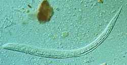
250px Strongyloides stercoralis larva

A parasite is a creature that lives on or in its host and feeds on or at the expense of that host. There is a detailed description of human parasites examples provided .

Human parasites are classified as endoparasites, which infect the body internally, and ectoparasites, which infect the skin externally.Humans can be infected by a number of parasitic worms. Flatworms, roundworms, and worms with thorny heads are among them (spiny-headed worm.

  1. Human whipworm
  2. Taenia saginata
  3. Threadworm
  4. Entamoeba histolytica
  5. Necator americanus
  6. Schistosoma mansoni
  7. Dracunculus medinensis
  8. Human pinworm
  9. Ancylostoma duodenale
  10. Fasciola hepatica
  11. Schistosoma japonicum
  12. Schistosoma haematobium
  13. Toxoplasma gondii
  14. Paragonimus westermani
  15. Entamoeba coli
  16. Cryptosporidium parvum
  17. Malaria parasite P. vivax
  18. Toxocara canis
  19. Crab louse
  20. Plasmodium malariae
  21. Plasmodium falciparum
  22. Trypanosoma brucei
  23. Plasmodium knowlesi
  24. Ascaris lumbricoides

Human whipworm

Whipworm is a parasitic roundworm that infects the human large intestine and causes trichuriasis.

Human parasites examples
Human whipworm from Wikipedia

It is frequently referred to as the whipworm because of its shape, which resembles a whip with broader “handles” at the back end.

Taenia saginata

Taenia saginata, sometimes known as the beef tapeworm, is a zoonotic tapeworm belongs to the genus Taenia and the order Cyclophyllidea.

Human parasites examples
Taenia saginata from Wikipedia

In humans, it causes taeniasis and cysticercosis, while in cattle, it causes taeniasis and cysticercosis.

Threadworm

Strongyloides stercoralis is a parasitic roundworm that causes strongyloidiasis in humans.

Human parasites examples
Threadworm from Wikipedia

Threadworm is its common name in the United States.

Entamoeba histolytica

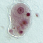
Entamoeba histolytica is a parasitic anaerobic amoebozoan belonging to the Entamoeba genus. E. histolytica, which causes amoebiasis in humans and other primates, is thought to infect about 35-50 million people worldwide.

800px Entamoeba histolytica
Entamoeba histolytica from Wikipedia

More than 55,000 people are estimated to die each year from E. histolytica infection.

Necator americanus

The New World hookworm, Necator americanus, is a species of hookworm. It belongs to the phylum Nematoda, which also includes other hookworms.

It’s a parasitic nematode that resides in human hosts’ small intestines.

Schistosoma mansoni

Schistosoma mansoni is a water-borne parasite of humans, and belongs to the group of blood flukes.

Schistosoma mansoni2
Schistosoma mansoni from Wikipedia

The adult lives in the blood vessels near the human intestine. It causes intestinal schistosomiasis. Clinical symptoms are caused by the eggs.

Dracunculus medinensis

Dracunculus medinensis is a parasitic nematode that causes dracunculiasis (guinea worm illness).

The disease is caused by the female, which may grow to be up to 80 cm long and is one of the longest nematodes that can infect humans. The male Guinea worm with the longest recorded length is only 4 cm.

Read more about Archaea Examples

Human pinworm

The parasitic nematode (roundworm) Because of the female’s long, pointed tail, Enterobius vermicularis is commonly referred to as the human pinworm. The popular names “seatworm” and “threadworm” are used in various locations .

Ancylostoma duodenale

Ancylostoma duodenale is a roundworm species belonging to the Ancylostoma genus. It’s a parasitic nematode worm that goes by the name of Old World hookworm. It reproduces and matures in the small intestines of humans, cats, and dogs.

Fasciola hepatica

Fasciola hepatica is a parasitic trematode of the class Trematoda, phylum Platyhelminthes, popularly known as the common liver fluke or sheep liver fluke. It infects the livers of a variety of mammals, including humans, and is spread over the world by sheep and cattle.

Schistosoma japonicum

Schistosoma japonicum is a significant parasite and one of the main schistosomiasis infecting agents. Because this parasite infects at least 31 species of wild animals, including 9 carnivores, 16 rodents, one primate (human), two insectivores, and three artiodactyls, it is classified as a real zoonosis.

Schistosoma haematobium

A urinary blood fluke, Schistosoma haematobium, is a digenetic trematode belonging to the Schistosoma (blood fluke) genus. Africa and the Middle East are home to this species. Schistosomiasis, the most common parasite infection in humans, is caused by it.

Toxoplasma gondii

Toxoplasmosis is caused by the parasitic protozoan Toxoplasma gondii, which is an obligate intracellular parasitic protozoan. T. gondii can infect almost any warm-blooded animal, but felids, such as domestic cats, are the only definitive hosts where the parasite can reproduce sexually.

Paragonimus westermani

Paragonimus westermani is the most common lung fluke that causes paragonimiasis in humans. The most prevalent human infections occur in eastern Asia and South America.

Entamoeba coli

Entamoeba coli is a non-pathogenic Entamoeba species that lives in the human gastrointestinal tract as a commensal parasite. E. coli is important in medicine because it can be mistaken for the pathogenic Entamoeba histolytica during microscopic examination of stained stool specimens.

Cryptosporidium parvum

Cryptosporidiosis is a parasite disease of the mammalian digestive tract caused by Cryptosporidium parvum, one of several species. Parvum Cryptosporidium C. parvum oocysts in immunofluorescence. Classification in science.

Malaria parasite P. vivax

Plasmodium vivax is a human disease and protozoal parasite. The parasite that causes recurrent malaria is the most common and widespread.

Toxocara canis

Toxocara canis is a parasitic helminth parasite that affects dogs and other canids all over the world. The term comes from the Greek words “toxon,” which means “bow or quiver,” and “caro,” which means “flesh.” They dwell in the final host’s small intestine.

Plasmodium malariae

The parasitic protozoan Plasmodium malariae causes malaria in humans. It’s one of numerous Plasmodium parasite species that infect other creatures as pathogens, including Plasmodium falciparum and Plasmodium vivax, which are responsible for the majority of malaria infections.

Crab louse

The crab louse, sometimes known as the pubic louse, is a parasitic insect that feeds solely on human blood. The crab louse lives in the pubic hair of most people.

Plasmodium falciparum

Plasmodium falciparum is a human unicellular protozoan parasite and the deadliest Plasmodium species that causes malaria. The parasite is spread by a female Anopheles mosquito bite and produces falciparum malaria, the most deadly form of malaria.

Trypanosoma brucei

The parasitic kinetoplastid Trypanosoma brucei belongs to the genus Trypanosoma. Tsetse fly species in Sub-Saharan Africa carry this parasite, which causes vector-borne illnesses in vertebrate animals, including humans. T. brucei causes African trypanosomiasis, also known as sleeping sickness, in humans.

Plasmodium knowlesi

The parasite Plasmodium knowlesi causes malaria in humans and other primates. It can be found all over Southeast Asia, and in Malaysia, it is the most common cause of human malaria. P. knowlesi, like other Plasmodium species, has a life cycle that necessitates the infection of a mosquito and a warm-blooded host.

Ascaris lumbricoides

Ascaris lumbricoides is a human “big roundworm” that can grow up to 35 cm in length. It is one of several Ascaris species. It is the most prevalent parasitic worm in humans and is an ascarid nematode of the phylum Nematoda.

Also Read: