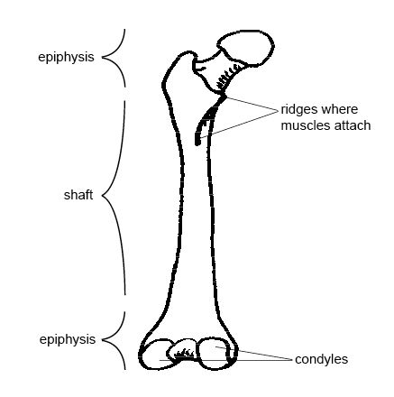
The femur, also known as the thigh bone, is the longest and strongest bone in the human body. It is located in the upper leg, connecting the hip to the knee joint. The femur consists of several important anatomical features, including the head, neck, shaft, and condyles. The head of the femur fits into the acetabulum of the pelvis, forming the hip joint, while the condyles articulate with the tibia to form the knee joint. The femur plays a crucial role in supporting body weight and facilitating movement. Understanding the anatomy of the femur is essential for medical professionals and individuals interested in learning about the skeletal system.
Key Takeaways
| Anatomical Feature | Description |
|---|---|
| Head | Rounded end of the femur that fits into the hip socket |
| Neck | Narrow section below the head |
| Shaft | Long, cylindrical portion of the femur |
| Condyles | Rounded projections at the bottom of the femur that articulate with the tibia |
| Hip Joint | Formed by the articulation of the femur and the pelvis |
| Knee Joint | Formed by the articulation of the femur and the tibia |
Understanding the Basics of Femur Anatomy

Definition of Femur Anatomy
The femur, also known as the thigh bone, is the longest and strongest bone in the human skeletal system. It is located in the lower limb and plays a crucial role in supporting the body’s weight and facilitating movement. The femur is a vital component of the hip joint and connects the pelvis to the knee joint.
The Femur: The Longest Bone in the Body
The femur extends from the hip joint to the knee joint and consists of several distinct parts. These include the femoral neck, femoral shaft, distal femur, proximal femur, femur head, and femoral condyles. The femoral neck is a narrow section that connects the femur head to the femoral shaft. The femoral shaft is the long, cylindrical portion of the bone that provides strength and stability. At the distal end of the femur, we find the femoral condyles, which are rounded projections that articulate with the tibia, forming the knee joint.
Location of the Femur in the Human Body
The femur is located in the upper leg, between the hip and the knee. It is positioned medially, or towards the midline of the body, and runs parallel to the tibia, the other major bone in the leg. The femur is angled slightly inward, which helps to distribute the body’s weight evenly and maintain balance. Its unique shape and structure allow for efficient movement and provide attachment points for various muscles.
The femur is a complex bone with a rich blood supply, supplied by the femoral artery, and innervation from the femoral nerve. It also contains bone marrow, which is responsible for producing new blood cells. The femur’s structure includes prominent features such as the femoral trochanters, which are bony projections that serve as attachment sites for muscles. The femur’s bone density is crucial for overall bone health and plays a role in preventing fractures.
To better understand the anatomy of the femur, it is helpful to refer to a femur diagram. This visual representation provides a clear depiction of the bone’s different parts and their relationships. By studying the femur’s anatomy, healthcare professionals can diagnose and treat various conditions, such as femur fractures or abnormalities in the femoral head.
In conclusion, the femur is a remarkable bone that serves as a vital component of the human skeletal system. Its unique structure and location enable us to perform essential movements and bear weight. Understanding the basics of femur anatomy is crucial for healthcare professionals and individuals interested in learning more about the human body’s intricate design.
Detailed Structure of the Femur
Proximal and Distal Femur: An Overview
The femur, also known as the thigh bone, is the longest and strongest bone in the human skeletal system. It plays a crucial role in supporting the weight of the body and facilitating movement in the lower limb. The femur is divided into two main parts: the proximal femur and the distal femur.
The proximal femur refers to the upper part of the bone, which connects to the hip joint. It consists of the femoral head, femoral neck, and greater and lesser trochanters. The femoral head is the rounded ball-like structure that articulates with the acetabulum of the pelvis, forming the hip joint. The femoral neck is a narrow section that connects the femoral head to the femoral shaft. The greater and lesser trochanters are bony projections that serve as attachment sites for various muscles.
On the other hand, the distal femur refers to the lower part of the bone, which connects to the knee joint. It consists of the femoral condyles, which are the rounded surfaces at the end of the bone that articulate with the tibia and patella. The distal femur also has a posterior surface called the popliteal surface, which is a smooth area that provides attachment for ligaments and muscles.
Femur Anatomy: Bones and Muscles Attachments
The femur bone is a complex structure that provides attachment points for several important muscles in the thigh and hip region. The muscles attached to the femur play a crucial role in various movements, such as walking, running, and jumping.
The medial and lateral condyles of the femur are important bony landmarks that provide stability to the knee joint. They are connected by the intercondylar fossa, a depression that accommodates the cruciate ligaments of the knee. The linea aspera is a ridge-like structure on the posterior surface of the femur that serves as an attachment site for muscles.
The femur also has a bony prominence called the greater trochanter, which is located on the lateral side of the proximal femur. It serves as an attachment site for the gluteus medius and minimus muscles. The lesser trochanter, located on the medial side of the proximal femur, provides attachment for the iliopsoas muscle.
Femur Anatomy: Diaphysis and Radiology Insights
The diaphysis of the femur refers to the long, cylindrical shaft of the bone. It is the main weight-bearing part of the femur and is responsible for transmitting forces from the hip to the knee joint. The diaphysis is composed of compact bone, which provides strength and support.
Radiologically, the femur can be visualized using various imaging techniques such as X-rays, CT scans, and MRI scans. These imaging modalities can help in diagnosing and evaluating conditions such as femur fractures, bone tumors, and bone density abnormalities. They can also provide insights into the structure and health of the femur bone, including the femoral head, femoral neck, and femoral shaft.
In summary, the detailed structure of the femur encompasses the proximal and distal regions, as well as the bones and muscles attachments. Understanding the anatomy of the femur is essential for comprehending its function, diagnosing injuries or conditions, and ensuring optimal bone health.
The Function and Importance of the Femur
How Does the Femur Work: A Look at its Function
The femur, also known as the thigh bone, is the longest and strongest bone in the human skeletal system. It plays a crucial role in supporting the body and facilitating movement. Let’s take a closer look at how the femur works and its function within the body.
The femur bone structure consists of several key components, including the femoral neck, femoral shaft, distal femur, proximal femur, and femur head. These different parts work together to provide stability and mobility to the hip joint and the entire lower limb.
One of the primary functions of the femur is to support the weight of the body. It acts as a pillar, transferring the forces generated during activities such as walking, running, and jumping. The femur’s length and shape are optimized to withstand these forces and distribute them evenly, reducing the risk of fractures or injuries.
The femur also plays a crucial role in muscle attachment. Various muscles, including the quadriceps and hamstrings, attach to different parts of the femur. These muscle attachments allow for movement and provide the necessary strength for activities that involve the lower limb.
Why is the Femur Important: Role in Body Support and Movement
The femur is of utmost importance in the human body due to its role in body support and movement. Without a properly functioning femur, our ability to walk, run, and perform daily activities would be severely compromised.
The femur’s strength and durability enable it to bear the weight of the body and provide stability to the hip joint. This stability is crucial for maintaining balance and preventing falls or injuries. Additionally, the femur’s length and shape contribute to the overall alignment of the lower limb, ensuring proper posture and gait.
Furthermore, the femur is essential for the transmission of forces generated by the muscles during movement. As muscles contract and relax, they exert forces on the femur, allowing for coordinated and controlled movement of the lower limb. This coordination is vital for activities that require precision and agility, such as dancing or playing sports.
Does the Femur Protect Organs: Understanding its Protective Role
While the primary function of the femur is to support body weight and facilitate movement, it also plays a protective role for vital organs. Although not directly involved in protecting organs, the femur indirectly contributes to their safety.
The femur’s location in the thigh provides a layer of protection for the femoral artery and femoral nerve, which are crucial for blood supply and nerve function in the lower limb. In addition, the femur’s strong structure acts as a shield for the bone marrow, which is responsible for producing red and white blood cells.
Moreover, the femur’s distal end forms the femoral condyles, which articulate with the tibia to form the knee joint. This joint is responsible for bearing weight and facilitating movement, protecting the delicate structures within the knee, such as ligaments and cartilage.
In summary, the femur’s function and importance in the human body cannot be overstated. It provides support, facilitates movement, and indirectly protects vital organs. Understanding the role of the femur helps us appreciate the intricate design of our skeletal system and the remarkable capabilities of the human body.
Growth and Development of the Femur
The femur, also known as the thigh bone, is the longest and strongest bone in the human skeletal system. It plays a crucial role in supporting the weight of the body and facilitating movement in the lower limb. Let’s explore the growth and development of the femur in more detail.
When Does the Femur Growth Plate Close: A Look at Bone Development
Bone development is a complex process that begins during fetal development and continues throughout childhood and adolescence. The growth plates, also known as epiphyseal plates, are responsible for the longitudinal growth of bones, including the femur. These growth plates are made up of cartilage and are located near the ends of long bones.
In the case of the femur, the growth plate is situated at the proximal end, near the hip joint. As a child grows, the growth plate gradually closes, signaling the end of longitudinal bone growth. The closure of the femur growth plate typically occurs between the ages of 14 and 18 in boys, and between 12 and 16 in girls. However, it’s important to note that the timing can vary from person to person.
Does Femur Length Determine Height: Exploring the Connection
Femur length is often used as an indicator of overall height. The length of the femur is influenced by genetic factors and can vary among individuals. While there is a correlation between femur length and height, it is not the sole determining factor. Other factors, such as the length of the tibia (shinbone) and the spine, also contribute to a person’s height.
It’s worth mentioning that variations in femur length can occur due to growth abnormalities or conditions such as dwarfism or gigantism. These conditions can affect the overall growth and development of the femur, leading to deviations from the average femur length for a given height.
When is Femur Length a Concern: Understanding Growth Abnormalities
In some cases, an abnormal femur length may raise concerns about a person’s growth and development. If a child‘s femur length is significantly shorter or longer than expected for their age, it may indicate a growth abnormality or underlying medical condition.
Short femur length can be associated with conditions like achondroplasia, a form of dwarfism, or skeletal dysplasia. On the other hand, excessively long femur length may be a sign of conditions like Marfan syndrome or gigantism.
If there are concerns about femur length or growth abnormalities, it is important to consult with a healthcare professional who can evaluate the individual’s overall growth and development, considering various factors such as family history, medical history, and physical examination.
In conclusion, the growth and development of the femur is a dynamic process that involves the closure of the growth plate and the influence of various factors on femur length. Understanding these aspects can provide valuable insights into an individual‘s growth and overall bone health.
The Femur in Different Species


Femur Anatomy in Dogs: A Comparative Look
The femur, also known as the thigh bone, is a crucial component of the skeletal system in various species, including dogs. It plays a vital role in providing support, stability, and mobility to the lower limbs. Let’s take a comparative look at the femur anatomy in dogs and explore its unique features.
In dogs, the femur bone structure is similar to that of humans and other mammals. It consists of several key components, including the femoral neck, femoral shaft, distal femur, proximal femur, femur head, and femoral condyles. These structures work together to facilitate movement and bear weight during various activities.
One notable difference in the femur anatomy of dogs is the presence of femoral trochanters. These bony projections serve as attachment points for muscles, allowing for efficient movement and agility. Additionally, the femur bone in dogs is slightly curved, which helps distribute forces evenly and enhances stability.
The femur length in dogs can vary depending on the breed and size of the dog. Larger breeds tend to have longer femurs to support their body weight, while smaller breeds have relatively shorter femurs. This variation in femur length is essential for maintaining balance and optimal movement.
Femur fractures can occur in dogs, just like in humans. These fractures can be caused by trauma, excessive strain, or underlying bone conditions. In severe cases, femur fractures may result in damage to surrounding structures such as the femoral artery and femoral nerve, leading to further complications.
Is the Femur Bone Good for Dogs: Understanding Canine Bone Health
The femur bone plays a crucial role in the overall health and well-being of dogs. It provides structural support, facilitates movement, and serves as an attachment site for muscles. Maintaining good femur bone health is essential for ensuring the overall mobility and quality of life for our canine companions.
Proper nutrition is vital for maintaining optimal femur bone density in dogs. A balanced diet rich in essential nutrients, including calcium, phosphorus, and vitamin D, helps promote healthy bone development and strength. Regular exercise and physical activity also play a significant role in maintaining femur bone health by stimulating bone remodeling and strengthening the surrounding muscles.
Regular veterinary check-ups are essential for monitoring the condition of a dog’s femur bone. X-rays and other diagnostic tests can help identify any potential issues, such as fractures, bone abnormalities, or signs of osteoporosis. Early detection and intervention can prevent further complications and ensure appropriate treatment.
In conclusion, understanding the femur anatomy in dogs and the importance of maintaining good femur bone health is crucial for ensuring the overall well-being and mobility of our furry friends. By providing proper nutrition, regular exercise, and veterinary care, we can help support their skeletal system and ensure they lead happy and active lives.
What is the Role of the Abdomen in Supporting the Femur?
The abdomen anatomy: core structure explained plays a crucial role in supporting the femur. The muscles of the abdomen, including the hip flexors, help stabilize the pelvis. This stability allows the femur, the thigh bone, to move and function efficiently during various activities such as walking, running, or even sitting. Proper functioning of the core muscles in the abdomen is essential for maintaining balance and preventing injuries to the hips and thighs.
Common Issues and Conditions Related to the Femur
The femur, also known as the thigh bone, is the longest and strongest bone in the human skeletal system. It plays a crucial role in supporting the weight of the body and facilitating movement in the lower limb. However, like any other bone, the femur is susceptible to various issues and conditions that can affect its structure and function.
Is Femur Bone Break: Understanding Fractures and their Causes
One of the most common issues related to the femur is a fracture, which occurs when the bone breaks due to excessive force or trauma. Femur fractures can be caused by various factors, including:
Trauma: High-impact accidents, such as car crashes or falls from a significant height, can result in femur fractures. The force exerted on the bone exceeds its strength, causing it to break.
Osteoporosis: Weakening of the bones due to osteoporosis can make the femur more prone to fractures, even with minimal trauma. Osteoporosis is a condition characterized by low bone density and increased risk of fractures.
Overuse or Stress: Repetitive stress on the femur, often seen in athletes or individuals who engage in high-impact activities, can lead to stress fractures. These fractures occur when the bone is unable to withstand the repetitive load placed upon it.
Femur fractures can vary in severity, ranging from hairline fractures to complete breaks. Treatment options for femur fractures depend on the location and severity of the fracture and may include immobilization, casting, or surgical intervention.
Why Femur Bone Pain: Common Causes and Symptoms
Femur bone pain can be caused by various conditions and injuries. Some common causes of femur bone pain include:
Osteoarthritis: Degenerative joint disease, such as osteoarthritis, can affect the hip joint where the femur connects. This can cause pain and discomfort in the femur region.
Muscle Strains: Straining or overexerting the muscles surrounding the femur can lead to pain in the thigh area. This can occur due to sudden movements, excessive exercise, or improper form during physical activities.
Bursitis: Inflammation of the bursae, small fluid-filled sacs that cushion the joints, can cause pain in the hip joint and radiate to the femur. Bursitis can be caused by repetitive motions or direct trauma to the hip area.
Femoral Stress Syndrome: Also known as “runner’s knee,” femoral stress syndrome is a condition characterized by pain in the front of the knee and along the femur. It is commonly seen in athletes who engage in activities that involve repetitive knee bending.
Symptoms of femur bone pain may include localized pain, swelling, tenderness, difficulty walking or bearing weight, and limited range of motion. It is essential to consult a healthcare professional for an accurate diagnosis and appropriate treatment based on the underlying cause of the pain.
In conclusion, the femur is a vital bone in the human body, and issues related to it can significantly impact mobility and overall well-being. Understanding the causes and symptoms of femur fractures and bone pain can help individuals seek timely medical attention and appropriate treatment for a faster recovery.
What is the relationship between femur anatomy and ankle anatomy?
The relationship between femur anatomy and ankle anatomy is essential for understanding the overall structure and function of the lower limb. The femur is the largest and strongest bone in the human body, while the ankle joint connects the foot to the leg. The femur provides support and stability to the body, transmitting forces from the upper body to the ankle joint. On the other hand, the ankle joint, comprising various bones and ligaments, allows for a range of movements, including flexion, extension, inversion, and eversion. To learn more about ankle anatomy and functions, “Explore ankle anatomy and functions”.
Conclusion
In conclusion, the femur is the longest and strongest bone in the human body. It plays a crucial role in supporting our weight and facilitating movement. The femur consists of various anatomical structures, including the head, neck, shaft, and condyles. These structures work together to provide stability and mobility to the hip and knee joints. Understanding the anatomy of the femur is essential for medical professionals, athletes, and individuals interested in learning about the human body. By appreciating the complexity and importance of the femur, we can better comprehend the remarkable design and functionality of our skeletal system.
Frequently Asked Questions
What is the role of the femur in the human skeletal system?
The femur, also known as the thigh bone, is the longest and strongest bone in the human body. It plays a vital role in supporting the body’s weight and facilitating movement, as it connects the hip joint to the knee.
How does the femur function?
The femur functions as a crucial component in the hip joint and the knee joint. It allows for a range of movements like walking, running, jumping, and squatting. Its length also contributes to the overall height of an individual.
What is the difference between distal and proximal femur?
The femur is divided into three parts: the proximal end, the shaft, and the distal end. The proximal end of the femur is closer to the body (the hip joint), featuring the femoral head and neck. The distal end is further from the body, closer to the knee, and consists of the femoral condyles and epiphysis.
Is the femur the longest bone in the human body?
Yes, the femur is the longest bone in the human body. It accounts for approximately one-quarter of a person’s total height.
Why is the femur important in the anatomy of dogs?
Just like in humans, the femur in dogs plays a crucial role in mobility. It allows for a range of motions and supports the dog‘s weight. Understanding its anatomy can help diagnose and treat conditions that affect a dog’s movement.
How can one identify the femur bone?
The femur bone can be identified by its location and structure. It is located in the upper part of the leg, extending from the hip to the knee. It is characterized by a rounded head that fits into the hip socket, a long shaft, and a distal end that connects to the knee.
When does the femur growth plate close?
The femur growth plate, also known as the epiphyseal plate, typically closes when an individual reaches the end of puberty – around 16-18 years for girls and 14-16 years for boys.
How does femur length influence height?
Femur length is a significant determinant of an individual‘s height. As the longest bone in the body, it accounts for approximately one-quarter of a person’s total height.
Are femur and femoral the same?
“Femur” refers to the bone itself, while “femoral” is an adjective used to describe anything related to the femur, such as the femoral artery, femoral nerve, or femoral neck.
How does a femur fracture affect the body?
A femur fracture can severely affect mobility, as the femur plays a crucial role in weight-bearing and movement. Depending on the location and severity of the fracture, it may also impact the function of the hip or knee joint. It’s a serious injury that requires immediate medical attention.
Also Read:
- Contractile vacuole in euglena
- Dna supercoiling vital mechanism for dna packaging
- Do chloroplasts have ribosomes
- Intracellular enzyme examples
- Do animal cells have chloroplasts
- Calvin cycle vs krebs cycle
- Cell wall and vacuoles
- Carbohydrates structure examples
- Lobster characteristics
- Hydrophobic tail

The TechieScience Core SME Team is a group of experienced subject matter experts from diverse scientific and technical fields including Physics, Chemistry, Technology,Electronics & Electrical Engineering, Automotive, Mechanical Engineering. Our team collaborates to create high-quality, well-researched articles on a wide range of science and technology topics for the TechieScience.com website.
All Our Senior SME are having more than 7 Years of experience in the respective fields . They are either Working Industry Professionals or assocaited With different Universities. Refer Our Authors Page to get to know About our Core SMEs.
